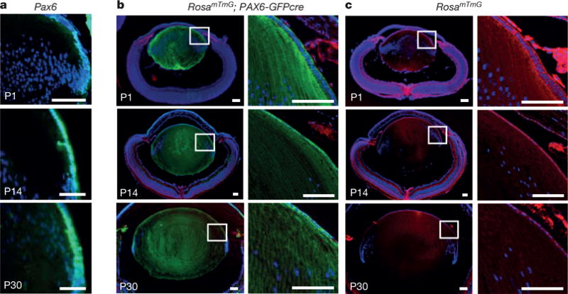
Figure 1. Lineage tracing of Pax6+ LECs in mice.

a, Pax6-directed GFP was expressed in mouse LEC nuclei at post-natal days P1, P14 and P30; a sagittal section of a P0-3.9-GFPcre mouse lens is shown. Blue and green represent DAPI and GFP, respectively. b, Lineage tracing of Pax6+ LECs in ROSAmTmG; P0-3.9-GFPcre mice at P1, P14 and P30 reveals that lens fibre cells express membrane GFP fluorescence; hence, PAX6+ LECs were able to generate lens fibre cells. c, As an additional control, the ROSAmTmG allele alone exhibits Tomato (red) staining at sites of non-recombination. All scale bars, 100 μm.
