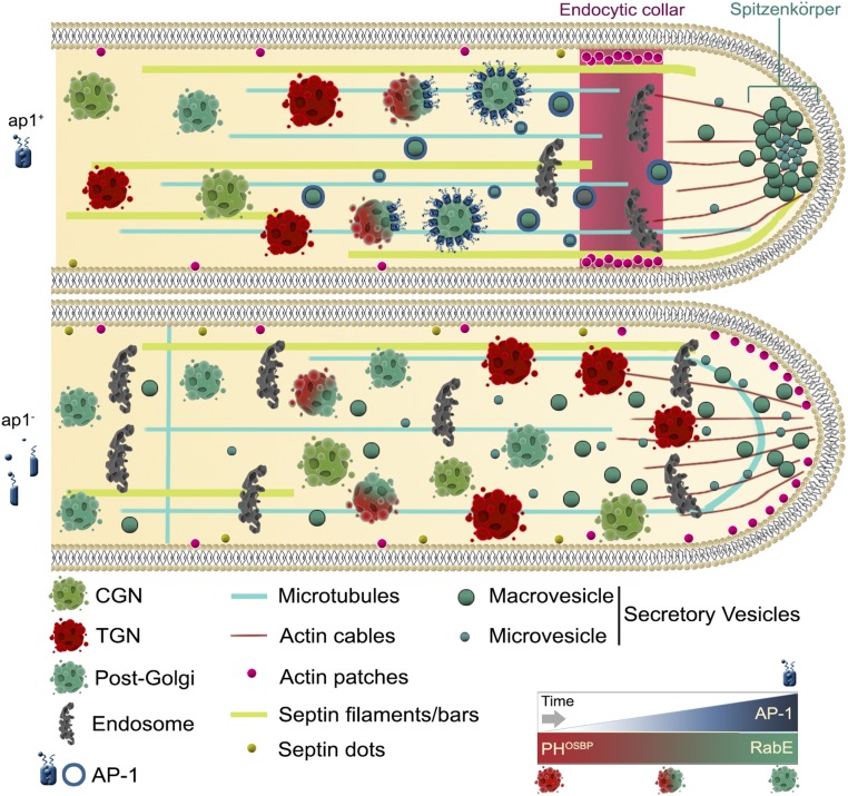
Figure 8.
Highly speculative scheme on the role of AP-1 in A. nidulans hyphal tip growth based on the herein described microscopic observation of fluorescent tagged protein markers. In an ap1+ background the late-Golgi progressively turns into post-Golgi RabE-containing SVs coated by AP-1 (also depicted in the lower right panel). In the absence of a functional AP-1 complex (ap1−), apparent accumulation of Golgi toward the fungal apex, mislocalization of endocytic collar associated actin patches toward the tip, failure of accumulation of SVs at the level of Spk, septin, and MT disorganization, and an overall enrichment in “sorting” endosomal and/or vacuolar structures are observed.

