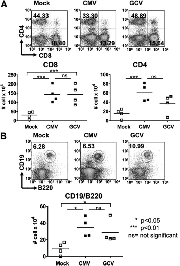
FIGURE 3.

Adaptive inflammatory infiltrates are induced in murine cytomegalovirus (MCMV)-infected allo-grafts but are not affected by ganciclovir (GCV) treatment. (A) Cells from uninfected (Mock, open squares), CMV-infected (CMV, closed squares), and CMV-infected, GCV-treated (GCV, hatched squares) allografts were stained for CD45+/CD8+ and CD45+/CD4+ surface markers, evaluated by flow cytometry and shown as cells per organ. CMV grafts showed induction of CD8+ and CD4+ infiltrates (P<0.01) compared with Mock grafts. No difference was observed in these T lymphocyte subsets between the CMV-infected allografts with and without GCV treatment (P>0.05, ns). (B) Cells from allografts of Mock, CMV, and GCV animals were stained for CD19+/B220+ markers and evaluated by flow cytometry. CMV-infected grafts showed greater CD19+/B220+ lymphocyte infiltrates (P<0.05) compared with Mock grafts, which was sustained in the GCV grafts compared with CMV grafts (P>0.05, ns) and remained elevated in the GCV grafts compared with the Mock grafts (P<0.05).
