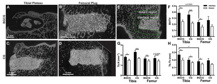
Figure 2.
Assessment of CG/nHA-PEUR and BGCG/nHA-PEUR implants by μCT at 16 weeks. (A – D) Representative μCT images of grafts. (A) BGCG/nHA-PEUR tibial plateau defect. (B) BGCG/nHA-PEUR femoral condyle plug defect. (C) CG/nHA-PEUR tibia plateau defect. (D) CG/nHA-PEUR femoral condyle plug defect. (E – H) μCT analysis of morphometric parameters of host bone outside the graft. (E) Representative μCT image showing the contours of the interface and distant regions used for analysis. The morphometric parameters (F) bone volume fraction (BV/TV), (G) trabecular number (Tb.N.), and (H) trabecular thickness (Tb.Th.) were calculated for all samples both the interface and distant region.

