Abstract
Purpose
In mice, retinal development continues throughout the postnatal stage accompanied by the proliferation of retinal precursor cells. Previous reports showed that during the postnatal stage microglia increase from postnatal day 0 (P0) to P7. However, how microglia are associated with retinal development remains unknown.
Methods
The involvement of microglia in retinal development was investigated by two approaches, microglial activation and loss, using lipopolysaccharide (LPS) and PLX3397 (pexidartinib), respectively.
Results
LPS injection at 1 mg/kg, intraperitoneally (i.p.) in the neonatal mice increased the number of retinal microglia at P7. 5-Bromo-2´-deoxyuridine (BrdU)-positive proliferative cells were increased by LPS treatment compared to the control group. The proliferative cells were mainly colocalized with paired box 6 (Pax6), a marker of retinal precursor cells. However, the depletion of microglia by treatment with PLX3397 decreased the BrdU-positive proliferative cells. Moreover, progranulin deficiency decreased the number of microglia and retinal precursor cells.
Conclusions
These findings indicated that microglia regulate the proliferation of immature retinal cells.
Introduction
Retinal development is initiated during the embryonic stage and progresses throughout the postnatal stage. Retinal development is essential for normal formation of the eye. Aniridia is a developmental disorder in which the loss of the iris and foveal hypoplasia are observed [1]. Aniridia is caused by haploinsufficiency of paired box protein 6 (Pax6; Gene ID: 5080, OMIM: 607108) which is associated with multipotency of retinal precursor cells [2,3]. During postnatal development, retinal precursor cells continue to proliferate although some differentiate into mature neurons. In the central retina, proliferation is limited by postnatal day 6 (P6) [4]. However, proliferative cells are observed at even P6 in the peripheral area [4]. Postnatal proliferation is thought to be essential for mature retinal cells that develop in a later stage. Rod photoreceptor cells, Müller glia, bipolar cells, and some amacrine cells show later development than other retinal cells [5].
A large number of microglia are observed during retinal development. Microglia have been reported to play a role in the formation of retinal blood vessels [6]. This report implies microglia are associated with angiogenesis. However, microglia are also present in the embryonic retina which is a retinal avascular stage [7,8]. Therefore, microglia are not only essential for angiogenesis during retinal development. Microglia are rich in the embryonic retina, but a subtle decrease occurs from the embryonic stage to P0 [9]. After P0, they are reactivated and migrate into the retina between P0 and P14. However, how repopulation of microglia works during the postnatal stage remains unclear.
Recently, it was suggested that microglia are associated with neurogenesis in the brain. It has been thought to be due to microglia-derived growth factors, such as insulin-like growth factor 1 (IGF-1) [10]. There are two types of microglia: proinflammatory (M1) and anti-inflammatory (M2). M2 microglia secrete various growth factors. It has been reported that systemic administration of lipopolysaccharide (LPS) to neonatal mice induces M2-like microglia which express transforming growth factor-β (TGF-β) in the subventricular zone (SVZ) and promote neurogenesis [11,12]. However, a recent report showed that neurogenesis is induced by microglia-secreted inflammatory cytokines rather than IGF-1 [13]. How microglia-derived factors contribute to neurogenesis remains controversial. A recent report showed that in zebrafish the delayed migration of macrophages into the retina results in microphthalmia [14].
Progranulin (Gene ID: 2896, OMIM: 138945) is highly expressed in microglia in the central nervous system. Our previous findings revealed that progranulin was increased at P9 from P1 in the retina, and progranulin deficiency caused abnormal retinal development [15]. In particular, photoreceptor differentiation, retinal ganglion cells (RGCs), and astrocytes are defective. Interestingly, the day of high progranulin expression matches the day of high microglial migration into the retina [9]. Another report showed that progranulin promoted microglial migration, suggesting that progranulin is a chemoattractant for microglia [16]. Recent reports showed progranulin could be associated with synaptic pruning and phagocytosis [17,18]. Various functions and states of microglia are regulated by progranulin. Taken together, progranulin may be associated with microglial migration or functions during retinal development, as well as RGCs, photoreceptor cells, and astrocytes.
However, it remains unclear how microglia are associated with retinal development in mice. In this study, we investigated how postnatal microglia play a role in retinal development in mice and the association with progranulin.
Methods
Animals
Maternal C57BL/6 mice (Japan SLC, Hamamatsu, Japan) and neonatal mice were maintained under a controlled lighting environment (12 h:12 h light-dark cycle). Neonatal mice were collected from the same litter. Grn−/− mice generated by Kayasuga et al. [19] were obtained from Riken BioResource Center (Tsukuba, Japan) and were backcrossed with C57BL/6J mice (Charles River Japan, Yokohama, Japan). Genotyping was performed according to the previous protocol [19]. Briefly, amplification was performed using a DNA thermal cycler (Takara Bio, Shiga, Japan) for 30 cycles. A cycle profile consisted of 30 s at 94 °C for denaturation, 30 s at 60 °C for annealing and 60 s at 72 °C for primer extension. All experiments were performed in accordance with the ARVO Statement for the Use of Animals in Ophthalmic and Vision Research, and the procedures were approved and monitored by the Institutional Animal Care and Use Committee of Gifu Pharmaceutical University and were performed after approval from the Bioethics and Biosafety Committee of Gifu Pharmaceutical University.
Immunostaining
The enucleated eyes were fixed in 4% paraformaldehyde for 24 h at 4 °C. The eyes were then cryoprotected in 25% sucrose for 24 h at 4 °C and embedded in optimal cutting temperature compound (Sakura Finetechnical Co., Ltd., Tokyo, Japan). The eyes were cut in transverse cryostat sections of 10 μm thickness and placed on glass slides (MAS COAT; Matsunami Glass Ind., Ltd., Osaka, Japan). Immunostaining was conducted in accordance with the methods described in detail [20]. Briefly, the sections were blocked with non-immune serum and incubated overnight with the primary antibody at 4 °C. The mouse-on-mouse (M.O.M.) immunodetection kit (Vector Labs, Burlingame, CA) was used for blocking and solvents. After overnight incubation with the primary antibody, the sections were incubated with the secondary antibody for 1 h. They were then counterstained and mounted.
For 5-bromo-2′-deoxyuridine (BrdU) staining, the retinal sections were pretreated for 30 min with 2 M hydrochloric acid (HCl) 2 M for 30 min. Then they were incubated with 0.3% Triton X-100 (Bio-Rad Labs, Hercules, CA) for 30 min. They were then treated with 0.1% trypsin (Wako Pure Chemical Industries, Ltd., Osaka, Japan) at 37 °C for 7 min.
For Pax6 staining, the retinal sections were pretreated with 0.3% Triton X-100 (Bio-Rad Labs) for 30 min. They were then treated with 0.1% trypsin (Wako Pure Chemical Industries, Ltd.) at 37 °C for 7 min. The following primary antibodies were used: mouse anti-Pax6 (1:300 dilution; Abcam, Cambridge, MA), mouse anti-Chx10 (1:200 dilution; SantaCruz, Dallas, TX), rabbit anti-Iba-1 (1:50 dilution; Wako Pure Chemical Industries, Ltd.), rabbit anti-CRX (1:20 dilution; SantaCruz), rabbit anti-Sox2 (1:200 dilution; Millipore, Bedford, MA), rabbit anti-cleaved caspase-3 (1:100 dilution; Cell Signaling Technology, Danvers, MA), rat anti-BrdU (1:200 dilution; Abcam), sheep anti-progranulin (1:20 dilution; R&D Systems, Minneapolis, MN), mouse anti-CD206 (1:50 dilution; Abcam), Alexa Fluor® 488 goat anti-mouse immunoglobulin G (IgG), Alexa Fluor® 546 goat anti-rat IgG, Alexa Fluor® 546 donkey anti-rabbit IgG, and Alexa Fluor® 647 donkey anti-sheep IgG (Invitrogen, Carlsbad, CA).
Images were acquired with a confocal microscope (FLUOVIEW FV10i; Olympus, Tokyo, Japan). For quantitative data, photographs were analyzed at 500 µm and the peripheral area from the optic nerve head. The number of BrdU- and Pax6-positive cells was counted within the area of the image (211.968 × 211.968 µm). The number of Iba-1-positive cells and cleaved caspase-3-positive cells was counted within the whole retina. Three retinal sections were analyzed per one eye.
Western blotting
Western blotting was performed according to our methods described in detail [20]. Briefly, mice retinas were lysed using a buffer containing protease and phosphatase inhibitors. The tissue was homogenized, and the cell lysate was centrifuged. The supernatant was used for the subsequent experiments. The protein concentration was measured using a protein assay kit (Thermo Scientific, Waltham, MA). Samples were analyzed with sodium dodecyl sulfate-polyacrylamide gel electrophoresis (SDS–PAGE) using 5–20% gradient gels (Wako Pure Chemical Industries, Ltd.), and the proteins were transferred onto a membrane. After blocking for 30 min at room temperature, the membranes were washed and then incubated with the primary antibody overnight at 4 °C. The following primary antibodies were used: mouse anti-Pax6 (1:1,000 dilution; Abcam), mouse anti-rhodopsin (1:1,000 dilution; Millipore), mouse anti-GS (1:1,000 dilution; Millipore), mouse anti-calbindin (1:1,000 dilution; Abcam), mouse anti-nestin (1:200 dilution; BD Biosciences, San Jose, CA), rabbit anti-Iba-1 (1:200 dilution; Wako Pure Chemical Industries, Ltd.), rabbit anti-GAPDH (1:1,000 dilution; Cell Signaling Technology), sheep anti-progranulin (1:200 dilution; R&D Systems), and mouse anti-β-actin (1:2,000 dilution; Sigma-Aldrich, St Louis, MO). After exposure to the primary antibody, the membranes were incubated with peroxidase goat anti-rabbit, goat anti-mouse, or rabbit anti-sheep IgG (Thermo Scientific) as the secondary antibody. The immunoreactive bands were made visible with ImmunoStar LD (Wako Pure Chemical Industries, Ltd.). The band intensity was measured using a Luminescent image analyzer LAS-4000 UV mini (Fujifilm, Tokyo, Japan). The protein band intensities were quantified using MultiGauge software Ver. 3.0 (Fujifilm) and normalized to the level of β-actin and GAPDH (for progranulin only).
LPS treatment
LPS treatment was performed following a previous report in which LPS treatment (1 mg/kg) for neonatal mice intraperitoneally (i.p.) at P3 increased the number of microglia in the brain at P7 [12]. Briefly, LPS (1 mg/kg; Sigma-Aldrich) was treated i.p. at P3. LPS 1 mg/ml in PBS (1X; 136.9 mM NaCl, 2.68 mM KCl, 10.14 mM Na2HPO4, 1.76 mM KH2PO4, pH 7.3) was prepared as a stock solution and an injected solution. The control group was treated with an equal amount of PBS. The injection was performed with a Nanopass 34-gauge needle (Terumo Corporation, Tokyo, Japan). To evaluate the proliferative cells, BrdU (50 mg/kg; Sigma-Aldrich) was injected i.p. from P3 to P7 at the same time. BrdU 10 mg/ml in PBS was prepared at time of use. At P7, the mice were euthanized by decapitation, and the eyes were enucleated. The eyelids with neonatal mice were cut not to damage eyes. Then, eyes were pluck out after the removal of tissues surrounding eyes.
PLX3397 treatment
PLX3397 (pexidartinib; Selleck Chemicals, Houston, TX) was treated at a dose of 0.25 and 1 mg/kg (i.p., twice daily) to neonatal mice from P0 to P7. PLX3397 250 mg/ml in 100% dimethyl sulfoxide (DMSO) was prepared as the stock solution. The stock solution was diluted with PBS for the injected solution (0.25 mg/ml in PBS plus 0.1% DMSO). The control group was treated with an equal amount of 0.1% DMSO in PBS. BrdU was injected similarly as described above. At P7, the mice were euthanized, and the eyes were enucleated.
Statistical analysis
The data are presented as the means ± standard error of the means (SEMs). The significance of the differences was determined with a two-tailed Student t test or Dunnett’s test (SPSS version 24; IBM, Armonk, NY). A p value of less than 0.05 was considered statistically significant.
Results
Iba-1-positive microglial change in the postnatal retina
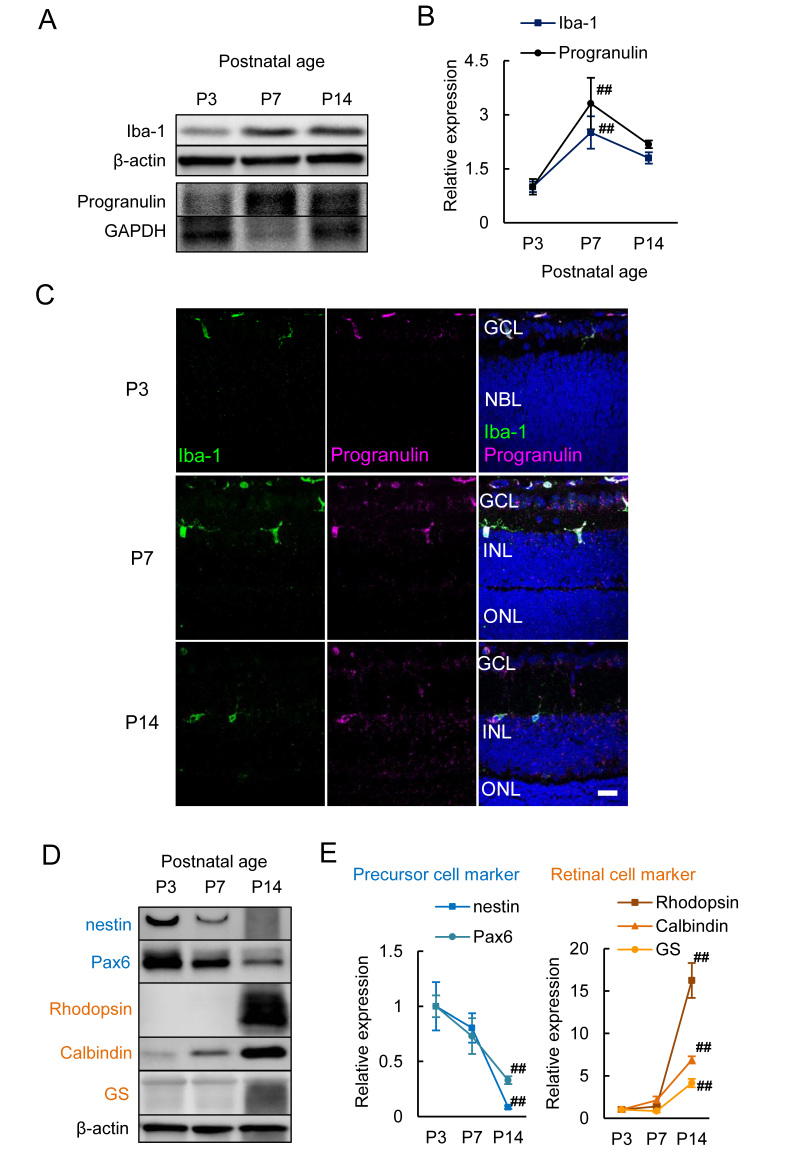
It has been reported that the number of microglia increase and peak at P7 [9]. To confirm this report, we investigated the expression of ionized calcium binding adaptor molecule 1 (Iba-1) as the marker of microglia in the context of retinal cell markers. Western blotting analysis showed that Iba-1 expression was increased to 250% at P7 compared with the expression at P3 (p=0.002, Figure 1A,B). The expression was decreased to 180% at P14 compared with the expression at P7 (Figure 1A,B). Immunostaining of Iba-1 also showed that the number of microglia peaked at P7 and localized at the inner retinal layer (Figure 1C). Thus, we confirmed that the expression of Iba-1 peaked at P7 during postnatal development. Moreover, we investigated the expression of retinal precursor cell markers and retinal cell markers to identify how the increase in microglia corresponded to retinal cell development. Retinal precursor cell markers (nestin and Pax6) were obviously decreased at P14 compared with P7 (nestin, p=0.003; Pax6, p=0.001; Figure 1D,E), and retinal cell markers (rhodopsin, calbindin, and glutamine synthetase [GS]) increased dramatically at P14 (rhodopsin, calbindin, and GS, p<0.0001, Figure 1D,E). These markers were expressed in photoreceptor cells, amacrine cells, and Müller glia, respectively [5,21]. These results indicated that microglia may contribute to any events from P3 to P7 that occur before postnatal differentiation of retinal cells. Progranulin is expressed in retinal astrocytes and microglia at the postnatal stage [15]. Progranulin expression showed a concerted increase that matched the expression of Iba-1, and microglia strongly expressed progranulin at P7 (p=0.001, Figure 1A,B).
Figure 1.
The expression of Iba-1 and progranulin in the postnatal retina. A, B: Western blotting results show the expression of Iba-1 and progranulin during postnatal development. Typical bands and quantitative data show the expression of Iba-1 and progranulin peaks at P7. C: Immunostaining of Iba-1 (green) and progranulin (magenta). Nuclei are stained with Hoechst 33342 (blue). Iba-1-positive microglia increase at P7 and express progranulin. D, E: Western blotting results show the expression of retinal precursor cell markers and retinal cell markers. Retinal precursor cell markers (nestin and Pax6) are decreased at P14 compared to P7. Retinal cell markers (rhodopsin, calbindin, and glutamine synthetase [GS]) are increased at P14. These markers indicate photoreceptor cells, amacrine cells, and Müller glia, respectively. Data are the mean ± standard error of the mean (SEM; n=4 to 6). ##p<0.01 versus P3 (Dunnett’s test). Scale bar=20 μm.
LPS treatment of neonatal mice increased the number of microglia and BrdU-positive proliferative cells
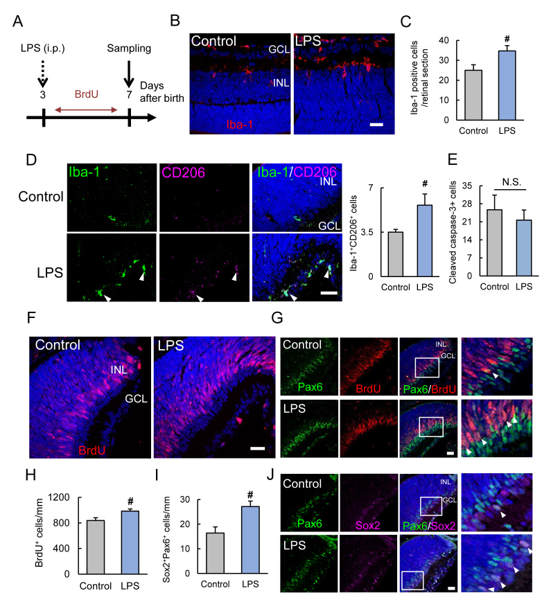
In light of the present findings indicating that microglia increased from P3 to P7, we hypothesized that microglia could promote the proliferation of retinal precursor cells continuing at the postnatal stage. First, we performed the treatment with LPS to investigate how the microglial increase contributed to the proliferation of retinal precursor cells. The treatment was performed based on the report in which systemic administration of LPS to neonatal mice promoted neurogenesis in the SVZ [12]. LPS at 1 mg/kg was intraperitoneally administered at P3, and BrdU was incorporated from P3 to P7 (Figure 2A). LPS treatment increased the number of microglia in the retina (p=0.036, Figure 2B,C). Some microglia expressed one of the M2 markers, CD206 [22], and LPS increased the number of CD206-positive microglia (p=0.047, Figure 2D). LPS treatment did not alter the number of cleaved caspase-3-positive cells (Figure 2E). LPS treatment increased the number of BrdU-positive proliferative cells in the peripheral area (p=0.035, Figure 2F,H). The increased BrdU-positive proliferative cells merged with a retinal precursor cell marker, Pax6 (Figure 2G). To clarify the increase in the retinal precursor cells by LPS treatment, Pax6 and Sox2 double-positive cells were analyzed [23]. LPS treatment increased the number of Pax6 and Sox2 double-positive cells (p=0.012, Figure 2I,J).
Figure 2.
Systemic LPS treatment increased the number of microglia and BrdU-positive proliferative cells. A: The lipopolysaccharide (LPS) treatment scheme. Intraperitoneal injection of LPS is performed at P3, and 5-bromo-2′-deoxyuridine (BrdU) is incorporated from P3 to P7. The retina was evaluated at P7. B, C: Immunostaining shows Iba-1-positive microglia (red). LPS treatment increases the number of microglia in the retina at P7. D: CD206, a marker of M2 microglia, is increased by LPS treatment. E: The number of cleaved caspase-3-positive cells is not changed by LPS treatment. F–H: Immunostaining shows BrdU-positive proliferative cells (red) in the peripheral retina. LPS treatment increases the number of BrdU-positive proliferative cells in the peripheral area. The increased BrdU-positive proliferative cells merge with the retinal precursor cell marker, Pax6. I, J: Pax6 (green) and Sox2 (magenta) double-positive cells are increased with LPS treatment. Data are the mean ± standard error of the mean (SEM; n=4 or 5). #; p<0.05 versus control (Student t test). Scale bar=20 μm.
PLX3397 treatment of neonatal mice decreased the number of microglia and BrdU-positive proliferative cells
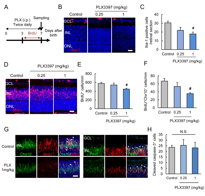
PLX3397, a colony-stimulating factor 1 receptor (CSF1R) inhibitor, is generally used for microglial depletion [24,25]. In this study, neonatal mice were treated with PLX3397 with an intraperitoneal injection (Figure 3A). The treatment with PLX3397 twice daily from P0 to P7 decreased the number of microglia (p=0.015, Figure 3B,C). PLX3397 also decreased the number of BrdU-positive proliferative cells in the retina at 500 μm from the optic nerve (p=0.021, Figure 3D,E). To determine that the proliferative cells were retinal precursor cells, we costained BrdU and the retinal precursor cell markers, Pax6 and Chx10, which is also known as visual system homeobox 2 (Vsx2) [26]. PLX3397 statistically significantly decreased the BrdU and Chx10 double-positive cells and tended to decrease BrdU and Pax6 double-positive cells (p=0.038, Figure 3F,G). PLX3397 did not alter the number of cleaved caspase-3-positive cells (Figure 3H).
Figure 3.
PLX3397 treatment decreased the number of microglia and BrdU-positive proliferative cells. A: PLX3397, a colony-stimulating factor 1 receptor (CSF1R) inhibitor, is treated to neonatal mice by intraperitoneal injection (twice daily) to deplete the microglia. B, C: The treatment decreases the number of Iba-1-positive microglia (red). D, E: PLX3397 decreases the number of 5-bromo-2′-deoxyuridine (BrdU)-positive proliferative cells (red). F, G: PLX3397 decreases the BrdU and retinal precursor cell markers, Chx10 or Pax6 (green) double-positive cells. The quantitative data show the decrease in the BrdU and Chx10 double-positive cells by PLX3397 treatment. H: PLX3397 does not alter the cleaved caspase-3-positive cells. Data are the mean ± standard error of the mean (SEM; n=4). #p<0.05 versus control (Dunnett’s test). Scale bar=20 μm.
Progranulin deficiency decreased the number of microglia and retinal precursor cells
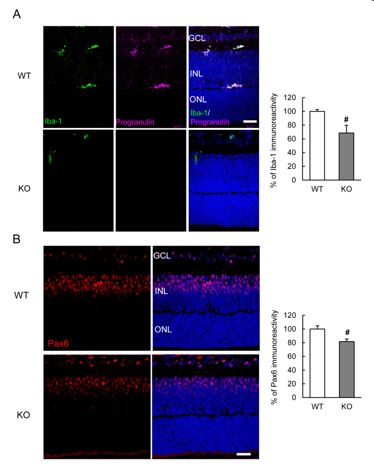
Progranulin has been reported to promote microglial migration, and it was suggested that progranulin is a chemoattractant for microglia [16]. We hypothesized that progranulin deficiency could decrease the microglial migration into the retina. At P7, progranulin was not observed in the Grn−/− retina although the expression was highly seen in the microglia of the wild-type (WT) mice (Figure 4A). The number of Iba-1-positive microglia was decreased in the retina of the Grn−/− mice at P7 (p=0.016, Figure 4A). To investigate the effect on retinal precursor cells under progranulin deficiency, Pax6 was immunostained. The Grn−/− retina showed a decrease in Pax6-positive retinal precursor cells (p=0.016, Figure 4B). These findings strengthened the association of microglia with retinal precursor cells.
Figure 4.
The loss of microglia and Pax6-positive retinal precursor cells in Grn−/− mice. A: Progranulin (magenta) is not observed in the Grn−/− retina at P7. The number of Iba-1-positive microglia (green) is decreased in Grn−/− mice. B: The retinal precursor cell marker, Pax6 (red), is decreased in Grn−/− mice. Data are the mean ± standard error of the mean (SEM; n=5 or 6). #p<0.05 versus wild type (Student t test). Scale bar=20 μm.
Discussion
In the present study, the aim was to determine how microglia are associated with postnatal retinal development. An increase in the microglia in the retina by systemic treatment with LPS enhanced the proliferation of retinal precursor cells and increased the number of retinal precursor cells (Figure 2F–J). LPS has broad effects through toll-like receptor 4 (TLR4) signaling, and TLR4 signaling is reported to be associated with the proliferation of neural precursor cells [27,28]. Therefore, we needed to show more clearly the association of microglia with the proliferation of retinal precursor cells. The decrease in microglia in the retina by PLX3397 treatment reduced the proliferation of retinal precursor cells (Figure 3D–G). However, these reagents (LPS and PLX3397) did not alter cell death because the cleaved caspase-3-positive cells were not changed between the control and reagents-treated groups (Figure 2E and Figure 3H). Microglia secrete various factors, such as inflammatory cytokines and growth factors [29]. Thus, microglia have a paradoxical role, and there are two types (M1 and M2). Some recent reports revealed that microglia are associated with neurogenesis in the SVZ and the hippocampal dentate gyrus [12,13]. Although it remains controversial whether M1 (inflammatory cytokines secreted) or M2 (growth factors secreted) microglia contribute to neurogenesis, microglial activation presumably promotes neurogenesis. It was reported that systemic administration of LPS to neonatal mice increased M2 microglia and promoted neurogenesis in the SVZ [12]. The present results also showed LPS increased M2 microglia (Figure 2D). It was suggested that M2 microglia could be associated with the proliferation of neural stem cells. However, M1 microglia may also be induced by LPS. M1 and M2 microglia exist on a spectrum (M2a, M2b, etc.) and cannot easily be placed into defined groups [30]. Moreover, it has been suggested that the polarization is doubtful, and a “multidimensional” stage is thought to exist [31]. Another report showed minocycline decreased the number of microglia and neural precursor cells through a decrease in some cytokines [13]. This result was supported by a recent report that neurogenesis is associated with microglia in the zebrafish retina [14]. Microglia can have multiple effects, such as synaptic maturation, although polarization remains controversial [32,33]. Further experiments are needed to reveal how the polarization of microglia is associated with neurogenesis or the proliferation of precursor cells although the present data show that LPS induces polarization to M2 microglia in neonatal mice.
Retinal precursor cells continue to proliferate at even P6 in the peripheral area [4]. BrdU-labeled proliferative cells merge with the marker of some retinal cells [34]. Rod photoreceptor cells, bipolar cells, and Müller glia proliferate by P6, P9, and P12, respectively. The number of proliferative cells is limited by P12. Most rod photoreceptor cells, bipolar cells, and Müller glia differentiate into mature cells by P12. Moreover, the separation of the inner nuclear layer (INL) and the outer nuclear layer (ONL) occurs at almost P6 [35]. In the present study, microglia increased at P7. This timing is almost the same as that of the proliferation and separation of the retinal layer. Therefore, microglia may be important for the proliferation of retinal cells and the formation of the retinal layer.
In this study, we also identified that progranulin may have the potential to promote microglial migration into the retina, because progranulin deficiency decreased the number of microglia in the retina. Progranulin may be associated with microglial migration during retinal development, because it was suggested progranulin had the potential to promote microglial migration [16]. Interestingly, progranulin expression was increased corresponding to Iba-1 expression (Figure 1A–C). Thus, progranulin surely regulates the microglial status in the retina. Another report showed the association of neuronal cells with progranulin. Progranulin increased the expression of fractalkine (CX3CL1) which is associated with the migration of monocytes in neurons [16,36]. Therefore, progranulin deficiency may decrease the invasion of microglia into the retina through the neurons. Moreover, the expression of progranulin was increased at P7 (Figure 1A,B). Recent reports showed progranulin could be associated with microglial functions, such as synaptic pruning and phagocytosis [17,18]. Taken together, progranulin can regulate the number of retinal microglia by accelerating the migration although progranulin may directly be associated with the proliferation of retinal cells and the formation of the retinal layer. Moreover, astrocytes and blood vessels spread across the retina after birth. It was reported that astrocytes and endothelial cells could have a potential effect against the proliferation of neural precursor cells [37,38]. Progranulin deficiency causes excessive activation of astrocytes in the postnatal stage [15]. Therefore, other cells except microglia might affect the proliferation of retinal precursor cells. In conclusion, these findings indicate that microglia, the resident myeloid cells except neuronal lineage, have a potent influence on the proliferation of precursor cells in the retina.
References
- 1.Gregory-Evans CY, Wallace VA, Gregory-Evans K. Gene networks: dissecting pathways in retinal development and disease. Prog Retin Eye Res. 2013;33:40–66. doi: 10.1016/j.preteyeres.2012.10.003. [DOI] [PubMed] [Google Scholar]
- 2.Glaser T, Jepeal L, Edwards JG, Young SR, Favor J, Maas RL. PAX6 gene dosage effect in a family with congenital cataracts, aniridia, anophthalmia and central nervous system defects. Nat Genet. 1994;7:463–71. doi: 10.1038/ng0894-463. [DOI] [PubMed] [Google Scholar]
- 3.Marquardt T, Ashery-Padan R, Andrejewski N, Scardigli R, Guillemot F, Gruss P. Pax6 is required for the multipotent state of retinal progenitor cells. Cell. 2001;105:43–55. doi: 10.1016/s0092-8674(01)00295-1. [DOI] [PubMed] [Google Scholar]
- 4.Close JL, Gumuscu B. Reh T a. Retinal neurons regulate proliferation of postnatal progenitors and Müller glia in the rat retina via TGF beta signaling. Development. 2005;132:3015–26. doi: 10.1242/dev.01882. [DOI] [PubMed] [Google Scholar]
- 5.Bassett EA, Wallace VA. Cell fate determination in the vertebrate retina. Trends Neurosci. 2012;35:565–73. doi: 10.1016/j.tins.2012.05.004. [DOI] [PubMed] [Google Scholar]
- 6.Checchin D, Sennlaub F, Levavasseur E, Leduc M, Chemtob S. Potential role of microglia in retinal blood vessel formation. Invest Ophthalmol Vis Sci. 2006;47:3595–602. doi: 10.1167/iovs.05-1522. [DOI] [PubMed] [Google Scholar]
- 7.Sapieha P. Eyeing central neurons in vascular growth and reparative angiogenesis. Blood. 2012;120:2182–94. doi: 10.1182/blood-2012-04-396846. [DOI] [PubMed] [Google Scholar]
- 8.Marín-Teva JL, Almendros A, Calvente R, Cuadros MA, Navascués J. Proliferation of actively migrating ameboid microglia in the developing quail retina. Anat Embryol (Berl) 1999;200:289–300. doi: 10.1007/s004290050280. [DOI] [PubMed] [Google Scholar]
- 9.Santos AM, Calvente R, Tassi M, Carrasco M-C, Martín-Oliva D, Marín-Teva JL, Navascués J, Cuadros MA. Embryonic and postnatal development of microglial cells in the mouse retina. J Comp Neurol. 2008;506:224–39. doi: 10.1002/cne.21538. [DOI] [PubMed] [Google Scholar]
- 10.Sato K. Effects of Microglia on Neurogenesis. Glia. 2015;63:1394–405. doi: 10.1002/glia.22858. [DOI] [PMC free article] [PubMed] [Google Scholar]
- 11.Chen Z, Jalabi W, Shpargel KB, Farabaugh KT, Dutta R, Yin X, Kidd GJ, Bergmann CC, Stohlman SA, Trapp BD. Lipopolysaccharide-Induced Microglial Activation and Neuroprotection against Experimental Brain Injury Is Independent of Hematogenous TLR4. J Neurosci. 2012;32:11706–15. doi: 10.1523/JNEUROSCI.0730-12.2012. [DOI] [PMC free article] [PubMed] [Google Scholar]
- 12.Pang Y, Dai X, Roller A, Carter K, Paul I, Bhatt AJ, Lin RC, Fan LW. Early Postnatal Lipopolysaccharide Exposure Leads to Enhanced Neurogenesis and Impaired Communicative Functions in Rats. PLoS One. 2016;11:e0164403. doi: 10.1371/journal.pone.0164403. [DOI] [PMC free article] [PubMed] [Google Scholar]
- 13.Shigemoto-Mogami Y, Hoshikawa K, Goldman JE, Sekino Y, Sato K. Microglia enhance neurogenesis and oligodendrogenesis in the early postnatal subventricular zone. J Neurosci. 2014;34:2231–43. doi: 10.1523/JNEUROSCI.1619-13.2014. [DOI] [PMC free article] [PubMed] [Google Scholar]
- 14.Huang T, Cuia J, Lia L, Hitchcockc PF, Lia Y. The role of microglia in the neurogenesis of zebrafish retina. Biochem Biophys Res Commun. 2012;421:214–20. doi: 10.1016/j.bbrc.2012.03.139. [DOI] [PMC free article] [PubMed] [Google Scholar]
- 15.Kuse Y, Tsuruma K, Mizoguchi T, Shimazawa M, Hara H. Progranulin deficiency causes the retinal ganglion cell loss during development. Sci Rep. 2017;7:1679. doi: 10.1038/s41598-017-01933-8. [DOI] [PMC free article] [PubMed] [Google Scholar]
- 16.Pickford F, Marcus J, Camargo LM, Xiao Q, Graham D, Mo JR, Burkhardt M, Kulkarni V, Crispino J, Hering H, Hutton M. Progranulin is a chemoattractant for microglia and stimulates their endocytic activity. Am J Pathol. 2011;178:284–95. doi: 10.1016/j.ajpath.2010.11.002. [DOI] [PMC free article] [PubMed] [Google Scholar]
- 17.Minami SS, Min S-W, Krabbe G, Wang C, Zhou Y, Asgarov R, Li Y, Martens LH, Elia LP, Ward ME, Mucke L, Farese RV, Jr, Gan L. Progranulin protects against amyloid β deposition and toxicity in Alzheimer’s disease mouse models. Nat Med. 2014;•••:1157–1161. doi: 10.1038/nm.3672. [DOI] [PMC free article] [PubMed] [Google Scholar]
- 18.Lui H, Zhang J, Makinson SR, Cahill MK, Kelley KW, Huang HY, Shang Y, Oldham MC, Martens LH, Gao F, Coppola G, Sloan SA, Hsieh CL, Kim CC, Bigio EH, Weintraub S, Mesulam MM, Rademakers R, Mackenzie IR, Seeley WW, Karydas A, Miller BL, Borroni B, Ghidoni R, Farese RV. Jr1, Paz JT, Barres BA, Huang EJ. Progranulin Deficiency Promotes Circuit-Specific Synaptic Pruning by Microglia via Complement Activation. Cell. 2016;165:921–35. doi: 10.1016/j.cell.2016.04.001. [DOI] [PMC free article] [PubMed] [Google Scholar]
- 19.Kayasuga Y, Chiba S, Suzuki M, Kikusui T, Matsuwaki T, Yamanouchi K, Kotaki H, Horai R, Iwakura Y, Nishihara M. Alteration of behavioural phenotype in mice by targeted disruption of the progranulin gene. Behav Brain Res. 2007;185:110–8. doi: 10.1016/j.bbr.2007.07.020. [DOI] [PubMed] [Google Scholar]
- 20.Kuse Y, Tsuruma K, Sugitani S, Izawa H, Ohno Y, Shimazawa M, Hara H. Progranulin promotes the retinal precursor cell proliferation and the photoreceptor differentiation in the mouse retina. Sci Rep. 2016;6:23811. doi: 10.1038/srep23811. [DOI] [PMC free article] [PubMed] [Google Scholar]
- 21.Osakada F, Ooto S, Akagi T, Mandai M, Akaike A, Takahashi M. Wnt signaling promotes regeneration in the retina of adult mammals. J Neurosci. 2007;27:4210–9. doi: 10.1523/JNEUROSCI.4193-06.2007. [DOI] [PMC free article] [PubMed] [Google Scholar]
- 22.Hu X, Leak RK, Shi Y, Suenaga J, Gao Y, Zheng P, Chen J. Microglial and macrophage polarization—new prospects for brain repair. Nat Rev Neurol. 2015;11:56–64. doi: 10.1038/nrneurol.2014.207. [DOI] [PMC free article] [PubMed] [Google Scholar]
- 23.Matsushima D, Heavner W, Pevny LH. Combinatorial regulation of optic cup progenitor cell fate by SOX2 and PAX6. Development. 2011;138:443–54. doi: 10.1242/dev.055178. [DOI] [PMC free article] [PubMed] [Google Scholar]
- 24.Liddelow SA, Guttenplan KA, Clarke LE, Bennett FC, Bohlen CJ, Schirmer L, Bennett ML, Münch AE, Chung WS, Peterson TC, Wilton DK, Frouin A, Napier BA, Panicker N, Kumar M, Buckwalter MS, Rowitch DH, Dawson VL, Dawson TM, Stevens B, Barres BA. Neurotoxic reactive astrocytes are induced by activated microglia. Nature. 2017;541:481–7. doi: 10.1038/nature21029. [DOI] [PMC free article] [PubMed] [Google Scholar]
- 25.Renee M, Elmore P, Najafi AR, Koike MA, Nazih N, Spangenberg EE, Rice RA, Kitazawa M, Matusow B, Nguyen H, West BL, Green KN. CSF1 receptor signaling is necessary for microglia viability, which unmasks a cell that rapidly repopulates the microglia- depleted adult brain. Neuron. 2015;82:380–97. doi: 10.1016/j.neuron.2014.02.040. [DOI] [PMC free article] [PubMed] [Google Scholar]
- 26.Livne-bar I, Pacal M, Cheung MC, Hankin M, Trogadis J, Chen D, Dorval KM, Bremner R. Chx10 is required to block photoreceptor differentiation but is dispensable for progenitor proliferation in the postnatal retina. Proc Natl Acad Sci USA. 2006;103:4988–93. doi: 10.1073/pnas.0600083103. [DOI] [PMC free article] [PubMed] [Google Scholar]
- 27.Okun E, Griffioen KJ, Mattson MP. Toll-like receptor signaling in neural plasticity and disease. Trends Neurosci. 2011;34:269–81. doi: 10.1016/j.tins.2011.02.005. [DOI] [PMC free article] [PubMed] [Google Scholar]
- 28.Shechter R, Ronen A, Rolls A, London A, Bakalash S, Young MJ, Schwartz M. Toll-like receptor 4 restricts retinal progenitor cell proliferation. J Cell Biol. 2008;183:393–400. doi: 10.1083/jcb.200804010. [DOI] [PMC free article] [PubMed] [Google Scholar]
- 29.Tang Y, Le W. Differential Roles of M1 and M2 Microglia in Neurodegenerative Diseases. Mol Neurobiol. 2016;53:1181–94. doi: 10.1007/s12035-014-9070-5. [DOI] [PubMed] [Google Scholar]
- 30.Murray PJ, Allen JE, Biswas SK, Fisher EA, Gilroy DW, Goerdt S, Hamilton JA, Ivashkiv LB, Lawrence T, Locati M, Mantovani A, Martinez FO, Mege JL, Mosser DM, Natoli G, Saeij JP, Schultze JL, Shirey KA, Sica A, Suttles J, Udalova I, van Ginderachter JA, Vogel SN, Wynn TA. Macrophage activation and polarization: nomenclature and experimental guidelines. Immunity. 2014;41:14–20. doi: 10.1016/j.immuni.2014.06.008. [DOI] [PMC free article] [PubMed] [Google Scholar]
- 31.Ransohoff RM. A polarizing question: do M1 and M2 microglia exist? Nat Neurosci. 2016;19:987–91. doi: 10.1038/nn.4338. [DOI] [PubMed] [Google Scholar]
- 32.Paolicelli RC, Bolasco G, Pagani F, Maggi L, Scianni M, Panzanelli P, Giustetto M, Ferreira TA, Guiducci E, Dumas L, Ragozzino D, Gross CT. Synaptic pruning by microglia is necessary for normal brain development. Science. 2011;333:1456–8. doi: 10.1126/science.1202529. [DOI] [PubMed] [Google Scholar]
- 33.Squarzoni P, Oller G, Hoeffel G, Pont-Lezica L, Rostaing P, Low D, Bessis A, Ginhoux F, Garel S. Microglia Modulate Wiring of the Embryonic Forebrain. Cell Reports. 2014;8:1271–9. doi: 10.1016/j.celrep.2014.07.042. [DOI] [PubMed] [Google Scholar]
- 34.Ogawa M, Saitoh F, Sudou N, Sato F, Fujieda H. Cell type-specific effects of p27KIP1 loss on retinal development. Neural Dev. 2017;12:17. doi: 10.1186/s13064-017-0094-1. [DOI] [PMC free article] [PubMed] [Google Scholar]
- 35.Skapek SX, Lin SCJ, Jablonski MM, Mckeller RN, Tan M, Hu N, Lee EY. Persistent expression of cyclin D1 disrupts normal photoreceptor differentiation and retina development. Oncogene. 2001;20:6742–51. doi: 10.1038/sj.onc.1204876. [DOI] [PubMed] [Google Scholar]
- 36.Ruitenberg MJ, Vukovic J, Blomster L, Hall JM, Jung S, Filgueira L, McMenamin PG, Plant GW. CX3CL1/fractalkine regulates branching and migration of monocyte-derived cells in the mouse olfactory epithelium. J Neuroimmunol. 2008;205:80–5. doi: 10.1016/j.jneuroim.2008.09.010. [DOI] [PubMed] [Google Scholar]
- 37.Sourial M, Doering LC. Astrocyte-Secreted Factors Selectively Alter Neural Stem and Progenitor Cell Proliferation in the Fragile X Mouse. Front Cell Neurosci. 2016;10:1–11. doi: 10.3389/fncel.2016.00126. [DOI] [PMC free article] [PubMed] [Google Scholar]
- 38.Nakagomi N, Nakagomi T, Kubo S, Nakano-Doi A, Saino O, Takata M, Yoshikawa H, Stern DM, Matsuyama T, Taguchi A. Endothelial cells support survival, proliferation, and neuronal differentiation of transplanted adult ischemia-induced neural stem/progenitor cells after cerebral infarction. Stem Cells. 2009;27:2185–95. doi: 10.1002/stem.161. [DOI] [PubMed] [Google Scholar]


