Madam,
Montgomery tube (MT) is placed after tracheal stenosis surgeries where it acts as a stent to prevent restenosis and also maintains the patency of the airway. It is a pliable, uncuffed, T-shaped tube made of silicone.[1] Various complications have been reported with MT in situ such as blockade by secretions, kinking of extra- and intra-tracheal lumen, migration, and inhalation. We report here an adverse event that occurred while replacing MT after surgery and emergency airway management to circumvent the situation.
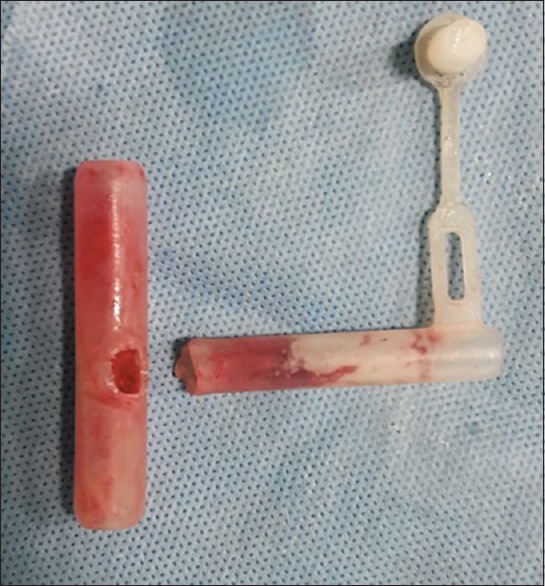
A 12-years-old male child, an operated case of laryngotracheal reconstruction for subglottic stenosis, was posted for excision of granulations over glottis. He had 10-mm external diameter MT in situ following previous surgery. To preserve spontaneous ventilation, inhalational induction was performed with oxygen and sevoflurane by connecting MT to the circuit. After administering 60 μg of fentanyl, direct laryngoscopy was done to assess air leak from glottic aperture. The upper end of MT and glottic aperture was completely occluded by the granulation tissue. There was no leak on performing positive pressure ventilation. Atracurium was used for muscle relaxation. After removing granulation tissues over MT, a surgeon wanted to replace the MT with a tracheostomy tube (TT). After providing 100% oxygen for 3 min, the surgeon using a curved forceps attempted removal of MT. While doing so, the extratracheal limb of the MT broke off from the rest of the intratracheal limb [Figure 1]. This created an emergency situation with incompletely removed granulation obstructing at glottis and a broken MT obstructing at tracheostomy stoma. Meanwhile, as the surgeon struggled to remove the broken tube, saturation of the child dropped to 89%. Immediately, a 5-mm internal diameter uncuffed TT was inserted into the opening of broken MT that was visible through tracheostomy stoma. Ventilation was possible through the TT. Another attempt was made to remove the MT after keeping rigid bronchoscope ready. The intratracheal limb of MT was then removed successfully through the tracheostomy stoma. The child made an uneventful recovery and shifted to postanesthesia care unit.
Figure 1.

Broken limbs of Montgomery T-tube after retrieval from the trachea
It is routine to replace MT with a TT or flexometallic tube before abolishing spontaneous breathing and proceeding to surgery. However, in this case, MT was adherent to granulation tissue, so tube change was attempted after releasing granulations. Airway compromise due to fracture of MT while attempted retrieval has not been reported previously. Restoring ventilation and oxygenation in such a scenario is of utmost importance. As the opening in broken MT was visible through tracheostomy stoma, a smaller size uncuffed TT was inserted into the MT by holding the upper limb of MT with forceps. Utmost care was taken to prevent distal migration of MT while inserting TT. Our plan B was to ventilate with facemask by occluding the tracheostomy stoma as the glottis was partially patent by now. On extreme situation, pushing the intratracheal MT into one of the bronchi was our last option.[2] Understanding the complexity and emergent nature of such situation and readiness with a plan of action are needed to prevent any undue complication.
Financial support and sponsorship
Nil.
Conflicts of interest
There are no conflicts of interest.
References
- 1.Guha A, Mostafa SM, Kendall JB. The Montgomery T-tube: Anaesthetic problems and solutions. Br J Anaesth. 2001;87:787–90. doi: 10.1093/bja/87.5.787. [DOI] [PubMed] [Google Scholar]
- 2.Pawar DK. Dislodgement of bronchial foreign body during retrieval in children. Paediatr Anaesth. 2000;10:333–5. doi: 10.1046/j.1460-9592.2000.00525.x. [DOI] [PubMed] [Google Scholar]


