FCR regimen associating fludarabine, cyclophosphamide and rituximab (FCR) is still considered the gold standard for the first line treatment of medically fit patients with active B-cell chronic lymphocytic leukemia (CLL) and without del(17p) and/or TP53 mutations.1 In addition, the persistence of detectable minimal residual disease (MRD) at the end of treatment correlates with both shorter response duration and lower survival, irrespective of the treatment used.2 Thus, achieving negative MRD (nMRD) is a valuable objective in younger patients with CLL.
A lower exposure of rituximab (RTX) with the conventional 375 mg/m2 has justified that patients with CLL receive first 375 mg/m2 and then 500 mg/m2 during the following infusions. Several arguments suggest that higher doses of RTX might be beneficial. A phase I study, using RTX as monotherapy, demonstrated a dose response relationship with 22%, 43% and 75% of objective response rates (ORR) for patients receiving 500–825 mg/m2, 1000–1500 mg/m2 and 2250 mg/m2, respectively.3 Similar results were also found in another phase II study.4 In pharmacokinetic (PK) studies, patients with CLL exhibited lower RTX exposure than lymphoma patients.5 The reason of the discrepancy remains unclear but could be related to a larger antigenic burden in patients with CLL. The influence of CD20 burden on RTX PK and response has already been suggested in a syngeneic murine model6 and in patients with diffuse large B-cell lymphoma (DLBCL).7 In CLL, CD20 burden affects RTX PK by increasing the antibody target-mediated elimination,8 but its influence on RTX exposure and outcomes remains to be investigated. We conducted therefore a randomized phase II study evaluating the effectiveness of higher doses of RTX associated with FC (Online Supplementary Figure S2).
This prospective randomized phase II study (clinicaltrials.gov identifier 01370772) has included 140 treatment-naive patients (aged 18–65 years) diagnosed with confirmed Binet stage C or active Binet stages A/B CLL without 17p deletion.9 Patients were stratified according to IGHV mutational status, FISH analysis (11q deletion or not) and randomly assigned to receive either 6 cycles of FCR (intravenous RTX 375 mg/m2 for the first course, D1 and 500 mg/m2 for the others, oral fludarabine 40 mg/m2/d D2-4, oral cyclophosphamide 250 mg/m2/d D2-4) every 28 days or Dense-FCR with an intensified RTX prephase (500 mg on D0, and 2000 mg on D1, D8 and D15) before the FCR starting at D22. The primary endpoint was the rate of CR with nMRD three months after the end of treatment. MRD was determined by flow cytometry in both peripheral blood (PB) and bone marrow (BM) at M9. nMRD was defined as the detection of less than one CLL cell per 10,000 leukocytes. The CD20 antigen burden was defined as the sum of CD20 antigenic targets estimated on both B-cells in PB (CD20cir) by using CD20-PE QuantiBRITE™ reagents and in the lymph nodes (CD20LN) by CT-scan using semi-automated accurate measurement technique.10 RTX exposure was assessed using a semi-mechanistic pharmacokinetic model.
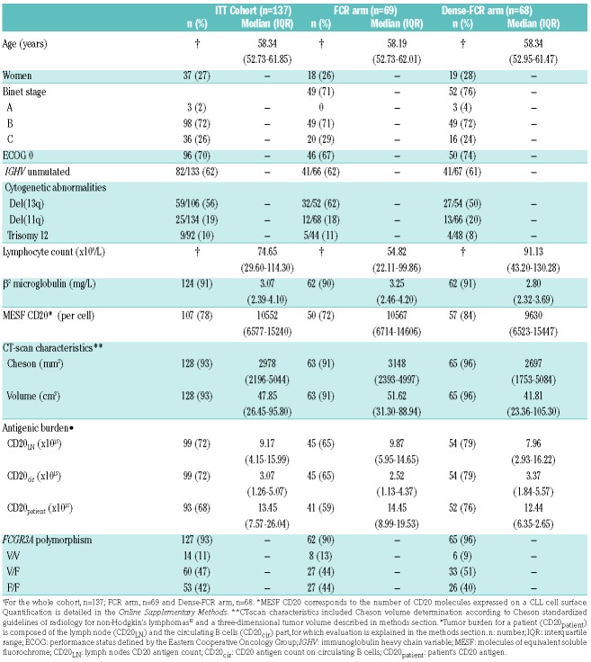
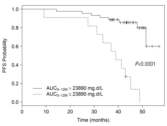
One hundred and forty patients were recruited, 69 patients in the FCR arm and 68 patients in the Dense-FCR arm. Both treatment groups were well-balanced with respect to stratification criteria, clinical, biological, and tumor burden parameters (Table 1). Grade 3/4 infusion-related reactions were reported in only two patients in the Dense-FCR arm leading to treatment discontinuation in one patient (Online Supplementary Table S1). Monitoring of lymphocyte counts before each RTX infusion demonstrated that 31%, 53% and 64% of patients had lymphocyte counts lower than 5.0×10⁹/L after 2500 mg (D8), 4500 mg (D15) and 6500 mg (D22), respectively (Online Supplementary Figure S3). The ORR was 94%, including 55% CR or CRi, 3% nPR, 36% PR, 2% progression and 4% not evaluable. No difference was observed according to treatment arm (Online Supplementary Figure S4). MRD determined in PB and in BM was assessed respectively in 113 (53 in FCR arm; 60 in Dense-FCR arm) and 102 patients (49 in FCR arm; 53 in Dense-FCR arm). Seventy-six patients (55%) had PB nMRD, 39 (57%) in the FCR arm and 37 (54%) in the Dense-FCR arm. Forty-seven patients (34%) had BM nMRD, 25 (36%) in the FCR arm and 22 (32%) in the Dense-FCR arm. Thirty-three patients (24%) achieved a CR with PB and BM nMRD, distributed into 16 patients (23%) in the FCR arm and 17 patients (25%) in the Dense-FCR arm. We concluded that no difference was observed according to our primary endpoint. Patients achieving CR with BM nMRD were more frequently stage Binet A/B with lower lymphocyte count (P=0.005) and had lower CD20cir (P=0.016), without any difference in the level of CD20 expression on CLL cells. Univariate logistic regression analysis showed no significant difference in CR with BM nMRD rate according to IGHV mutational status, cytogenetic abnormalities or tumor burden (Online Supplementary Table S2). In the Dense-FCR arm, lymphodepletion determined between D0 and D22 correlated with CD20cir (P=0.007). On multivariable analysis, Binet stage A/B (P=0.008) and lymphocyte count at D0 < 29.63×10⁹/L (P=0.001) were significantly associated with a superior likelihood of achieving CR with BM nMRD. With a median follow up of 42.7 months, median progression-free survival (PFS) was not reached (71% at 4 years) with no difference between treatment arms. Unmutated IGHV status (P=0.008), high lymphocyte count at D0 (>74.65×10⁹/L, P=0.006), high CD20 expression level (MESF CD20>9169, P<0.001) and tumor burden (Cheson> 3535 mm2, P=0.01; volume> 51.4 cm3, P=0.002) were negatively associated with PFS in univariate analysis (Online Supplementary Table S3). In multivariable analysis, the lymphocyte counts at D0 >74.65×10⁹/L (P=0.010), the level of CD20 expression on CLL cells (<9169, P=0.023) and tumor burden (either Cheson >3535; P=0.001) or volume>51.4 cm3, P=0.005) were associated with a lower PFS. In total, 86% of patients were alive at 4 years without difference according to treatment arm (Online Supplementary Figure S5). PK population (n=118) did not differ from the whole population (Online Supplementary Table S4), and for 93 of those patients MRD was available. RTX AUC0-12M was significantly higher in the Dense-FCR arm as compared to the FCR arm. CR BM nMRD patients had significantly higher RTX AUC0–12M compared to non-responder patients in the whole population and according to treatment arm (Figure 1). The optimal RTX AUC0–12M cut-off of 23890 mg.d/L allows two groups of patients with significantly different PFS to be separated (38.9 months vs. NA; log-rank test, P<0.0001, Figure 2).
Table 1.
Patient characteristics.
Figure 1.
Rituximab AUC0–12M according to response in PK population (n=93). Rituximab AUC0–12M in the pharmacokinetic cohort (A), in standard arm (B) and in Dense-FCR arm (C). Other patients refer to patients achieving complete response (CR) with detectable minimal residual disease (MRD) and no-CR patients whatever the MRD. CR: complete response; nMRD: negative minimal residual disease; RTX AUC0–12M: area under the curve of rituximab concentration from treatment onset to 6 months (M12) after the end of the treatment.
Figure 2.
PFS of PK population according to RTX AUC0–12M. PFS of the cohort according to RTX AUC0–12M. RTX AUC0–12M, area under the curve of rituximab concentration from treatment onset to 6 months (M12) after the end of the treatment.
In our study, we demonstrated a significant increase in RTX exposure in patients receiving the intensified RTX regimen, but this did not translate into increased rate of CR with BM nMRD, Binet stage A/B and low lymphocyte count before treatment being the only factors affecting our primary endpoint. Calculation of the number of patients for this study was based on a 35% rate of CR with PB and BM nMRD with FCR treatment. We assumed an increase of 15% of CR with nMRD by using high doses of RTX and a rate of 10% of patients not assessable for response. We observed however a significant drop-out (20% during treatment course and up to 33% for PB and BM nMRD assessment), but the lack of any difference between the two arms suggests that more patients evaluable for the primary end-point would not have changed our final results. Similar results were also observed in a study where patients received 3 RTX infusions per cycle of chemotherapy (FC).
PFS was not influenced by treatment arm but was affected by high CD20 antigen burden assessed by lymphocyte count, CD20 expression level and tumor volume, questioning the role of CD20 antigenic mass on rituximab exposure. All patients in CR with BM nMRD exhibited higher exposure of RTX than non-responder patients and patients with higher RTX AUC also exhibited higher PFS. We have previously reported extended results of our PK data in this population.8 We demonstrated an increased rituximab ‘consumption’ (target-mediated elimination) correlating with a higher amount of baseline CD20 and FCGR3A-158VV genotype, which were associated with lower rituximab concentrations in early treatment cycles. Only 32% of the inter-individual variability in the elimination rate was explained by circulating CD20 antigen suggesting that CD20 antigenic mass was not the main factor explaining fast RTX clearance observed in patients with CLL. The reasons of this consumption remain undetermined but could be related to the CD20 internalization observed in vitro.11 This internalization was not observed with type II anti-CD20 mAbs suggesting a potential advantage obinutuzumab in patients with CLL. Recently, we demonstrated in patients with DLBCL treated with immuno-chemotherapy that tumor burden influenced RTX exposure and patient’s outcome.7 We then proposed a nomogram providing a rational scheme for increasing the RTX dose in patients according to tumor burden in order to achieve RTX exposures that have a better chance of prolonging the duration of response. CLL and DLBCL seem, therefore, completely different models for RTX PK. In patients with CLL, RTX elimination is fast, not significantly influenced by CD20 antigenic mass and cannot be corrected by higher doses of RTX, while in patients with DLBCL, tumor metabolic volume is the main factor influencing RTX exposure and increasing doses of RTX should increase RTX exposure and improve outcome.
Supplementary Material
Footnotes
Funding: this study was funded by FILO group and F. Hoffman-La Roche Ltd (Basel, Switzerland). This work was supported by the French Higher Education and Research Ministry under the program ‘Investissements d’avenir’ Grant Agreement: LabEx MAbImprove ANR-10-LABX-53-01
Information on authorship, contributions, and financial & other disclosures was provided by the authors and is available with the online version of this article at www.haematologica.org.
References
- 1.Hallek M, Fischer K, Fingerle-Rowson G, et al. Addition of rituximab to fludarabine and cyclophosphamide in patients with chronic lymphocytic leukaemia: a randomised, open-label, phase 3 trial. Lancet. 2010;376(9747):1164–1174. [DOI] [PubMed] [Google Scholar]
- 2.Bottcher S, Ritgen M, Fischer K, et al. Minimal residual disease quantification is an independent predictor of progression-free and overall survival in chronic lymphocytic leukemia: a multivariate analysis from the randomized GCLLSG CLL8 trial. J Clin Oncol. 2012;30(9):980–988. [DOI] [PubMed] [Google Scholar]
- 3.O’Brien SM, Kantarjian H, Thomas DA, et al. Rituximab dose-escalation trial in chronic lymphocytic leukemia. J Clin Oncol. 2001;19(8):2165–2170. [DOI] [PubMed] [Google Scholar]
- 4.Byrd JC, Murphy T, Howard RS, et al. Rituximab using a thrice weekly dosing schedule in B-cell chronic lymphocytic leukemia and small lymphocytic lymphoma demonstrates clinical activity and acceptable toxicity. J Clin Oncol. 2001;19(8):2153–2164. [DOI] [PubMed] [Google Scholar]
- 5.Li J, Zhi J, Wenger M, et al. Population pharmacokinetics of rituximab in patients with chronic lymphocytic leukemia. J Clin Pharmacol. 2012;52(12):1918–1926. [DOI] [PubMed] [Google Scholar]
- 6.Dayde D, Ternant D, Ohresser M, et al. Tumor burden influences exposure and response to rituximab: pharmacokinetic-pharmacodynamic modeling using a syngeneic bioluminescent murine model expressing human CD20. Blood. 2009;113(16):3765–3772. [DOI] [PubMed] [Google Scholar]
- 7.Tout M, Casasnovas O, Meignan M, et al. Rituximab exposure is influenced by baseline metabolic tumor volume and predicts outcome of DLBCL patients: a Lymphoma Study Association report. Blood. 2017;129(19):2616–2623. [DOI] [PubMed] [Google Scholar]
- 8.Tout M, Gagez AL, Lepretre S, et al. Influence of FCGR3A-158V/F genotype and baseline CD20 antigen count on target-mediated elimination of rituximab in patients with chronic lymphocytic leukemia: a study of FILO Group. Clin Pharmacokinet. 2017;56(6):635–647. [DOI] [PubMed] [Google Scholar]
- 9.Hallek M, Cheson BD, Catovsky D, et al. Guidelines for the diagnosis and treatment of chronic lymphocytic leukemia: a report from the International Workshop on Chronic Lymphocytic Leukemia updating the National Cancer Institute-Working Group 1996 guidelines. Blood. 2008;111(12):5446–5456. [DOI] [PMC free article] [PubMed] [Google Scholar]
- 10.Nougaret S, Jung B, Aufort S, Chanques G, Jaber S, Gallix B. Adrenal gland volume measurement in septic shock and control patients: a pilot study. Eur Radiol. 2010;20(10):2348–2357. [DOI] [PubMed] [Google Scholar]
- 11.Beers SA, French RR, Chan HT, et al. Antigenic modulation limits the efficacy of anti-CD20 antibodies: implications for antibody selection. Blood. 2010;115(25):5191–5201. [DOI] [PubMed] [Google Scholar]
- 12.Cheson BD, Horning SJ, Coiffier B, et al. Report of an international workshop to standardize response criteria for non-Hodgkin’s lymphomas. NCI Sponsored International Working Group. J Clin Oncol. 1999;17(4):1244. [DOI] [PubMed] [Google Scholar]
Associated Data
This section collects any data citations, data availability statements, or supplementary materials included in this article.


