Abstract
Localized scleroderma (LS) is a sclerotic skin disorder rarely reported in the veterinary literature. We herein report the first case of a linear LS-like skin lesion in a cat. A 1-year-old castrated male Himalayan cat was presented with a 1-month history of an alopecic, indurated, serpiginous, branched skin lesion on the dorsal cervical to scapular area. The cat had no history of trauma, although a topical spot-on endectocide had been applied near the lesion. Histopathological examination revealed a focal area of hyperplastic dermal collagen with the absence of pilosebaceous units. The cutaneous lesion remained unchanged during a 2-year follow-up period. Clinical and histopathological similarities of this skin lesion with those of the linear form of LS in humans were considered.
Keywords: cat, collagen, linear scleroderma
Localized scleroderma (LS), also referred to as morphea, is a heterogeneous group of sclerotic skin disorders reported in humans [6]. Although no widely accepted classifications of LS in humans have been established [6], the most recent guidelines proposed classification of this disease group into limited, linear, generalized, deep, and mixed forms [4]. Histopathologically, LS in humans is characterized by overproduction of dermal collagen and other extracellular matrix proteins that replace the subcutaneous adipose tissue [4]. Similar skin conditions in companion animals have rarely been reported in the veterinary literature [1, 2, 7]. In this case report, we describe a linear form of LS-like disease in a young cat.
A 1-year-old castrated male Himalayan cat that was kept indoors was presented with a 1-month history of a nonpruritic, alopecic, raised skin lesion on the dorsal cervical to scapular area. The owner had adopted the cat 5 months prior to the initial visit. The cat was current on vaccinations against feline herpesvirus, feline calicivirus, and feline parvovirus, although the injection sites of these vaccinations were unknown. The owner mentioned that the lesion had started in the dorsal cervical area and gradually extended cranially. Before the owner noticed the skin lesion, the cat had received a spot-on form of 10% fipronil/12% (S)-methoprene (Frontline Plus™; Boehringer Ingelheim, Ingelheim, Germany) three times in the interscapular area before the initial presentation. The owner also declared that the cat had no history of trauma before development of the skin lesion.
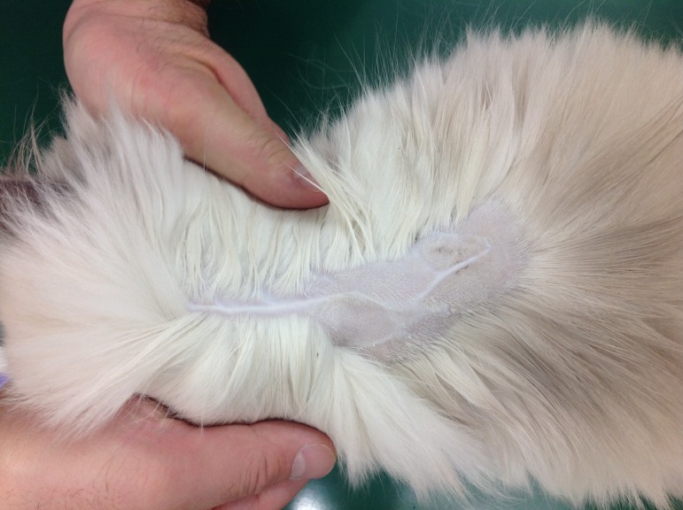
General physical examination was unremarkable except for the skin lesion. Dermatological examination revealed a moderately firm, alopecic, shiny, well circumscribed, linear-shaped, serpiginous, branched skin lesion of approximately 85 mm in length and 3 mm in width over the dorsal cervical and interscapular region (Fig. 1). On palpation, the lesion was raised and indurated and was not adhered to the underlying tissue. The results of a complete blood count, serum chemistry, thoracic and abdominal radiography, and abdominal ultrasonography were unremarkable. An antinuclear antibody (ANA) test was not performed because owner consent was not obtained. The initial differential diagnoses included LS, scar tissue, linear granuloma, collagen hamartoma, burn, vascular disorders, and cicatricial alopecia.
Fig. 1.
Clinical appearance of the skin lesion.
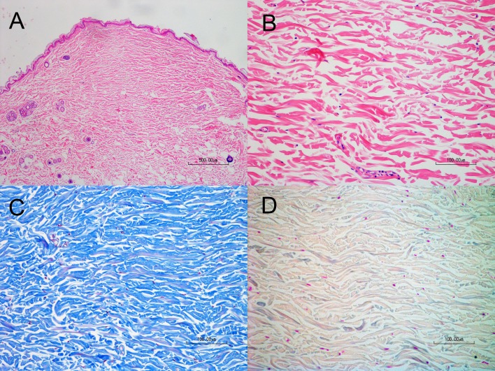
A 6-mm punch biopsy sample was obtained from the caudal end of the skin lesion, fixed in 10% neutral-buffered formalin, routinely processed, embedded in paraffin, sectioned at 5-µm intervals, stained with hematoxylin and eosin, and examined by light microscopy (Fig. 2A and 2B). The epidermis was normal. The dermis was expanded by abundant fibrous connective tissue in which pilosebaceous units were absent. The lesion slightly impinged on the panniculus; however, replacement of the panniculus by the expanding dermal collagen was not evident. Neither inflammatory infiltrates nor infectious agents were observed in the biopsied sample. Masson trichrome staining confirmed the proliferation of dermal collagen (Fig. 2C). Colloidal iron staining did not detect mucin in the sample (Fig. 2D). These histopathological findings were consistent with those recognized in morphea-like diseases in dogs and cats.
Fig. 2.
Histopathological findings. (A) Lower and (B) higher magnifications of hematoxylin and eosin staining. (C) Masson trichrome staining. (D) Colloidal iron staining.
Because spontaneous recovery of feline morphea-like diseases has been described in a previous report [1], the present cat remained under follow-up observation without any treatment except for changing of the endectocide applied to the area to selamectin (Revolution™; Zoetis, Parsippany, NJ, U.S.A.) and ensuring that all future vaccination sites were in the hind limbs. However, the lesion remained stable and did not improve during a 2-year follow-up period.
Morphea-like diseases characterized by patchy sclerotic areas with or without raised skin have also been reported in two cats [1, 7]. These skin lesions developed at the age of 3 to 7 years and resolved within 130 days after disease onset. Both cats were male (one intact and one castrated), although a sex predilection of feline morphea-like disease could not be established because of the limited number of cases. The clinical manifestations in previous feline cases resembled a plaque type, limited form of LS in humans. In contrast, the clinical and histopathological manifestations in the present cat resembled those in the later stage of a linear form of human LS and have not been reported in the veterinary literature. The skin lesions in the linear form of human LS usually resolve with residual hyperpigmentation, although the lesions in LS “en coup de sabre”, which is thought to be a variant of the linear form of LS that occurs in childhood, reportedly stabilize after 2 to 10 years [6].
The linear form of LS usually develops in young people and is characterized by linear-arranged sclerotic confluent areas that may follow Blaschko’s lines [6]. Conversely, the lesion in the present case did not appear to follow Blaschko’s lines because the lesion was present along the dorsal median line. Histopathologically, the hyperplastic dermal collagen in the present case was consistent with reports of LS in humans and companion animals, whereas the absence of inflammatory cell infiltration in the skin lesion was not consistent with the corresponding human disease, in which abundant inflammatory cell infiltration is recognized at least in the early stage of the disease [6]. Inflammatory cell infiltration was mild or unremarkable in previous feline cases of morphea-like diseases [1, 7]. Because feline skin is covered by a dense hair coat, owners and veterinarians are unlikely to find the early skin lesions of LS in cats. No previous reports have described positive ANA tests in feline morphea-like diseases [1, 7], although human patients with the linear form of LS are likely to show a positive ANA test result [6]. The similarities and differences between human and feline LS cases indicate a complex pathophysiologic basis of this condition, warranting further investigation.
Scar tissue, cicatricial alopecia, and collagen hamartoma were three major differential diagnoses for the skin lesion in the present case. Scar tissue and cicatricial alopecia develop secondary to a variety of insults including physical, chemical, or thermal injury; severe bacterial furunculosis; neoplasia; sterile nodular panniculitis; and rarely other immune-mediated diseases [3, 5]. However, no such history or histological lesions were evident through an interview with the cat owner and examination of biopsied samples. A collagen hamartoma could not be completely ruled out, although the gross appearance of the skin lesion in the present case did not resemble this disease category.
A previous report described the possible association of a topical spot-on endectocide with the development of morphea-like disease in a cat. The lesion developed within a few days after the application of a spot-on topical drug containing praziquantel and emodepside and resolved after minoxidil therapy. The LS-like skin lesion in the present case also developed after the use of a spot-on endectocide. Further accumulation of similar cases is needed to determine the association of spot-on medications and the development of LS-like skin diseases in cats.
REFERENCES
- 1.Bensignor E., Carlotti D. N., Pin D.1998. Morphea-like lesion in a cat. J. Small Anim. Pract. 39: 538–540. doi: 10.1111/j.1748-5827.1998.tb03701.x [DOI] [PubMed] [Google Scholar]
- 2.Doelle M., Linder K. E., Boche J., Jagannathan V., Leeb T., Linek M.2016. Initial characterization of stiff skin-like syndrome in West Highland white terriers. Vet. Dermatol. 27: 210–e53. doi: 10.1111/vde.12316 [DOI] [PubMed] [Google Scholar]
- 3.Gross T. L., Ihrke P. J., Walder E. J., Affolter V. K.2005. Skin Diseases of the Dog and Cat, 2nd ed., Blackwell, Oxford. [Google Scholar]
- 4.Kreuter A., Krieg T., Worm M., Wenzel J., Moinzadeh P., Kuhn A., Aberer E., Scharffetter-Kochanek K., Horneff G., Reil E., Weberschock T., Hunzelmann N.2016. German guidelines for the diagnosis and therapy of localized scleroderma. J. Dtsch. Dermatol. Ges. 14: 199–216. doi: 10.1111/ddg.12724 [DOI] [PubMed] [Google Scholar]
- 5.Maxie G.2015. Jubb, Kennedy & Palmer’s Pathology of Domestic Animals-Vol. 1, Elsevier, Amsterdam. [Google Scholar]
- 6.Moinzadeh P., Hunzelmann N., Kreuter A., Krieg T.2016. Localized scleroderma: a review. J. Scleroderma Relat. Disord. 1: 286–297. doi: 10.5301/jsrd.5000215 [DOI] [Google Scholar]
- 7.Seixas G., Taboada P.2012. Morphea-like lesion following topical endectocide application in a cat. Vet. Dermatol. 23: 244–e50. doi: 10.1111/j.1365-3164.2012.01039.x [DOI] [PubMed] [Google Scholar]


