ABSTRACT
This is a case report of a bite by an Opisthoglyphous snake Thamnodynastes pallidus (Linnaeus, 1758) in an undergraduate herpetologist observed at the Universidade Federal da Paraiba (Rio Tinto, PB, Brazil). The female victim was bitten in her left hand between the index finger and the middle finger and presented symptoms of local envenomation such as bleeding, itching, pain in the wound and swelling. The patient was first seen at the University and afterwards at home during the 36 hours following the incident, when the symptoms disappeared. This is the first case report of an accident by T. pallidus in a human being in Brazil.
KEYWORDS: Ophidic accident, Snake bites, Human envenomation, Venom
INTRODUCTION
In Brazil, approximately 26,000 cases of snakebite were reported in 2016, of which the majority (81%) was caused by venomous snakes (Elapids and viperids) and 6% were caused by species considered non-venomous, such as colubrids 1 . Although the number of accidents caused by non-venomous species seems low, reports of human poisoning with clinical manifestations caused by Opistoglyphous colubrids have increased considerably over the years, and little information on the composition and biological activities of these venoms is available 2 .
The Dipsadinae is one of the most diverse snakes’ subfamily worldwide. It has about 700 species distributed in South America and Ocidental Indias 3 , 4 , with 258 species occurring in Brazil 5 . Despite the dipsadinae snakes being considered “non-venomous”, most snakes of this subfamily possess an opistoglyphous (rear-fanged) dentition and present a mild or high toxicity in their venom 6 , 7 . One of these Opisthoglyphous snake is Thamnodynastes pallidus (Linnaeus, 1758). This snake is small (reaching 580 mm of total length) and viviparous, presenting semi arboreal habits in forest environments 8 . Also, it feeds mainly on amphibians and insect larvae 9 , 10 and presents nocturnal activity 10 . The species is distributed throughout Bolivia, Colombia, Ecuador, Guyana, Peru, Suriname, Venezuela and Brazil 9 , 11 . In Brazil, it occurs in the States of Para, Rondonia, Acre, Mato Grosso do Sul, Paraiba, Pernambuco, Alagoas Bahia and Sergipe 8 , 11 - 14 .
In this report, we describe a snakebite caused by Thamnodynastes pallidus in an undergraduate herpetologist during the management of the species.
CASE REPORT
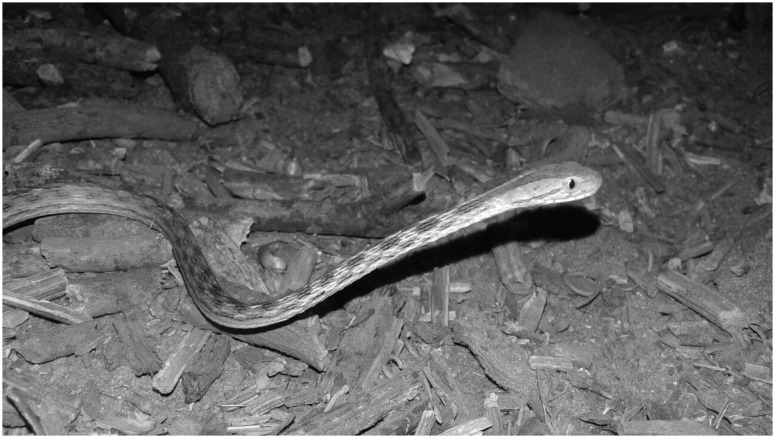
In January 7th 2018, at 16:30 h, an ophidic accident occurred during a contention activity at the Laboratory of Animal Ecology of Universidade Federal da Paraíba (UFPB), Rio Tinto municipality, Paraiba State, Northeast Brazil. A female undergraduate researcher (21 years old, 57 kg, 1.57 m) was bitten in her left hand between the index finger and the middle finger by a female specimen of T. pallidus (20 g, 548 mm of total length) (Figure 1). The animal was captured at 19:39 h in December 18th 2017 in a survey inventory of an Atlantic Forest patch of Reserva Biológica Guaribas (-6.806826°S, −35.087114°W; DATUM WGS84), located in Rio Tinto, PB (the specimen was collected under license SISBIO 59536-1). Since the capture, the animal was placed in a translucent plastic box (30X20X40 cm) with a water container, a tree branch and sawdust. In January 6th 2018, this animal gave birth to five hatchlings and the accident occurred during the transfer of the animals to individual boxes. The contact of the fangs with the researcher's hand lasted about 50 s, until another researcher removed the snake.
Figure 1. The snake Thamnodynastes pallidus responsible for the accident.
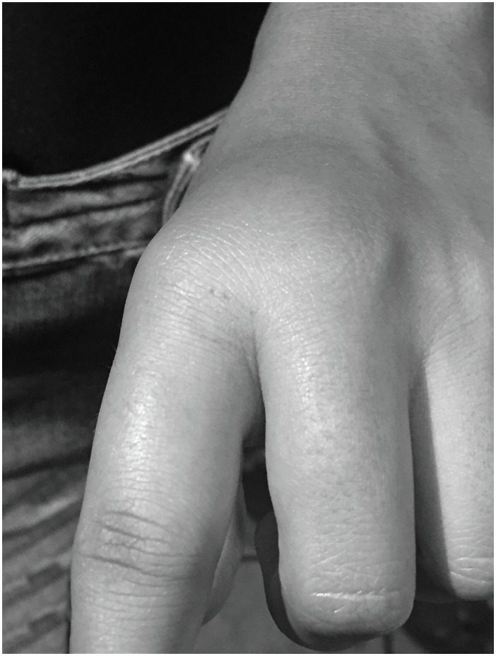
The injury marks were evident a few minutes after the bite; the lesion presented a small bleeding, accompanied by redness around the wound, swelling, itch and pain of small intensity radiated through all her left hand. The edema reached its maximum size (6 X 4.5 cm) a few minutes after the bite (Figure 2). One hour later (at 17:33 h) the patient presented migraine. As the patient did not present serious health complications and the symptoms diminished four hours after the bite, she was not taken to the hospital. She went home where she applied only ice on the injured area and was kept in observation during the first 24 h. No medication was used during the period of intoxication. After 36 h, the symptoms had completely disappeared.
Figure 2. Swelling of the victim's hand bitten by a Thamnodynastes pallidus specimen.
DISCUSSION
Here we present the second documented case of an ophidic accident caused by the “non-venomous” snake Thamnodynastes pallidus, and the first case in Brazil. Despite the genus Thamnodynastes is mentioned in literature as a potential agent of snakebite accidents 2 , 7 , 15 , only one other snakebite record by T. pallidus was well-documented in Venezuela by Diaz et al. 16 . In both cases, the bites were in the hand when people were handling the animal and the contact occurred for more than 40 seconds. This indicates an aggressive behavior of T. pallidus when handled. In the case presented by Diaz et al. 16 the snake was a male and in our report it was a female, showing that this aggressiveness may be independent of the specimen sex. Hands and arms are the most frequently bitten parts of the body in accidents with “non-venomous” snakes 2 .
The symptoms of envenomation by Thamnodynastes species that are described in literature can include edema, radiating pain, ecchymotic lesions, high local temperature, excessive salivation with metallic taste, strong headache and also hemorrhagic and proteolytic effects 15 , 17 . However, in both documented cases with T. pallidus the victims symptoms are not severe and they completely disappeared within 36 h, so that the T. pallidus’ venom can be seen as less dangerous compared to other dipsadinae snakes such as Phalotris and Philodryas 2 . The recommended treatment in T. pallidus accidents, like in other cases of bite by “non-venomous” dipsadinae snakes, is with non-hormonal painkillers and antipyretic ice over the swollen area 15 , some liquid intake, resting and observation.
It is important to emphasize that, in cases of envenomation by any species, the recommendation is to look for medical care in a close unity and keep the snake for identification in order to expedite the correct treatment 15 . Herein, a snake specialist already acquainted with non-venomous snake bites monitored the victim.
Snake bites cause lesions that can often result in secondary infections 18 . These infections result from propitious conditions to the growth of microorganisms at the site of injury, these microorganisms are often associated with the buccal flora of snakes 19 . No sign of secondary infection caused by the bite of T. pallidus was observed in this case corroborating the report by Diaz et al. 16 Relevant secondary infections caused by colubrids are more difficult to occur because the venom of these animals has no proteolytic action 18 .
Opisthoglyphous snakes are generally treated with negligence and commonly classified as non-dangerous snakes, both for their limited capacity of injecting venom and for the low risk of their toxin's effects in humans 16 , 20 . However, many cases of envenomation by Opisthoglyphous snakes have already been reported in the literature, some with serious clinical manifestations and even with fatal outcomes 2 , 7 , 15 . Our report reinforces the precaution and care that researches and students in herpetology must have in handling many of the snakes considered “non-venomous”.
ACKNOWLEDGEMENTS
PFA thanks CNPq for the PIBIC undergraduate grant, WMS thanks CAPES and FAPESQ for the current master's degree scholarship and RCF thanks FAPESB for her current PhD scholarship. We thank I. Pedrosa and two anonymous reviewers for their critical comments on the previous drafts. We thank Willianilson Pessoa for the snake picture. FGRF thanks the financial support from CNPq (Universal Grant N° 404671/2016-0).
Footnotes
FUNDING
This work is partially funded by CNPq Universal, grant N° 404671/2016-0.
REFERENCES
- 1.Brasil. Ministério da Saúde Sistema de Informação de Agravos de Notificação. Acidente por animais peçonhentos. [[cited 2018 June 11]]. Available from: http://tabnet.datasus.gov.br/cgi/tabcgi.exe?sinannet/cnv/animaisbr.def.
- 2.Salomão MG, Albolea ABP, Almeida-Santos SM. Colubrid snakebite: a public health problem in Brazil. Herpetol Rev. 2003;34:307–312. [Google Scholar]
- 3.Hedges SB, Couloux A, Vidal N. Molecular phylogeny, classification, and biogeography of West Indian racer snakes of the tribe Alsophiini (Squamata, Dipsadidae, Xenodontinae) Zootaxa. 2009;2067:1–28. [Google Scholar]
- 4.Zaher H, Grazziotin FG, Cadle JE, Murphy RW, Moura-Leite JC, Bonatto SL. Molecular phylogeny of advanced snakes (Serpentes, Caenophidia) with an emphasis on South American xenodontines: a revised classification and descriptions of new taxa. Pap Avulsos Zool. 2009;49:115–153. [Google Scholar]
- 5.Costa HC, Bérnils RS. Repteis do Brasil e suas unidades federativas: lista de espécies. Herpetol Bras. 2018;7:11–57. [Google Scholar]
- 6.Peichoto ME, Tavares FL, Santoro ML, Mackessy SP. Venom proteomes of South and North American opisthoglyphous (Colubridae and Dipsadidae) snake species: a preliminary approach to understanding their biological roles. Comp Biochem Physiol Part D Genomics Proteomics. 2012;7:361–369. doi: 10.1016/j.cbd.2012.08.001. [DOI] [PubMed] [Google Scholar]
- 7.Prado-Franceschi J, Hyslop S. South America colubrid envenomations. Toxin Rev. 2002;21:117–158. [Google Scholar]
- 8.Pereira GA, Filho, Alves RR, Vieira WL, França FG. Serpentes da Paraíba: diversidade e conservação. João Pessoa: edição dos autores; 2017. [Google Scholar]
- 9.Cunha OR, Nascimento FP. Ofídios da Amazônia. As cobras da região leste do Pará. Belém: Museu Paranaense Emilio Goeldi; 1993. [Google Scholar]
- 10.Vitt LJ, Vangilder LD. Ecology of a snake community in northeastern Brazil. Amphib Reptil. 1983;4:273–296. [Google Scholar]
- 11.Nóbrega RP, Montingelli GG, Trevine V, Franco FL, Vieira GH, Costa GC, et al. Morphological variation within Thamnodynastes pallidus (Linnaeus, 1758)(Serpentes: Dipsadidae: Xenodontinae: Tachymenini) Herpetol J. 2016;26:165–176. [Google Scholar]
- 12.Bernarde PS, Albuquerque S, Barros TO, Turci LC. Serpentes do estado de Rondônia, Brasil. Biota Neotrop. 2012;12:1–29. [Google Scholar]
- 13.Silva MV, Souza MB, Bernarde PS. Riqueza e dieta de serpentes do Estado do Acre, Brasil. Rev Bras Zoocien. 2010;12:165–176. [Google Scholar]
- 14.Silva NJ, Jr, Cintra CE, Silva HLR, Costa MC, Souza CA, Pachêco AA, Jr., et al. Herpetofauna, Ponte de Pedra Hydroelectric Power Plant, states of Mato Grosso and Mato Grosso do Sul, Brazil. Check List. 2009;5:518–525. [Google Scholar]
- 15.Puorto G, França FO. Serpentes não peçonhentas e aspectos clínicos dos acidentes. In: Cardoso JL, França HW, Málaque CM, Haddad V Jr., editors. Animais peçonhentos no Brasil: biologia, clínica e terapêutica dos acidentes. 2ª ed. São Paulo: Sarvier; 2009. pp. 125–131. [Google Scholar]
- 16.Diaz F, Navarrete LF, Pefaur J, Rodrigues-Acosta A. Envenomation by neotropical opistoglyphous colubrid Thamnodynastes cf. pallidus Linneé, 1758 (Serpentes: Colubridae) in Venezuela. Rev Inst Med Trop Sao Paulo. 2004;46:287–290. doi: 10.1590/s0036-46652004000500011. [DOI] [PubMed] [Google Scholar]
- 17.Ching AT, Paes Leme AF, Zelanis A, Rocha MM, Furtado MF, Silva DA, et al. Venomics profiling of Thamnodynastes strigatus unveils matrix metalloproteinases and other novel proteins recruited to the toxin arsenal of rear-fanged snakes. J Proteome Res. 2012;11:1152–1162. doi: 10.1021/pr200876c. [DOI] [PubMed] [Google Scholar]
- 18.Silva PR, Vilela RV, Possa AP. Infecções secundarias em acidentes ofídicos: uma avaliação bibliográfica. Estud Vida Saude. 2016;43:17–26. [Google Scholar]
- 19.Andrade JG, Pinto RN, Andrade AL, Martelli CM, Zicker F. Estudo bacteriológico de abscessos causados por picada de serpente do gênero Bothrops. Rev Inst Med Trop São Paulo. 1989;31:363–367. doi: 10.1590/s0036-46651989000600001. [DOI] [PubMed] [Google Scholar]
- 20.Hess PL, Squaiella-Baptistão CC. Toxinas animais: serpentes da família Colubridae e seus venenos. Estud Biol Ambiente Divers. 2012;34:135–142. [Google Scholar]


