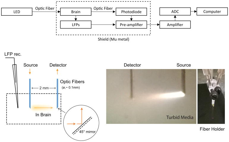
Fig. 1.
Simultaneous electrophysiology and IOS recordings with transmission measurement. The schematic illustration of simultaneous LFP and IOS recordings is shown in the top panel. The LED light is coupled into the optical fiber to targeted brain sites and transmitted through the sampled tissue. The detection fiber of the transmission probe collects the light to a photodiode. Both photodiode and LFP recording signals from the same site were ×10 pre-amplified respectively, then amplified and digitized. The animal and recording system were enclosed in a full-band electromagnetic shield during data collection. (Left bottom)Schematic representation of transmission measurement for the target area’s tissue-level neural activity using the minimally-invasive fiber pair. The mirror-coating of the 45° tips that allows light to turn 90° between source emission and detector collection with the parallel fiber-pair configuration is illustrated. (Middle bottom) Lighting (full-band wavelengths) path is demonstrated, captured in the turbid liquid of a tissue-simulating phantom. The fiber pair was assembled on a holder, shown in the right bottom panel, which was calibrated to ensure 2-mm separation and equal length.

