Figure 8.

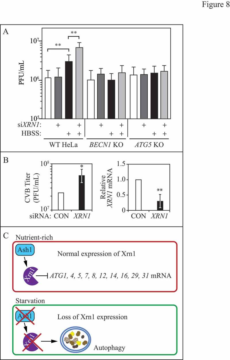
XRN1 depletion enhances virus infection in mammalian cells. (A) PV titers (plaque-forming units/ml; PFU) were determined by plaque assay in WT, BECN1 or ATG5 CRISPR KO HeLa cells transfected with scrambled control or XRN1-targeted siRNA. Cells were starved in HBSS for 1 h prior to infecting with PV at a multiplicity of infection of 0.1, and remained in starvation medium until harvested at 6 h post infection. Results shown are the mean of 3 independent experiments. (B) CVB titers (PFU/ml) as determined by plaque assay in HeLa cells (left). HeLa cells underwent plaque assays with supernatants from CVB-infected HBMECs expressing either control or XRN1-targeted siRNA. Results shown are the mean of 4 independent experiments. RT-qPCR KD of XRN1 mRNA levels in HBMECs (right). Results shown are the mean of 4 independent experiments. (C) Schematic representation of the model by which Xrn1 functions as a negative autophagy regulator in yeast.
