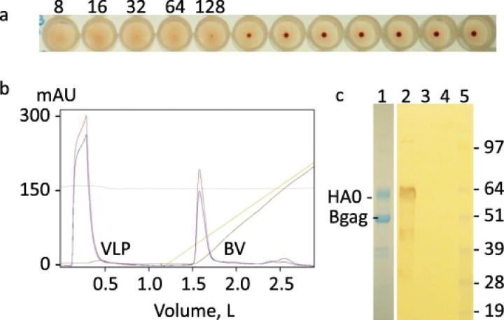
Fig. 2.

Preparation and characterization of multi-clade H5N1 VLPs from 2 L culture of Sf9 cells. (a) HA assay of Sf9 culture supernatant. Dilutions of VLPs are indicated. (b) Ion exchange chromatography profile of purification of VLPs. Location of VLP and rBV peaks are shown. (c) SDS-PAGE staining (left panel, lane 1) and western blot of purified VLPs (right panel, lanes 2-5). Western blot was done using H5-specific mouse IgG2a monoclonal antibody (H5N1) IT-003- 005M6. Lanes 1 and 2, purified VLPs; lanes 3 and 4 negative control; lane 5, protein molecular weight marker SeeBlue Plus 2. Positions of H5 HA0 and of Bgag proteins are indicated.
