Abstract
Many conventional studies on molecular hydrogen have not examined cell migration ability and the relationship between apoptosis and the cytoskeleton. Here we investigated the influence of hydrogen-occluding silica microparticles (H2-silica) on cell migration motility and changes of the cytoskeleton (F-actin) in normal human esophageal epithelial cells (HEEpiCs). As the results, cell migration was promoted, and formation of microvilli was activated in the 100 ppm (low concentration) scratched group. After performing a wound healing assay, cells exhibited migration after 48 hours and 72 hours for both 10 ppm and 100 ppm groups, suggesting that the wound-repairing effects could be attributed to the antioxidant ability of H2-silica. In scratched groups, high levels of activated caspase-3 were relatively expressed and presented a tendency to increase the observed Bax/Bcl-2 ratio at more than 300 ppm groups. The above-mentioned results show that H2-silica induced apoptosis in HEEpiCs, especially in the scratched cells. Toxicity may cause an exaggerated apoptosis. Furthermore, since the ratio of fascin/tubulin in the 100, 300, and 600 ppm groups tended to increase in both the scratched and the non-scratched control groups, H2-silica was thought to be able to promote fascin action on normal cells and may be have a proliferative effect.
Keywords: molecular hydrogen, hydrogen water, hydrogen-occluding-silica microparticle, normal human esophageal epithelial cells, cell migration, cytoskeleton, filopodia, microvillus, apoptosis-inducing effect, reactive oxygen species
INTRODUCTION
Hydrogen is the most abundant and simplest chemical element in the universe and the human body. The molecular weight of hydrogen is very small, it has a large diffusion capacity, and it has no toxicity or selective neutralizing to toxic free radicals.1,2,3 In 2007, Ohsawa et al.4 showed that inhaled hydrogen gas (2%) produced a selectively protective effect against hydroxyl radicals generated by shock caused by cerebral post-ischemic reperfusion injury in animal experiments. They concluded that molecular hydrogen has antioxidant effects and antiapoptotic properties.4 Many previous studies in hydrogen medicine have shown that molecular hydrogen has antioxidant, antiapoptotic, and preventive effects against oxidative stress in vitro and in vivo, and in clinical research.5,6,7,8 However, the use of hydrogen gas has severe handling restrictions in hospitals, medical facilities, and laboratories due to its explosive and other physicochemical properties. Therefore, in the present study, we used hydrogen-occluding-silica microparticles (H2-silica), which are silsesquioxane-based compounds with hydrogen interstitially embedded in a matrix of caged silica.9
In our previous study, the effect of H2-silica on cell migration was measured, and the fact was found that H2-silica is more effective in inhibiting migration of cancer cells than normal cells.10 It suggests that H2-silica can inhibit the metastasis of cancer cells in vitro, it also suggests that damaged normal cells would be restored and wound-healing effect would appear at the same time.
Several previous studies on hydrogen medicine reported the relationship between wound repair in normal human epithelial cells and hydrogen water (HW) in vitro.11,12 In another study, we used HW to treat patients with ulcer pressure in a clinical study.13 Meanwhile, in an in vitro study, we examined whether HW has a reactive oxygen species (ROS)-scavenging ability and exerts wound-repair effects as well as promoting type I collagen production in fibroblasts.13 However, none of these studies used western blot analysis to explore signaling pathways involved and did not estimate the cell migration ability, and consequently did not focus on the relationship between apoptosis and the cytoskeleton.
Cell migration depends on the depolymerization and remodeling of the cytoskeleton, in particular the actin fiber, as well as the fascin that is an actin bundling protein.14 Therefore, in the present study, we investigated whether H2-silica affects the reconstitution and the expression level of these proteins upon healing process from scratching on normal cells.
Apoptosis refers to the autonomic and orderly death of cells that is controlled by diverse genes. During apoptosis, the cell morphology undergoes a series of characteristic changes. In recent years, the cytoskeleton has been found to change in response to factors including degradation, cohesion, and uneven distribution of cytoskeleton network structure. Many studies on the biological activity and function of cytoskeletal proteins, dynamic changes in cytoskeleton morphology, and related regulatory factors have shown that changes to the apoptotic cytoskeleton are the basis of the morphological changes of apoptosis, and changing the cell skeletal structure could induce apoptosis.15,16,17 Thus, the relation to apoptosis was also examined.
We observed the influence of H2-silica on the behavior of cell migration, including the formation of filopodia and microvillus, and changes to the cytoskeleton (F-actin). Proliferation-promoting effects on epithelial cells damaged by wound healing assay, apoptosis-inducing effects, and changes to the cytoskeleton accompanying apoptosis were investigated, and western blot analysis was performed to explore the signal pathway.
MATERIALS AND METHODS
Cell culture
Normal HEEpiCs were purchased from ScienCell Research Laboratories (CA, USA) via Cosmo Bio Co., Ltd. (Tokyo, Japan). HEEpiCs were grown in epithelial cell medium-2 (EpiCM-2, ScienCell, San Diego, CA, USA) in a humidified atmosphere with 5% CO2 in air at 37°C. Once the cells reached 80% confluence, as observed under an inverted microscope (TCM400FLR, LaboMed America Inc., Fremont, CA, USA), they were passaged with 0.25% (w/v) trypsin and 0.03% (w/v) trypsin inhibitor (Gibco, Darmstadt, Germany). HEEpiCs were seeded at a density of 4.0 × 10⁵ cells per well in 6-well plates (BD Biosciences, Franklin Lakes, NJ, USA) for each assay. Cell density was changed in accordance with well numbers of the microplates used in the experiments.
Preparation and administration of H2-silica
H2-silica was manufactured as silica hydride “Microcluster,” kindly supplied from New-1-Ten-Rin Enterprise Co., Ltd. (Changhua, Taiwan, China). Prior to the administration of H2-silica, basic parameters such as pH, temperature, and oxidization reduction potential were measured. Dulbecco's Modified Eagle Medium was mixed with H2-silica at concentrations of 10, 40, 70, 100, 300, 600, 900, or 1200 ppm and applied to HEEpiCs that were previously incubated for 72-96 hours. Assays were performed after further culturing for 72 hours post-scratching.
Wound healing assay to confirm wound repair rate
The wound healing assay used in this study consisted of two methods. One method was performed using the tip of a 200 μL micropipette to make a straight scratch on a confluent monolayer of HEEpiCs in a 6-well plate in order to form a wound. The cells were then rinsed twice with Dulbecco's phosphate buffered saline (D-PBS (−)) before adding an EpiCM-2 culture medium containing H2-silica. Scratches were made with the pipette tip at an angle of around 30° to keep the scratch width limited. This allowed imaging of both wound edges using the 40× objective lens of a microscope. The H2-silica was then added to the EpiCM-2 culture medium at different concentrations (0, 10, 100, 300, 600, and 1200 ppm). Once the cells reached ~90% confluence, generally after 72 hours, wound closure in the area was photographed using an inverted phase microscope (CK2, Olympus, Tokyo, Japan) at 40× magnification. The wound closure areas were randomly selected so as to be evaluated as statistically significant differences and calculated (mm2) in order to show the closure of the wounds at each time period.18,19 The second method used a cell scraper (99010, TPP Techno, Trasadingen, Switzerland) for cells cultured in T25 flask (BD Biosciences). The cell scraper was 13 mm wide with a fixed blade and was scratched back and forth six times at random areas of the bottom of each T25 flask.
Crystal violet staining to assess cell morphological changes
Crystal violet staining was executed as previously described.3,20 First, the media in 6-well plates were removed, and HEEpiCs were washed with D-PBS (−), fixed in 99% methanol, and stained for 5 minutes with 0.5% crystal violet (Wako Pure Chemical Industries, Osaka, Japan) dissolved in methanol/water. Subsequently, excess staining dye was removed, and the wells were rinsed thoroughly with running water until no additional stain leached from the wells. To determine cell morphological changes, we took photographs in more than 20 fields per well using a 400× objective.
Alexa 488 phalloidin and 4′,6-diamidino-2-phenylindole (DAPI) staining to detect the cytoskeleton
HEEpiCs were harvested and fixed using cold 2% formaldehyde and 0.002% saturated picric acid in 0.1 M phosphate buffer, pH 8.0, for 4 hours followed by overnight immersion in buffer containing 30% sucrose. The cells were stored at −70°C until used. To stain for F-actin, tissue sections were incubated with Alexa-488-conjugated phalloidin (1:100 in 1% bovine serum albumin (BSA), Invitrogen, Carlsbad, CA, USA) for 20 minutes at room temperature, followed by incubation with 1 g/mL DAPI for nuclear staining (Sigma-Aldrich, St. Louis, MO, USA). Then, 1 mL of cold PBS (–) was added to each well and stained cells were examined with a fluorescence microscope and photographed with a camera (MT5310H, Meiji Techno Co., Ltd., Tokyo, Japan). Individual images generated from the green and blue channels were superimposed to generate the composite figures. Computerized morphological analysis was performed using ImageJ software (https://ImageJ.nih.gov/ij). To clearly show the microvilli of the HEEpiCs, look-up-table (LUT) method of ImageJ were used. To elevate the pixel qualitatively, the obtained photo was treated with a fast Fourier transform (FTT) bandpass filter of ImageJ to form a pseudo-image.
Western blot assays to detect activated caspase-3, Bax/Bcl-2, α-tubulin, and fascin
As previously described,10 HEEpiCs treated with H2-silica were seeded at approximately 1 × 10⁶ cells per T25 flask and further cultured for 72-96 hours. Cells were collected using a small rubber scraper (TR9000, TrueLine, Nashville, TN, USA), centrifuged at 5000 r/min at 4°C for 5 minutes, then washed twice in ice-cold PBS (–). Thereafter, the cell pellet was homogenized in 60 μL of cold lysis buffer (150 mM NaCl, 10 mM Tris–HCl (pH 7.4), 1% Triton X-100, 1% NP-40, 5 mM EDTA (pH 8.0), and 1/200 vol. of Protease Inhibitor Cocktail Set III (Calbiochem, CA, USA, #539134)). Following centrifugation at 14,000 r/min at 4°C for 5 minutes, 7 μL of the supernatant was mixed with 1/2 volume of 4× NuPAGE LDS sample buffer (Invitrogen) and 1/4 volume of 10× NuPAGE antioxidant (Invitrogen), and run on a 13.5% acrylamide sodium dodecyl sulfate polyacrylamide gel electrophoresis (SDS-PAGE) gel. Additional procedures were the same as reported in our previous study.10 Anti-Bax (BioLegend, CA, USA, #625901), anti-Bcl-2 (eBioscience, CA, #141028), anti-α-tubulin, and anti-fascin-1 (Santa Cruz Biotechnology Inc., Dallas, TX, USA, #SC-21743) were utilized as the primary antibodies, and Jackson Peroxidase AffiniPure Goat Anti-Mouse IgG (H+L) (#115-035-003) 2.0 mL was utilized as the secondary antibody. Finally, both the ratio of the intensity of activated caspase-3 versus native caspase-3 and the ratio of the intensity of Bax versus Bcl-2 were used to evaluate the occurrence of apoptosis in HEEpiCs. The intensities of protein bands were measured using the column average plot function of the ImageJ software. The ratio of the intensity of fascin versus α-tubulin was used to evaluate the occurrence of apoptosis and cytoskeleton in HEEpiCs.
Statistical analysis
Conventional statistical methods were used to calculate the results obtained from the wound healing assay, crystal violet staining, and western blot analysis. Data were expressed as the mean ± SD and P-values below 0.05 were regarded to be statistically significant. In addition, the repeated ANOVA analysis was used for measuring longitudinal data. All data were analyzed statistically using Excel Statistical 2012 Software for Windows (Social Survey Research Information Co., Ltd., Tokyo, Japan).
RESULTS
Cell migration rate of HEEpiCs to cause ability of closure to care wound area
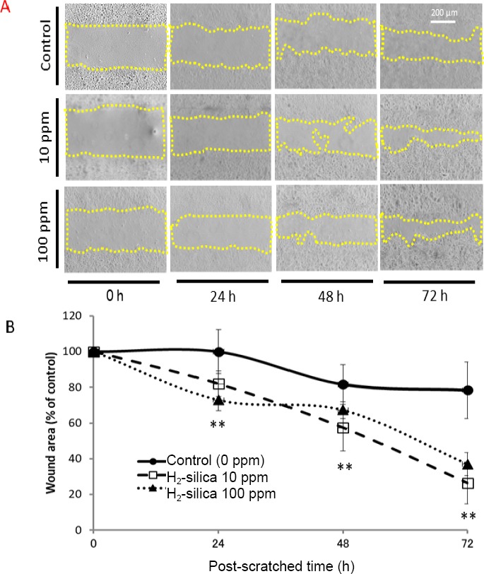
Cell migration is associated with cellular developmental and proliferative processes. In order to access the influence of H2-silica on cell migration of HEEpiCs, we performed a wound healing assay. As shown in Figure 1, images of wounds at 0, 24, 48, and 72 hours after scratching showed protrusions characteristic of migration after 24 hours for H2-silica at 10 ppm and 100 ppm, and eventual healing of the wound after 72 hours.
Figure 1.

Time-lapse changes to cell migration of HEEpiCs after the wound healing assay.
Note: (A) Images of wounds in HEEpiCs at 0, 24, 48, and 72 hours (h) post-scratching. Bar: 200 μm (40× magnification). (B) Rates of wounded area relative to the start time of scratch shows the relationship between duration and hydrogenoccluding silica microparticles (H2-silica) at 0, 10, and 100 ppm. Data were expressed as the mean ± SD and evaluated with the repeated ANOVA analysis. **P < 0.01.
Changes to the end edge (non-wound edge) 72 hours after wound healing assay
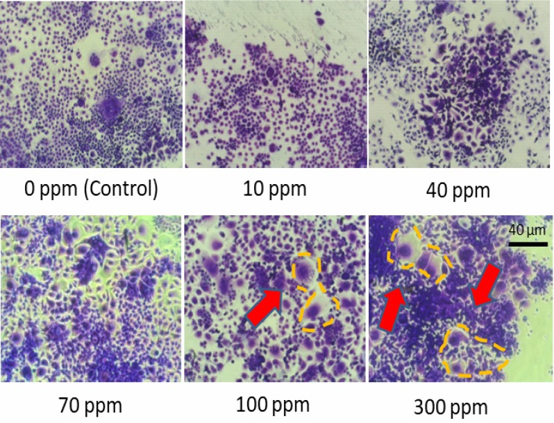
According to the wound healing assay, scratching will cause the wound edge that was designated as the leading edge, and the non-wound edge that was designated as end edge, existing the opposite outside. We observed filopodia and microvilli at the wound edge, and an apoptosis-like phenomenon at the non-wound edge. In the present study, cell death phenomena occurred severely at the end edge 72 hours after performing the wound healing assay and according to the concentration of administrated H2-silica. Figure 2 shows that H2-silica caused significant apoptosis-like features on HEEpiCs in a dose-dependent manner at lower concentrations. Huang et al.21 reported that if cells were seeded at a very low density, the bystander effect and apoptosis induction was dramatically reduced. H2-silica may result in an apoptotic bystander effect, and the reason may be related to the density of cultured cell.
Figure 2.

Changes to the end edge (non-wound edge) 72 hours after the wound healing assay adding hydrogen-occluding silica microparticles (H2-silica).
Note: A more obvious appearance of apoptosis-like morphological changes was observed with increasing concentrations of hydrogen-occluding silica microparticles (H2-silica). Bar: 40 μm (200× magnification).
Immunofluorescent phalloidin/4’,6-diamidino-2-phenylindole(DAPI) staining of HEEpiCs.
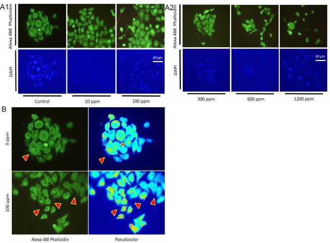
Phalloidin stain showed a clear increase in microvilli, confirmed to be extended from filopodia of HEEpiCs. Compared with the 0 ppm H2-silica group, the 100 ppm H2-silica group showed an increase in microvilli. DAPI staining showed the number of cell nuclei tended to decrease with the concentration of H2-silica at more than 300 ppm of H2-silica, indicating cell death such as apoptosis occurred. However, a proliferation phenomenon was observed in the 100 ppm scratched group, but a growth inhibitory effect was observed in the high concentration group (≥300 ppm) (Figure 3A). It should be emphasized that numerous protrusions caused by actin filament-bounding protein, fascin, such as microvilli, appeared along all sides of the cell periphery of HEEpiCs in 100 ppm group (indicated by the arrowheads in Figure 3B. As for the formation of microvilli, a certain unique change is seen together with filopodia (Figure 3B). Filopodia is more developed in the 100 ppm group than in the 0 ppm group. Observation with an electron microscope in the future is expected to make the microvilli appear more outstanding than one using in visual quality.
Figure 3.
Immunofluorescent phalloidin/4’,6-diamidino-2-phenylindole (DAPI) staining of HEEpiCs
Note: (A1, 2) The actin cytoskeleton was visualized by staining with Alexa 488 phalloidin (green, the upper panel). Cell nuclei were counterstained with DAPI (blue, the lower panel). The hydrogen-occluding silica microparticles (H2-silica) concentrations were 0 (control), 10, 100, 300, 600, and 1200 ppm. Scale bar: 50 μm (400× magnification). (B) To present the features of Alexa 488 phalloidin more sharply, LUT (look-up table) images in pseudocolor treated with ImageJ were obtained to quantitatively evaluate the number of microvilli.
Western blot analysis to detect activated caspase-3, Bax/Bcl-2, α-tubulin, and fascin
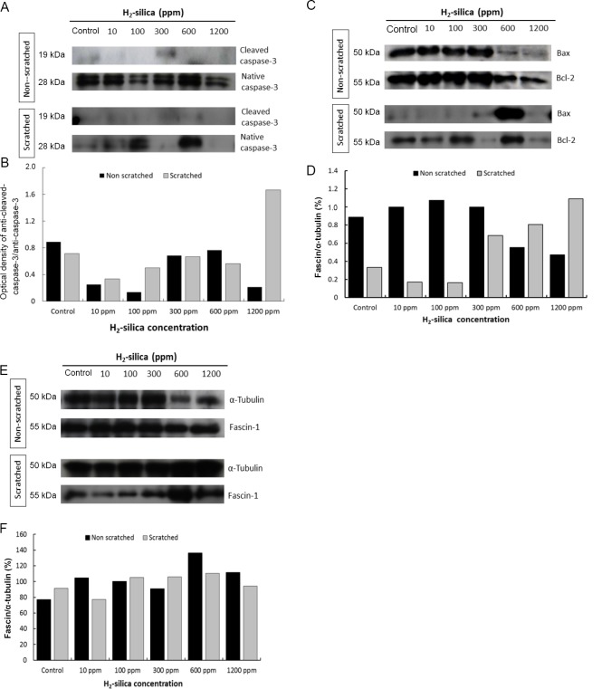
Western blotting was carried out in the H2-silica groups at concentrations of 0 (control), 10, 100, 300, 600, and 1200 ppm. Expression of uncleaved caspase-3 and activated caspase-3 was first measured (Figure 4A). Caspase-3 is regarded as a landmark of apoptotic pathways. The 17 kDa bands that corresponded to processed and activated caspase-3 were expressed in non-scratched HEEpiCs after 72 hours (Figure 4A) in 10 ppm, 300 ppm, and 600 ppm. On the other hand, in every group, 72 hours post-scratch HEEpiCs showed an increase in activated caspase-3 expression compared with the control group (Figure 4B), especially in 1200 ppm. These results suggested that H2-silica promoted apoptosis in the scratched HEEpiCs.
Figure 4.
Western blotting analysis of caspase-3, Bax/Bcl-2, α-tubulin, and fascin.
Note: (A, C, E) Immuno-detected bands for activated caspase-3 (19 kDa) and native caspase-3 (28 kDa) (A), Bax (19 kDa) versus Bcl-2 (28 kDa) (C), and fascin-1 (55 kDa) versus α-tubulin (50 kDa) (E) for the non-scratched group and the scratched group at 0 ppm (control), 10, 100, 300, 600, and 1200 ppm concentrations of H2-silica in HEEpiCs. The ratios (%) of band optical densities and expression levels of activated caspase-3 versus native caspase-3 (B), Bax versus Bcl-2 (D), and α-tubulin/fascin-1 (F) for the non-scratched group and the scratched group at 0 (control), 10, 100, 300, 600, and 1200 ppm concentrations of H2-silica in HEEpiCs were analyzed by densitometry via the ImageJ software, respectively. H2-silica: Hydrogen-occluding silica microparticles.
Second, Bax/Bcl-2 ratio was examined as another index of cell survival or mortality. These members of the Bcl-2 family include regulator proteins that adjust the degree of cell death (apoptosis). In the non-scratched group, there was a gradual decrease compared with the control group. Compared with the control group, the scratched group presented a tendency to increase the observed Bax/Bcl-2 ratio at more than 300 ppm group (Figure 4C, D).
Then, we used fascin to determine the signaling pathways involved in cytoskeleton expression using α-tubulin as an internal control. Fascin is a 55 kDa actin crosslinking protein and represents a family of actin-bundling proteins including sea urchin fascin and HeLa 55 kDa actin-bundling protein.22,23 It was concluded that fascin is associated with the formation of filopodia in coelomocytes. The expression of fascin/tubulin is increased in HEEpiCs at a lower concentration (10 ppm for non-scratched and 100 ppm for scratched) of H2-silica (Figure 4E, F), a little greater in non-scratched than scratched HEEpiCs. A remarkable difference of Fascin/Tubulin ratio was not appeared between the scratched groups and non-scratched groups.
DISCUSSION
In this in vitro study, several experimental methods were used to verify the biological effects of H2-silica on cell migration behavior of HEEpiCs.24,25,26
First, cell migration and filopodia formation were used as indicators. After performing a wound healing assay, cells exhibited characteristic migration protrusions after 24 hours and showed wound healing after 72 hours for both of 10 ppm and 100 ppm H2-silica groups (Figure 1), suggesting that the wound-repairing effects could be attributed to the antioxidant ability of H2-silica.
In addition, an apoptotic bystander phenomenon was observed at the non-scratched side with administration of a low concentration (100 ppm) of H2-silica (Figure 2). The reason may be related to the density of cultured cell. Huang et al reported that if cells were seeded at a very low density, the bystander effect and apoptosis induction was dramatically reduced.21
On the other hand, the cytoskeleton of eukaryotic cells is composed of microfilaments, microtubules, and intermediate diameter fibers. At the stage of cell division, microtubules are involved in determining the spindle combination and location, and the dynamic instability of the microtubule influences cell replication and apoptosis. Most microfilaments are concentrated directly under the cell membrane. It has the role of resisting tension, maintaining cell shape, and forming cytoplasmic protrusions, such as filopodia and microvilli.27,28
Phalloidin staining showed a clear increase in microvilli that was confirmed by filopodia in HEEpiCs. Compared with the control group (0 ppm), microvilli increased after administration of 100 ppm of H2-silica. A proliferation phenomenon was observed in HEEpiCs by DAPI staining in the 100 ppm group, but a growth suppression effect was observed in the high concentration groups (300 ppm or more). This suggested that wounds can be healed to some extent by H2-silica administration.
Second, apoptosis-like features of HEEpiCs and dose-dependently magnificently appeared 72 hours after H2-silica administration, suggesting that the injured cells induced apoptosis by the scratching-wound, resulting in autophagy and phagocytosis had not been promoted by H2-silica.
By the apoptosis assay using western blot, expression of native caspase-3 and anti-activated caspase-3 was detected. A 17 kDa band corresponding to cleaved and activated caspase-3 was expressed but equal levels with the control (0 ppm) in each non-scratched group after 72 hours. Each group of scratched HEEpiCs showed equal levels of caspase-3 expression compared with the control group lower, in 1200 ppm elevated expression of activated caspase-3, suggesting that H2-silica promotes cellular apoptosis of HEEpiCs (Figure 4A, B).
Using the Bax/Bcl-2 ratio as another index of apoptosis, non-scratched cells showed that H2-silica inhibited Bax expression and could inhibit apoptosis of HEEpiCs. But, Bax/Bcl-2 ratio in scratched cells increased at more than 300 ppm groups. Bax/Bcl-2 and caspase-3 are involved in cell apoptosis of HEEpiCs at high concentrations of H2-silica in scratched cells.
Caspase-3 is the downstream regulating protein of Bcl-2 and the initial factor of apoptosis. Overexpression of Bcl-2 effectively suppresses caspase-3 activity and the occurrence of cell apoptosis. It is possible to induce cell apoptosis through cytoskeletal changes and cytoskeletal changes can be regarded to precede cell apoptosis.29,30
In our previous studies, fascin exerted an important function in cell migration and filopodia formation.10 Since microfilaments exert important actions in processes such as cell migration and pseudopodia formation, formation of filopodia and microvilli can be used as an indicator of cell proliferation. In this study, cell migration was promoted, and formation of microvilli was activated in the 100 ppm (low concentration) scratched group, where a proliferation effect was seen. Furthermore, since the ratio of fascin/tubulin in the 100, 300, and 600 ppm groups slightly increased in both the scratched and the non-scratched control groups, H2-silica was thought to be able to promote fascin action and have a proliferative effect on normal cells.
The high concentrations of H2-silica used in this experiment, like other strongly antioxidant reagents, have a harmful effect on KYSE-70 and HEEpiCs, highlighting the difficulty of antioxidant handling in clinical practice. H2-silica is not only portable and easy to store but also safer than the H2-generating apparatus which is expensive, difficult to manipulate, and has the potential to explode.26 H2-silica can generate large amounts of hydrogen when it comes in contact with aqueous liquid, resulting in the scavenging of ROS.31,32,33,34 Kato et al.9 used H2-silica to investigate the suppressive efficacy against melanogenesis in HMV-II human melanoma cells and levodopa (L-DOPA)-tyrosinase reaction. They found that H2-silica has the potential to prevent melanin production against ultraviolet A and serves as a skin-lightening ingredient for supplements or cosmetics.9
The skin consists of an outer squamous epithelium, named the epidermis, and an inner connective tissue, named the dermis. Cell death in the epithelial tissues exhibits physiologic and pathological processes. Molecules involved in cell death that have a physiological meaning in mammals are called the caspase family and are called proteases that promote cell death. As well known, it is due to the action of apoptosis which usually occurs in interdigit-regions of mitte hands and feet of human fetus at the embryogenetic stage and then fingers and toes are formed one by one. In addition, a fetal eyelids form an opening also by the process of apoptosis.35 The results obtained from our study seem to have the same mechanism that the formation of filopodia and microvilli increased with scratched esophageal epithelial cells repaired. Toxicity may cause an exaggerated apoptosis.
Finally, we should pay attention to the antioxidant effect, convenience, and inexpensiveness of H2-silica, but on the other hand it should also be recognized as having its toxicity. Indeed, high concentrations of H2-silica have been found to be toxic to HEEpiCs. Toxicity may be due to the attribute of silica particles. Maybe this is because HW without silica particles exhibits no cytotoxicity even at the correspondingly high dissolved-hydrogen concentrations, and, additionally because hydrogen has the smallest molecular weight and is able to diffuse quickly. Here we want to emphasize that apoptosis may be partially induced by toxins. In addition, we will add caspase-14 in the next research, because caspase-14 is clearly required for normal skin development. And caspase-14 is nonapoptotic, almost exclusively expressed in the suprabasal epidermal cells.36,37
Footnotes
Conflicts of interest
The authors declare no conflict of interest.
Financial support
This work was supported by a Grant-in-Aid for Scientific Research (KAKENHI No. 26350681 to QL) from the Ministry of Education, Culture, Sports, Science and Technology of Japan.
Copyright license agreement
The Copyright License Agreement has been signed by all authors before publication.
Data sharing statement
Datasets analyzed during the current study are available from the corresponding author on reasonable request.
Plagiarism check
Checked twice by iThenticate.
Peer review
Externally peer reviewed.
Open peer review report
Reviewer: Sheng Chen, Zhejiang University, China.
Comments to authors: In the present study, authors employed hydrogen-occluding silica microparticles for the wound healing assay, expressions of cytoskeleton molecular, and apoptosis signals. They found low concentration of H2-silica could promote the proliferation and apoptosis of normal human esophageal epithelial cells. But the high concentration H2-silica could be toxic to those cells. Generally, the author well performed the experiments, but there are still major issues that should be addressed.
Funding: This work was supported by a Grant-in-Aid for Scientific Research (KAKENHI No. 26350681 to QL) from the Ministry of Education, Culture, Sports, Science and Technology of Japan.
REFERENCES
- 1.Sun X, Nakao A. Amsterdam: Springer; 2015. Hydrogen Molecular Biology and Medicine. [Google Scholar]
- 2.Huang CS, Kawamura T, Toyoda Y, Nakao A. Recent advances in hydrogen research as a therapeutic medical gas. Free Radic Res. 2010;44:971–982. doi: 10.3109/10715762.2010.500328. [DOI] [PubMed] [Google Scholar]
- 3.Ishiyama M, Tominaga H, Shiga M, Sasamoto K, Ohkura Y, Ueno K. A combined assay of cell viability and in vitro cytotoxicity with a highly water-soluble tetrazolium salt, neutral red and crystal violet. Biol Pharm Bull. 1996;19:1518–1520. doi: 10.1248/bpb.19.1518. [DOI] [PubMed] [Google Scholar]
- 4.Ohsawa I, Ishikawa M, Takahashi K, et al. Hydrogen acts as a therapeutic antioxidant by selectively reducing cytotoxic oxygen radicals. Nat Med. 2007;13:688–694. doi: 10.1038/nm1577. [DOI] [PubMed] [Google Scholar]
- 5.Ohta S. Recent progress toward hydrogen medicine: potential of molecular hydrogen for preventive and therapeutic applications. Curr Pharm Des. 2011;17:2241–2252. doi: 10.2174/138161211797052664. [DOI] [PMC free article] [PubMed] [Google Scholar]
- 6.Yokota T, Kamimura N, Igarashi T, Takahashi H, Ohta S, Oharazawa H. Protective effect of molecular hydrogen against oxidative stress caused by peroxynitrite derived from nitric oxide in rat retina. Clin Exp Ophthalmol. 2015;43:568–577. doi: 10.1111/ceo.12525. [DOI] [PubMed] [Google Scholar]
- 7.Ohta S. Molecular hydrogen as a novel antioxidant: overview of the advantages of hydrogen for medical applications. Methods Enzymol. 2015;555:289–317. doi: 10.1016/bs.mie.2014.11.038. [DOI] [PubMed] [Google Scholar]
- 8.Li Q, Yu P, Zeng Q, et al. Neuroprotective effect of hydrogen-rich saline in global cerebral ischemia/reperfusion rats: up-regulated tregs and down-regulated miR-21, miR-210 and NF-kappaB expression. Neurochem Res. 2016;41:2655–2665. doi: 10.1007/s11064-016-1978-x. [DOI] [PMC free article] [PubMed] [Google Scholar]
- 9.Kato S, Saitoh Y, Miwa N. Inhibitions by hydrogen-occluding silica microcluster to melanogenesis in human pigment cells and tyrosinase reaction. J Nanosci Nanotechnol. 2013;13:52–59. doi: 10.1166/jnn.2013.6848. [DOI] [PubMed] [Google Scholar]
- 10.Li Q, Tanaka Y, Miwa N. Influence of hydrogen-occluding-silica on migration and apoptosis in human esophageal cells in vitro. Med Gas Res. 2017;7:76–85. doi: 10.4103/2045-9912.208510. [DOI] [PMC free article] [PubMed] [Google Scholar]
- 11.Li Q, Tanaka Y, Saitoh Y, Miwa N. Effects of platinum nanocolloid in combination with gamma irradiation on normal human esophageal epithelial cells. J Nanosci Nanotechnol. 2016;16:5345–5352. doi: 10.1166/jnn.2016.12362. [DOI] [PubMed] [Google Scholar]
- 12.Xiao L, Miwa N. Hydrogen-rich water achieves cytoprotection from oxidative stress injury in human gingival fibroblasts in culture or 3D-tissue equivalents, and wound-healing promotion, together with ROS-scavenging and relief from glutathione diminishment. Hum Cell. 2017;30:72–87. doi: 10.1007/s13577-016-0150-x. [DOI] [PubMed] [Google Scholar]
- 13.Li Q, Kato S, Matsuoka D, Tanaka H, Miwa N. Hydrogen water intake via tube-feeding for patients with pressure ulcer and its reconstructive effects on normal human skin cells in vitro. Med Gas Res. 2013;3:20. doi: 10.1186/2045-9912-3-20. [DOI] [PMC free article] [PubMed] [Google Scholar]
- 14.Edwards RA, Bryan J. Fascins, a family of actin bundling proteins1. Cell Motil Cytoskeleton. 1995;32:1–9. doi: 10.1002/cm.970320102. [DOI] [PubMed] [Google Scholar]
- 15.Costanzo A, Fausti F, Spallone G, Moretti F, Narcisi A, Botti E. Programmed cell death in the skin. Int J Dev Biol. 2015;59:73–78. doi: 10.1387/ijdb.150050ac. [DOI] [PubMed] [Google Scholar]
- 16.Monier B, Suzanne M. The morphogenetic role of apoptosis. Curr Top Dev Biol. 2015;114:335–362. doi: 10.1016/bs.ctdb.2015.07.027. [DOI] [PubMed] [Google Scholar]
- 17.Horbay R, Bilyy R. Mitochondrial dynamics during cell cycling. Apoptosis. 2016;21:1327–1335. doi: 10.1007/s10495-016-1295-5. [DOI] [PubMed] [Google Scholar]
- 18.Pinco KA, He W, Yang JT. Alpha4beta1 integrin regulates lamellipodia protrusion via a focal complex/focal adhesion-independent mechanism. Mol Biol Cell. 2002;13:3203–3217. doi: 10.1091/mbc.02-05-0086. [DOI] [PMC free article] [PubMed] [Google Scholar]
- 19.Kramer N, Walzl A, Unger C, et al. In vitro cell migration and invasion assays. Mutat Res. 2013;752:10–24. doi: 10.1016/j.mrrev.2012.08.001. [DOI] [PubMed] [Google Scholar]
- 20.Gillies RJ, Didier N, Denton M. Determination of cell number in monolayer cultures. Anal Biochem. 1986;159:109–113. doi: 10.1016/0003-2697(86)90314-3. [DOI] [PubMed] [Google Scholar]
- 21.Huang X, Lin T, Gu J, et al. Cell to cell contact required for bystander effect of the TNF-related apoptosis-inducing ligand (TRAIL) gene. Int J Oncol. 2003;22:1241–1245. [PubMed] [Google Scholar]
- 22.Matsudaira P. Actin crosslinking proteins at the leading edge. Semin Cell Biol. 1994;5:165–174. doi: 10.1006/scel.1994.1021. [DOI] [PubMed] [Google Scholar]
- 23.Otto JJ. Actin-bundling proteins. Curr Opin Cell Biol. 1994;6:105–109. doi: 10.1016/0955-0674(94)90123-6. [DOI] [PubMed] [Google Scholar]
- 24.Ishibashi T. Molecular hydrogen: new antioxidant and anti-inflammatory therapy for rheumatoid arthritis and related diseases. Curr Pharm Des. 2013;19:6375–6381. doi: 10.2174/13816128113199990507. [DOI] [PMC free article] [PubMed] [Google Scholar]
- 25.Kato S, Matsuoka D, Miwa N. Antioxidant activities of nano-bubble hydrogen-dissolved water assessed by ESR and 2,2’-bipyridyl methods. Mater Sci Eng C Mater Biol Appl. 2015;53:7–10. doi: 10.1016/j.msec.2015.03.064. [DOI] [PubMed] [Google Scholar]
- 26.Li Q, Asada R, Tanaka Y, Miwa N. Fundamental insight into the methodology of hydrogen water in biological studies. J Nanosci Nanotech. 2017;17:5134–5138. [Google Scholar]
- 27.Mooseker MS, Tilney LG. Organization of an actin filament-membrane complex. Filament polarity and membrane attachment in the microvilli of intestinal epithelial cells. J Cell Biol. 1975;67:725–743. doi: 10.1083/jcb.67.3.725. [DOI] [PMC free article] [PubMed] [Google Scholar]
- 28.Bretscher A, Reczek D, Berryman M. Ezrin: a protein requiring conformational activation to link microfilaments to the plasma membrane in the assembly of cell surface structures. J Cell Sci. 1997;110:3011–3018. doi: 10.1242/jcs.110.24.3011. [DOI] [PubMed] [Google Scholar]
- 29.Bursch W, Ellinger A, Gerner C, Frohwein U, Schulte-Hermann R. Programmed cell death (PCD). Apoptosis, autophagic PCD, or others? Ann N Y Acad Sci. 2000;926:1–12. doi: 10.1111/j.1749-6632.2000.tb05594.x. [DOI] [PubMed] [Google Scholar]
- 30.Pawlak G, Helfman DM. Cytoskeletal changes in cell transformation and tumorigenesis. Curr Opin Genet Dev. 2001;11:41–47. doi: 10.1016/s0959-437x(00)00154-4. [DOI] [PubMed] [Google Scholar]
- 31.Stephanson CJ, Stephanson AM, Flanagan GP. Antioxidant capability and efficacy of Mega-H silica hydride, an antioxidant dietary supplement, by in vitro cellular analysis using photosensitization and fluorescence detection. J Med Food. 2002;5:9–16. doi: 10.1089/109662002753723179. [DOI] [PubMed] [Google Scholar]
- 32.Stephanson CJ, Flanagan GP. Antioxidant capacity of silica hydride: a combinational photosensitization and fluorescence detection assay. Free Radic Biol Med. 2003;35:1129–1137. doi: 10.1016/s0891-5849(03)00495-7. [DOI] [PubMed] [Google Scholar]
- 33.Stephanson CJ, Flanagan GP. Differential metabolic effects on mitochondria by silica hydride using capillary electrophoresis. J Med Food. 2004;7:79–83. doi: 10.1089/109662004322984743. [DOI] [PubMed] [Google Scholar]
- 34.Kato S, Miwa N. The hydrogen-storing microporous silica ‘Microcluster’ reduces acetaldehyde contained in a distilled spirit. Mater Sci Eng C Mater Biol Appl. 2016;69:117–121. doi: 10.1016/j.msec.2016.06.068. [DOI] [PubMed] [Google Scholar]
- 35.Sharov AA, Weiner L, Sharova TY, et al. Noggin overexpression inhibits eyelid opening by altering epidermal apoptosis and differentiation. EMBO J. 2003;22:2992–3003. doi: 10.1093/emboj/cdg291. [DOI] [PMC free article] [PubMed] [Google Scholar]
- 36.Denecker G, Ovaere P, Vandenabeele P, Declercq W. Caspase-14 reveals its secrets. J Cell Biol. 2008;180:451–458. doi: 10.1083/jcb.200709098. [DOI] [PMC free article] [PubMed] [Google Scholar]
- 37.Lippens S, Hoste E, Vandenabeele P, Declercq W. Cell death in the skin. In: Reed J, Green D, editors. Apoptosis Physiology and Pathology. Cambridge University Press; 2011. pp. 323–338. [Google Scholar]


