Abstract
Objectives:
The present study aimed to evaluate flexural strength of hydrogen plasma-treated polypropylene fibers-reinforced polymethyl methacrylate (PMMA) polymer composite.
Materials and Methods:
One control group with no fiber reinforcement and 9 polymer composite test groups with varying fiber weight percentage (2.5, 5, and 10 Wt%) and aspect ratio (3/220, 6/220, and 12 mm/220 μm) were prepared. Flexural strength was measured using Instron.
Results:
All hydrogen plasma-treated polypropylene fiber-reinforced test groups obtained significantly higher flexural strength characteristics. Among the test groups, 6 mm long fibers reinforced in 10 Wt% showed superior flexural strength.
Conclusion:
Hydrogen plasma treatment on polypropylene fiber has a significant role in enhancing the adhesion between PMMA polymer matrix and the polypropylene fibers and thereby the flexural strength.
Keywords: Flexural Strength, hydrogen plasma treatment, polymethyl methacrylate, polypropylene fiber
INTRODUCTION
Polymethyl methacrylate (PMMA) is a widely-accepted biomaterial in a variety of dental and medical applications. It is commonly employed in dental rehabilitation procedures such as maxillofacial reconstruction, partial and complete denture fabrication, fixed and removable prosthesis, and other such procedures due to many advantageous properties that include esthetics and easy processing techniques.[1] The primary objective of rehabilitation procedures is to provide the function and esthetics. The function is highly depending on the mechanical behavior of the restorative material used for its fabrication.[2] The synthetic polymeric materials employed for the reconstruction procedures are brittle in nature and thus possess inferior mechanical behaviors. PMMA is a thermoplastic synthetic polymer lacks flexural characteristics required for the long-lasting performance as a rehabilitating material, especially in the fabrication of denture base, craniofacial reconstruction procedures, and similar situations. To improve the mechanical characteristics of conventional PMMA biomaterial, a polymer composite can be prepared by fiber reinforcement.[3] The success of the polymer composite is directly related to the interaction and adhesion between polymeric matrix and the fibers incorporated.[4] Fiber surface if modified by a coating or interlayer, an interface between interlayer and the matrix will be developed which influences the mechanical properties of the fiber-reinforced polymer composite.[5]
Plasma surface modification technique is an effective tool to remove the impurity on the surface of the fibers to improve the wetting of fibers into the polymeric matrix. Thus, the adhesion between polymer and the fibers enhanced, turn to the production of polymer composite having superior mechanical properties.[6] Polypropylene fiber is extensively used in the field of surgery as it is highly biocompatible; possess low density, high strength, and relatively inert.[7] The present study aimed to prepare a modified PMMA composite by plasma-treated polypropylene fiber reinforcement and to evaluate the flexural strength of the modified PMMA.
MATERIALS AND METHODS
Materials used
Heat-activated acrylic resin in powder-liquid form from Dental Products of India (DPI), pure polypropylene fiber yarns obtained from Walter Enterprises, Mumbai. India. Type II and III gypsum products, modeling wax for the preparation of the wax pattern supplied by Hindustan Private Ltd., cold mold seal as the separating medium, supplied by DPI.
Characterization of the polypropylene fibers
Characterization of the fibers obtained from the manufacturers, performed using scanning electron microscope (SEM) The SEM used was JOEL analytical SEM, JSM 6380 LA model. The diameter of the fibers obtained verified using SEM.[8]
Plasma treatment on polypropylene fiber
Polypropylene fiber yarns were vacuum treated for 1 h in the plasma reactor to remove impurities present in it. The plasma treatment on the fibers was carried out for 10 min using hydrogen as plasma carrier gas. The pressure inside the plasma reactor was maintained at 2 Pa and the energy was 8.6518e-17 J. The plasma surface modification performed on the poly propylene fiber surface expected the improved adhesion between polypropylene fiber and the PMMA matrix.
Preparation of the wax pattern
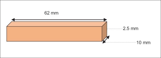
The specimens for characterization and flexural strength assessment were made with the help of wax patterns. The pattern (rectangular shape 62 mm × 10 mm × 2.5 mm) had the profile of the standard specimens [Figure 1] under consideration.[8]
Figure 1.

Schematic diagram of the sample profile for flexural strength analysis
Preparation of the mold
Wax pattern was prepared (modeling wax cut into the desired dimension for rectangular form) and invested in the dental flask using Type II and III gypsum products. The invested dental flask was kept for dewaxing after 1 h, and the mold obtained was cleaned using soap solution. A thin layer of cold mold seal was then applied to the mold as separating medium and allowed to dry.[8]
Preparations of the samples
Heat cure polymer powder and liquid (2.4 mg: 1 ml) mixed in a porcelain jar and allowed to reach dough consistency for the control group. For the reinforced groups, plasma-treated polypropylene fiber (220 μm diameter) of varying lengths (3, 6, 12 mm) and weight percentages (2.5, 5, 10 Wt%) were taken and impregnated in the monomer liquid for 5 min, and then, the polymer powder mixed and allowed to reach the dough consistency. The dough was then kneaded and packed into the mold, the closed flask was kept in a hydraulic press apparatus, and pressure of 1400 Psi was given for 30 min to dissipate the entire monomer into the polymer allowed bench curing. Flask with clamp was then transferred into the water bath in the acrylizer unit. Initially, the temperature was elevated to 72°C and maintained for 90 min; then to 100°C and maintained for 60 min. This allowed complete polymerization of the samples. The flask was permitted to cool in the same water bath to room temperature, the acrylic resin specimens were retrieved after deflasking. The specimens obtained were cleaned from stone particles and polished using sandpaper 80, 120, 150 grits. Each specimen visually inspected calibrated, polished using pumice.[8]
Measurement of flexural strength
All the prepared samples were tested for flexural strength using Instron 3366, universal testing machine. Specimens were placed in a position where its two edges supported from the lower side and the load was given in the middle of the specimen from an upper side (3 points bending). Using the digital micrometer attached to the machine, the specimen dimension was measured and recorded into the computer. Test carried out at room temperature using a cross head speed of 1 mm/min. The loading was continued up to the failure of the test sample. Flexural strength values were recorded directly from the computer connected to it. Six rectangular specimens were tested for each test groups and the mean value for each test group is reported.[8]
Scanning electron microscope analysis of the fractured specimen
Fractured specimens were subjected to SEM analysis to understand the fiber and matrix interaction. The mode of failure was also comprehended from the SEM by analyzing the fiber fracture, matrix fracture, fiber delamination/debonding, or fiber pullout.
Sample size and statistical analysis
One set of the control group with no fiber reinforcements and another set of fiber-reinforced test groups with varying fiber weight percentage and aspect ratio were considered for the present study. Six specimens were prepared in each groups; thus, the total number of sample n = 60. From the pilot study, the standardized effect size (signal/noise) obtained is 2. Therefore, six samples were chosen for each subgroup.
Null hypothesis (H0) = No effect of hydrogen plasma treatment on adhesion between polypropylene fiber and PMMA matrix, Alternate hypothesis (H1) = hydrogen plasma treatment can significantly change the interaction and adhesion between the polypropylene fiber and the PMMA matrix, thus the flexural properties of the prepared polymer composite. One way analysis of variance followed by Tukey Kramer multiple comparison tests was used for analyzing the data obtained.
RESULTS
From Table 1, it can be observed that all fiber-reinforced polymer composite test groups shown the significant increase in the flexural strength compared to control group having no fiber. Among the fiber-reinforced test groups, 6 mm long plasma-treated polypropylene fiber in 10 weight percentage showed higher flexural strength.
Table 1.
Flexural strength in MPa (mean±SD)

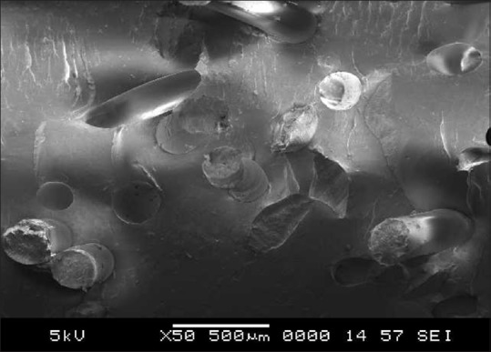
From Figure 2, the observations are made that at lower weight percentages, flexural failures were mainly due to fiber fracture indicates the strong bonding between fiber and the matrix after plasma treatment.
Figure 2.

Scanning electron microscope of fractured surface of fiber-reinforced test groups (lower weight percentage)
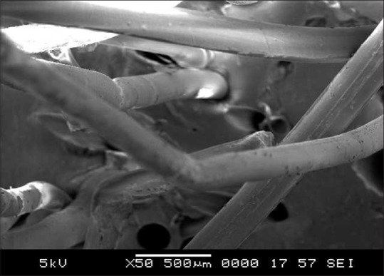
Figure 3 represents the fractured surface of the samples at higher weight percentage and demonstrates that flexural failure was mainly due to fiber pull out and delamination. It indicates less interaction between treated fiber and polymer matrix above optimum level.
Figure 3.

Scanning electron microscope of fractured surface of fiber-reinforced test groups (higher fiber weight percentage)
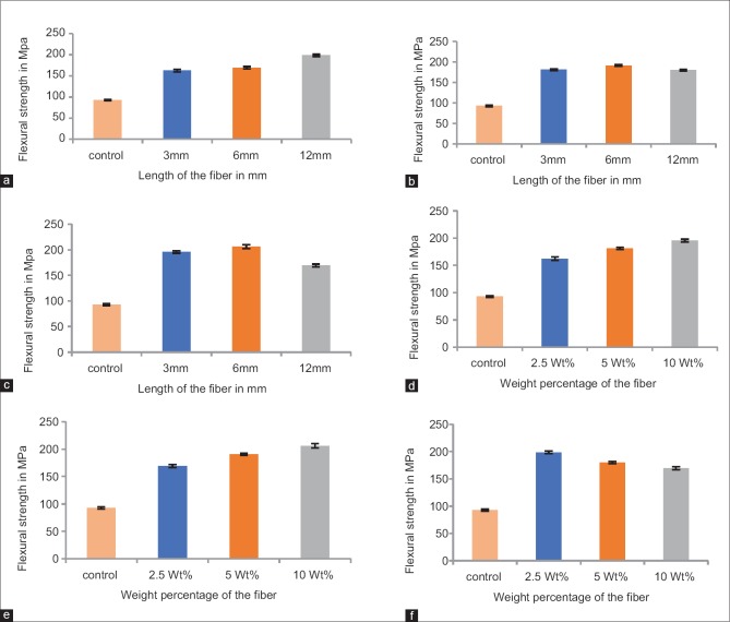
From Figure 4.c and Figure 4.f, it can be observed that increase in the fiber weight percentages and fiber lengths after a certain limit inversely affect the flexural strength of the prepared polymer composite.
Figure 4.
Flexural strength of the control group and plasma-treated polypropylene fiber-reinforced test group by varying fiber Wt% and fiber length. (a) Flexural strength of the plasma treated polypropylene fiber-reinforced polymethyl methacrylate at 2.5. (b) Flexural strength of the plasma-treated polypropylene fiber-reinforced polymethyl methacrylate at 5 Wt%. (c) Flexural strength of the plasma-treated polypropylene fiber-reinforced polymethyl methacrylate at 10 Wt%. (d) Flexural strength of the 3mm long plasma-treated polypropylene fiber-reinforced polymethyl methacrylate. (e) Flexural strength of the 6mm long plasma treated polypropylene fiber-reinforced polymethyl methacrylate. (f) Flexural strength of the 12mm long plasma-treated polypropylene fiber-reinforced polymethyl methacrylate
DISCUSSION
Flexural loading is an important parameter for the dental prosthetic materials as it imitates situations they undergo in the oral environment.[9] The present study utilized three-point bending test to measure the flexural strength of the prepared polymer composite. Rodrigues et al. reported that three-point bending is preferred over biaxial flexural test or similar flexural test as the three-point bending test gives lower standard deviation, lower coefficient of variation, and less complex crack distribution, these contribute easy calculations. In addition to all these, flexural strength analysis is widely used for comparative purposes because the specimen fabrication and the load application are quite simple. It describes the modulus of elasticity or the rigidity of a material. Relatively high modulus of elasticity is expected for dental restorative materials as it experiences large masticatory forces inside the mouth.[10]
In a polymer composite, the amount of load carried by each component depend on the product of average stress in matrix and its cross-sectional area added with the product of average stress in fiber and its cross-sectional area divided by the total cross-sectional area, the basic composite theory termed as the “rule of averages.”[11] Therefore, adhesion between the fiber and polymer matrix plays a very important role in improving the mechanical behavior.[12] Under stress, the fibers detach from the matrix, if not adhered properly with the matrix, thus decreases the strength characteristics.[13] A study conducted by Choksi R H revealed that as filler volume increases, the flexural property decreases.[14] This could be due to the improper adhesion between the filler and the matrix. In the present study, the fiber-reinforced test group showed improved flexural strength than the control group [Table 1] may be due to the good bonding between fiber and the matrix [Figures 2 and 3] due to the hydrogen plasma treatment on fibers. When the fibers intersect microcrack, they bridge the gap between two surfaces of the crack and under loading, the fibers apply a force opposing the crack propagation, thus the strength of the prepared composite increases.[15] In addition to the amount of reinforcing agent and bonding, there are several other factors which control the flexural strength of the polymer composite such as fiber orientation, diameter, length, quality and properties of the fiber and the matrix, and the processing conditions.[4,10,16,17] Whereas, the polymer alone if under stress, crack generates and propagates easily due to the brittle nature of the thermoplastic PMMA matrix.[18]
On analyzing the Figure 4.a–4.c, the flexural strength increased with increasing fiber length (P < 0.001) except for 10 Wt%. This agrees with the report by Omid et al. suggesting the effect of length of the fiber in enhancing the mechanical properties of a polymer composite.[19] On considering the fiber length with different fiber weight percentage [Figure 4.d–4.f], the flexural strength increased (P < 0.001) with increasing fiber weight percentage. Mechanical properties of fibers are much higher than those of resins; therefore, higher mechanical properties are expected with higher fiber volume fraction in a fiber-reinforced polymer composite. However, in practice, there is a limit to this since the fiber incorporated needed to be fully coated with the resin to be effective.[12,13]
In the present study, at 5 Wt% [Figure 4.b], there was no significant change in the flexural strength value between 3 mm and 12 mm long fiber (P > 0.05), supports the study conducted by Karacaer et al., that the addition of optimum fiber length required to get better mechanical properties coupled with easy and cost-effective reinforcing procedure.[20] Incorporation of 12 mm long fiber, significantly decreased the flexural strength from 2.5 to 10 Wt% (P < 0.001), suggest that the transfer of load from fiber to matrix cannot be achieved effectively when fiber to fiber distance if less and if each fiber covered by less matrix material.[13]
Increase in the fiber volume percentage sometimes compromises the flexibility of the rehabilitating materials. Therefore, optimum fiber volume fraction is preferred not only because of the flexural strength aspect but also for the flexibility required for easy adaptation of prosthesis and thereby patient comfort.[21]
Hydrogen plasma treatment on polypropylene fiber found as an effective method of enhancing the flexural properties of fiber-reinforced PMMA by improving the adhesion between the fiber and the matrix. However, the time of plasma exposure on the fiber, the type of plasma employed on the fiber, biological, physical, and other mechanical properties need to be studied further to employ this technique commercially.
CONCLUSION
Null hypothesis rejected and alternate hypothesis accepted indicates that hydrogen plasma treatment is an effective method to improve the interaction and adhesion between polypropylene fiber and the PMMA matrix
Fiber weight percentage, fiber length, and adhesion between fiber and the matrix play an important role in flexural characteristics of the hydrogen plasma-treated polypropylene fiber-reinforced PMMA polymer composite.
Financial support and sponsorship
Nil.
Conflicts of interest
There are no conflicts of interest.
REFERENCES
- 1.Maller US, Karthik KS, Maller SV. Maxillofacial prosthetic materials-past and present trends. JIADS. 2010;1:25–30. [Google Scholar]
- 2.De Lucena SC, Gomes SG, Da Silva WJ, Del Bel Cury AA. Patients’ satisfaction and functional assessment of existing complete dentures: Correlation with objective masticatory function. J Oral Rehabil. 2011;38:440–6. doi: 10.1111/j.1365-2842.2010.02174.x. [DOI] [PubMed] [Google Scholar]
- 3.Cha YH, Kim KS, Kim DJ. Evaluation on the fracture toughness and strength of fiber reinforced brittle matrix composites. KSME Int J. 1998;12:370–9. [Google Scholar]
- 4.Prasanna GV, Subbaiah KV. Modification, flexural, impact, compressive properties and chemical resistance of natural fibers reinforced blend composites. Malays Polym J. 2013;8:38–44. [Google Scholar]
- 5.Cech V, Babik A, Knob A, Palesch E. 13th International Conference on Plasma Surface Engineering, 2012, September 10-14 in Garmisch-Partenkirchen, Germany. 2012:51–5. [Google Scholar]
- 6.Chu PK, Chen JY, Wang LP, Huang N. Plasma surface modification of biomaterials: A review journal. Mater Sci Eng. 2012;36:143–206. [Google Scholar]
- 7.Scheidbach H, Tamme C, Tannapfel A, Lippert H, Köckerling F. In vivo studies comparing the biocompatibility of various polypropylene meshes and their handling properties during endoscopic total extraperitoneal (TEP) patchplasty: An experimental study in pigs. Surg Endosc. 2004;18:211–20. doi: 10.1007/s00464-003-8113-1. [DOI] [PubMed] [Google Scholar]
- 8.Mathew M, Shenoy K, Ravishankar KS. Flexural strength of polypropylene fiber reinforced PMMA. Int J Pharm Sci Invent. 2017;6:21–5. [Google Scholar]
- 9.Qasim SB, Al Kheraif AA, Ramakrishaniah R. An investigation into the impact and flexural strength of light cure denture resin reinforced with carbon nanotubes. World Appl Sci J. 2012;18:808–12. [Google Scholar]
- 10.Rodrigues Junior SA, Zanchi CH, Carvalho RV, Demarco FF. Flexural strength and modulus of elasticity of different types of resin-based composites. Braz Oral Res. 2007;21:16–21. doi: 10.1590/s1806-83242007000100003. [DOI] [PubMed] [Google Scholar]
- 11.Altan C. Preparation and Characterization of Glass Fiber Reinforced Poly (Ethylene Terephthalate), M.Sc Thesis. 2004, the Graduate School of Natural and Applied Sciences of Middle East Technical University. 2004:1–123. [Google Scholar]
- 12.Gupt P, Nagpal A, Samra RK, Verma R, Kaur J, Abrol S, et al. A comparative study to check fracture strength of provisional fixed partial dentures made of autopolymerizing polymethylmethacrylate resin reinforced with different materials: An in vitro study. J Indian Prosthodont Soc. 2017;17:301–9. doi: 10.4103/jips.jips_79_17. [DOI] [PMC free article] [PubMed] [Google Scholar]
- 13.Rezvani MB, Atai M, Hamze F. Effect of fiber diameter on flexural properties of fiber-reinforced composites. Indian J Dent Res. 2013;24:237–41. doi: 10.4103/0970-9290.116696. [DOI] [PubMed] [Google Scholar]
- 14.Choksi RH, Mody PV. Flexural properties and impact strength of denture base resins reinforced with micronized glass flakes. J Indian Prosthodont Soc. 2016;16:264–70. doi: 10.4103/0972-4052.176532. [DOI] [PMC free article] [PubMed] [Google Scholar]
- 15.Etcheverry M, Barbosa SE. Glass fiber reinforced polypropylene mechanical properties enhancement by adhesion improvement. Materials (Basel) 2012;5:1084–113. doi: 10.3390/ma5061084. [DOI] [PMC free article] [PubMed] [Google Scholar]
- 16.Ribeiro DG, Pavarina AC, Machado AL, Giampaolo ET, Vergani CE. Flexural strength and hardness of reline and denture base acrylic resins after different exposure times of microwave disinfection. Quintessence Int. 2008;39:833–40. [PubMed] [Google Scholar]
- 17.Vojvodic D, Matejicek F, Schauperl Z, Mehulic K, Bagic-Cukovic I, Segovic S. Flexural strength of E-glass fiber reinforced dental polymer and dental high impact strength resin. Strojarstvo. 2008;50:221–30. [Google Scholar]
- 18.Nagpal A, Rawat M, Verma PR, Samra RK, Verma R, Kaur J. Effect of different concentrations of glass fibers on transverse strength of four different brands of heat cure denture base resins – A comparative study. Indian J Dent Sci. 2014;1:37–40. [Google Scholar]
- 19.Omid T, Venus MM, Farahnaz S, Asghar AA. Effect of glass fiber length on flexural strength of fiber-reinforced composite resin. World J Dent. 2012;3:131–5. [Google Scholar]
- 20.Karacaer O, Polat TN, Tezvergil A, Lassila LV, Vallittu PK. The effect of length and concentration of glass fibers on the mechanical properties of an injection- and a compression-molded denture base polymer. J Prosthet Dent. 2003;90:385–93. doi: 10.1016/S0022391303005183. [DOI] [PubMed] [Google Scholar]
- 21.Singh K, Aeran H, Kumar N, Gupta N. Flexible thermoplastic denture base material for aesthetical removable partial denture framework. J Clin Diagn Res. 2013;7:2372–3. doi: 10.7860/JCDR/2013/5020.3527. [DOI] [PMC free article] [PubMed] [Google Scholar]


