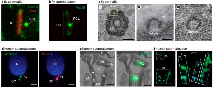
Figure 3.
Sperm centrioles in flies (a–c) and mammals (d–f). (a) Fluorescent microscope picture of fly spermatid with DC and PCL labeled by the centriolar protein Poc1B and Ana1. (b) Fluorescent microscope picture of fly spermatozoon with Poc1B faintly labeling the DC and intensely labeling the PCL. In (a,b) the scale bar is 1 μm, Poc1B is genetically tagged by GFP, and Ana1 is genetically tagged by tdTomato. (c) Electron microscope picture of fly spermatozoon centrioles. (i) A section in a plane that has a cross-section of the DC and longitudinal section of the PCL (yellow box). (ii) Zoom in the PCL boxed in (i). (iii) A cross-section of the PCL depicting the wall and central tubule. In (c), the scale bar is 0.1 μm. (d) Fluorescent microscope picture of a human spermatozoon with CENTE1/2 or POC1B labeling the DC and PCL. The scale bar is 1 μm. CENTE1/2 and Poc1B are labeled by specific antibodies. (e) Correlative light and electron microscopy picture of human spermatozoon centrioles. On the left, electron microscopy section depicting POC1B labeling of the PC that is found near the nucleus, and the DC that is attached to the axoneme. Ax, axoneme; M, mitochondria; mp, tail midpiece; N, nucleus; ne, neck; O, outer dance fibers; S, striated columns; V; vault; CA; centriole adjunct. (f) Stochastic Optical Reconstruction Microscopy (STORM) super-resolution fluorescent microscopy picture of bovine spermatozoon centrioles. On the left, a picture depicting POC5 labeling of the PC and the DC. On the right, zooming in on the DC identifies two major rods (marked as “1” and “2”) and one minor rod (marked as “3”) labeled by POC5. In (f), the scale bar is 0.1 μm. Panels (a–c) are from Khire 2016 [15]. Panels (d–f) are from Fishman [9].

