Figure 4.
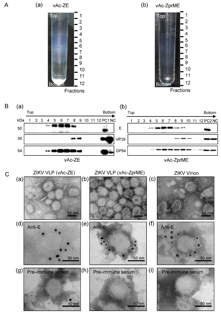
Characterization of baculovirus-expressed ZIKV VLPs from culture supernatants. (A) Culture supernatants of vAc-ZE (a) and vAc-ZprME (b) infected Sf9 cells were concentrated and layered onto 10–60% sucrose gradients and subjected to centrifugation. Twelve fractions were taken from top to bottom. (B) Western blot analysis of purified sucrose gradient fractions of culture supernatants of Sf9 cells infected with vAc-ZE (a) and vAc-ZprME (b) using the indicated antibodies. PC1: positive control 1 (vAc-ZE infected Sf9 cells); PC2: positive control 2 (vAc-ZprME infected Sf9 cells); NC: negative control (vAc-hsp70-egfp infected Sf9 cells). (C) Electron micrographs of negative staining and immunogold labeling of VLPs and ZIKV. (a,b) Negative staining of purified ZIKV VLPs from the E antigen-enriched fractions from the sucrose gradient fractions of culture supernatants of Sf9 cells infected with vAc-ZE (a) and vAc-ZprME (b); (c) Negative staining of purified ZIKV virions; (d–f) IEM of ZIKV VLPs (vAc-ZE) (d) ZIKV VLPs (vAc-ZprME) (e) and ZIKV virions (f) using anti-ZIKV-E-specific polyclonal antibody as primary antibody; (g–i) IEM of ZIKV VLPs (vAc-ZE) (g), ZIKV VLPs (vAc-ZprME) (h) and ZIKV virions (i) using rabbit pre-immune serum as primary antibody.

