Abstract
Background
Lipomas are very common benign tumors of mature fatty tissue that can occur in any part of the body. However, lipomas of the parietal peritoneum are extremely rare.
Case presentation
A 36-year-old man presented with urinary frequency for 6 months. On computerized tomography of the abdomen and pelvis, a well-defined fatty mass measuring 20 × 11 × 6.5 cm in size, was found in the lower abdominal cavity. We performed a laparoscopic parietal-peritoneum-preserving excision of the mass. The patient was discharged without complications on post-operative day 6.
Conclusions
To our knowledge, a laparoscopic excision with preservation of the parietal peritoneum for a giant parietal peritoneal lipoma has never been reported. Herein, we report a case of a giant lipoma of the parietal peritoneum successfully managed by laparoscopy.
Keywords: Giant lipoma, Laparoscopy, Parietal peritoneum, Urinary frequency
Background
Lipomas are very common benign tumors of mature fatty tissue that can occur in any part of the body. There are a few reports of giant lipoma of mesentery or omentum [1–4]. However, lipomas of the parietal peritoneum are extremely rare. And there have been no reports of extremely large-sized lipomas of the parietal peritoneum. We describe a case of a giant lipoma of the parietal peritoneum causing urinary frequency secondary to external compression of the bladder. This tumor was successfully managed by laparoscopic excision with preservation of the parietal peritoneum.
Case presentation
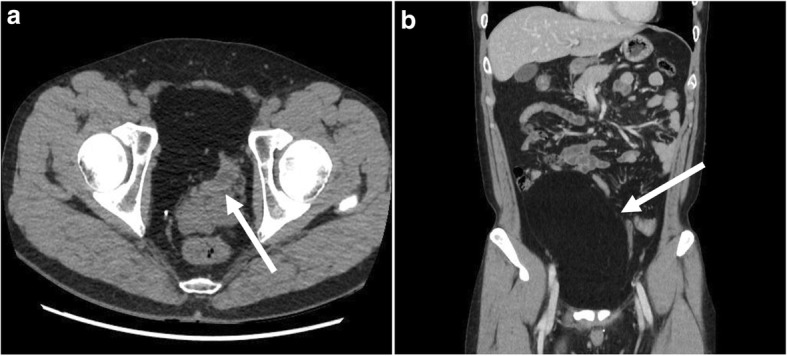
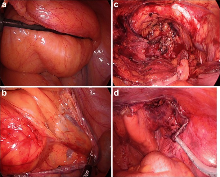
A 36-year-old man presented with urinary frequency for 6 months. He had no significant urologic abnormality and no palpable abdominal mass on physical examination. He denied abdominal pain, vomiting, anorexia, or bowel disturbances. There were no specific laboratory abnormalities. The abdomen and pelvis computed tomography scans showed a 20 × 11 cm, well-defined, fatty mass in the abdominal cavity. A mass was located between the abdominal wall muscles and the peritoneum and compressed bladder (Fig.1a, b). We performed surgery, firstly. The reasons are as follows: (1) the mass was just beneath the abdominal wall, (2) the patient had symptom (urinary frequency), and (3) the mass was considered benign from well-demarcate mass with homogenous features on CT scan. We performed a laparoscopic mass excision with preservation of the parietal peritoneum. Two 11-mm ports were inserted, one supra-umbilically, and the other in the left lower abdomen. A 5-mm port was inserted in the right lower abdomen. A huge, freely mobile, soft mass in the external peritoneal layer with no connection to other organs was seen in the lower abdomen (Fig.2a). After demarcating the mass, we excised the parietal peritoneum through the marked line with a monopolar instrument. Next, we dissected the mass from the peritoneum (Fig. 2b). The mass which was excised completely, was placed in a large plastic endopouch-type bag, and extracted through the extended left port site. Finally, the preserved peritoneum was fixed to the abdominal wall using a fixing material with a closed suction drain (Fig. 2c, d). The operative time was 90 min, with no estimated blood loss. The resected specimen size was 22 × 16 × 7.5 cm3, and the weight was 942 g. The pathological diagnosis was reported benign lipoma with clear resection margin. The patient was discharged without complications on post-operative day 6.
Fig. 1.

Contrast-enhanced abdomen-pelvis computed tomography scans. a Axial view. A mass compressed bladder (arrow). b Coronal view. A well-defined homogenous fatty mass measuring 20 × 11 cm in size (arrow)
Fig. 2.
Laparoscopic view. a A soft huge mass in the external peritoneal layer was seen in the lower abdomen and was free from other organs. b Dissection between mass and peritoneum. c Operative field after mass excision. d Preserved peritoneum was fixed to abdominal wall using fixation device
Discussion and conclusions
In 2006, the first case of a lipoma of the parietal peritoneum was reported by Barut et al [5]. Since then, only 5 more cases have been reported (Table 1). In previous reports, all the patients presented with abdominal pain. Three cases were presented with right quadrant abdominal pain mimicking appendicitis, and the largest lipoma had a diameter of 6.3 cm [6–10]. Our patient presented with urinary frequency caused by external compression of the bladder. A huge and heavy lipoma measuring 22 × 16 × 7.5 cm3 and 942 g disturbed the filling capacity of the bladder. In 4 previously reported cases, because the lipomas were small in size, they performed a laparoscopic excision of the lipoma and its associated peritoneum [6, 8–10]. In our case, we performed a peritoneal-preserving excision of the lipoma to reduce the pain that we anticipated might be caused by peritoneal resection. Since our patient’s lipoma was large in size, we dissected between the peritoneum and the lipoma, and the peritoneum was preserved with fixation around the abdominal wall. The fixation device (Protack™, Medtronic) which is often used in laparoscopic hernioplasties, was useful for fixation. To our knowledge, a laparoscopic excision with preservation of the parietal peritoneum for a giant parietal peritoneal lipoma has never been reported. This procedure is feasible for decreasing postoperative pain and better cosmetic results.
Table 1.
Reports regarding the treatment of a lipoma of the parietal peritoneum in the literature
| Reference (year) | Age (years) | Sex | Presentation | Surgical procedure | Maximum diameter (cm) |
|---|---|---|---|---|---|
| Barut et al. [4] (2006) | 67 | Female | Abd pain, nausea vomiting | Open | 6 |
| Bunker et al. [5] (2013) | 34 | Female | Abd pain | Laparoscopy | – |
| Bang et al. [6] (2014) | 75 | Male | Abd pain, palpable mass | Open | 4.5 |
| Shrestha et al. [7] (2014) | 32 | Male | Abd pain, loss of appetite | Laparoscopy | 3 |
| Sathyakrishna et al. [8] (2014) | 21 | Female | Abd pain | Laparoscopy | – |
| Salgaonkar et al. [9] (2016) | 79 | Male | Abd pain | Laparoscopy | 6.3 |
| Present case (2018) | 36 | Male | Urinary frequency | Laparoscopy | 22 |
In conclusion, this case highlights the fact that a giant lipoma of the parietal peritoneum can be an unusual cause of urinary disturbances. Laparoscopic procedures are feasible for the excision of a huge lipomas of the parietal peritoneum, and concomitant preservation of the peritoneum is useful for reducing postoperative pain.
Availability of data and materials
All data generated or analyzed during this study are included in this published article and its supplementary information files.
Abbreviations
- A-P CT
Abdominal and pelvic computed tomography
Authors’ contributions
All authors participated in the management of the patient in this case report. YK assisted in diagnosing field to the patient. DHR and JWC are surgeons, who operated on the patient, supervised and contributed to the case report. HC and YX researched and analyzed the data. HC was a major contributor in writing the manuscript, collected all references. All authors read and approved the final manuscript.
Ethics approval and consent to participate
Not applicable.
Consent for publication
Written informed consent was obtained from the participant for publication of this article and any accompanying tables/images. A copy of the written consent is available for review by the Editor of this journal.
Competing interests
The authors declare that they have no competing interests.
Publisher’s Note
Springer Nature remains neutral with regard to jurisdictional claims in published maps and institutional affiliations.
Footnotes
This work was supported by the intramural research grant of Chungbuk National University in 2015.
Contributor Information
Hanlim Choi, Email: sglim2729@gmail.com.
DongHee Ryu, Phone: +82-43-269-6034, Email: dhryu@chungbuk.ac.kr.
Jae-Woon Choi, Email: jwchoi@chungbuk.ac.kr.
Yanjie Xu, Email: xuyanjie1208@gmail.com.
Yook Kim, Email: sixtwin@hanmail.net.
References
- 1.Yang TW, Tsuei YW, Kao CC, Kuo WH, Chen YL, Lin YY. Torsion of a Giant Antimesenteric lipoma of the ileum: a rare cause of acute abdominal pain. Am J Case Rep. 2017;18:589–592. doi: 10.12659/AJCR.903574. [DOI] [PMC free article] [PubMed] [Google Scholar]
- 2.Kshirsagar AY, Nangare NR, Gupta V, Vekariya MA, Patankar R, Mahna A, et al. Multiple giant intra abdominal lipomas: A rare presentation. Int J Surg Case Rep. 2014;5(7):399–402. doi: 10.1016/j.ijscr.2014.04.002. [DOI] [PMC free article] [PubMed] [Google Scholar]
- 3.Cha JM, Lee JI, Joo KR, Choe JW, Jung SW, Shin HP, et al. Giant mesenteric lipoma as an unusual cause of abdominal pain: a case report and a review of the literature. J Korean Med Sci. 2009;24(2):333–336. doi: 10.3346/jkms.2009.24.2.333. [DOI] [PMC free article] [PubMed] [Google Scholar]
- 4.Shiroshita H, Komori Y, Tajima M, Bandoh T, Arita T, Shiraishi N, Kitano S, et al. Laparoscopic examination and resection for giant lipoma of the omentum: a case report and review of related literature. Surg Laparosc Endosc Percutan Tech. 2009;19(5):e217–ee20. doi: 10.1097/SLE.0b013e3181b97edd. [DOI] [PubMed] [Google Scholar]
- 5.Barut I, Tarhan OR, Cerci C, Ciris M, Tasliyar E. Lipoma of parietal peritoneum: an unusual cause of abdominal pain. Ann Saudi Med. 2006;26:388–390. doi: 10.5144/0256-4947.2006.388. [DOI] [PMC free article] [PubMed] [Google Scholar]
- 6.Bunker DL, Ilie VG, Halder TK. Torsion of an Abdominal-Wall pedunculated lipoma: a rare differential diagnosis for right iliac Fossa pain. Case Rep Surg. 2013;2013:587380. doi: 10.1155/2013/587380. [DOI] [PMC free article] [PubMed] [Google Scholar]
- 7.Bang CS, Kim YS, Balik GH, Han SH. A case of lipoma of parietal peritoneum causing abdominal pain. Korean J Gastroenterol. 2014;63:369–372. doi: 10.4166/kjg.2014.63.6.369. [DOI] [PubMed] [Google Scholar]
- 8.Shrestha BB, Karmacharya M. Torsion of a lipoma of parietal peritoneum: A rare case mimicking acute appendicitis. J Surg Case Rep. 2014;2014(6). 10.1093/jscr/rju062. [DOI] [PMC free article] [PubMed]
- 9.Sathyakrishna BR, Boggaram SG, Jannu NR. Twisting lipoma presenting as appendicitis-a rare presentation. J Clin Diagn Res. 2014;8(8):ND07. doi: 10.7860/JCDR/2014/9663.4728. [DOI] [PMC free article] [PubMed] [Google Scholar]
- 10.Salgaonkar HP, Behera RR, Katara AN, Bhandarkar DS. Laparoscopic excision of a lipoma of parietal peritoneum. J Min Access Surg. 2016;12:196–197. doi: 10.4103/0972-9941.178515. [DOI] [PMC free article] [PubMed] [Google Scholar]


