

 Usnic acid, a dibenzofuran, was originally isolated from lichens producing secondary metabolites, and is well known as an antibiotic, but is also endowed with several other interesting properties.
Abstract
Usnic acid, a dibenzofuran, was originally isolated from lichens producing secondary metabolites, and is well known as an antibiotic, but is also endowed with several other interesting properties. Thus, the goal of this paper is the design of new usnic acid derivatives and evaluation of their antimicrobial activity. All newly synthesized compounds possess good antibacterial activity with MIC ranging from 1.02–50.93 × 10⁻² mmol mL–1 and MBC from 2.05–70.57 × 10⁻² mmol mL–1. The most sensitive bacterial species was Staphylococcus aureus, while Pseudomonas aeruginosa and Escherichia coli were the most resistant among the ATCC strains, and MRSA was the most resistant among all tested bacteria (ATCC and clinical isolates). Their antifungal activity was very strong (MIC = 0.35–7.53 × 10⁻² mmol mL–1 and MFC = 0.70–15.05 × 10⁻² mmol mL–1) – better than those of reference compounds and usnic acid itself. The most sensitive fungal species was Trichoderma viride, while Penicillium versicolor var. cyclopium appeared to be the most resistant. It should be mentioned that in general most of the compounds showed weaker antibacterial activity, but better antifungal properties than usnic acid itself. The results allow us to conclude that the title compounds are good lead compounds for novel more active antibacterial drugs. On the other hand, these compounds are very promising as antifungals.
1. Introduction
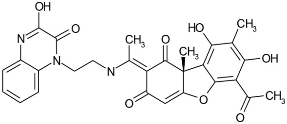
Natural products continue to attract interest as leads for new compounds possessing biological activity as promising precursors of pharmaceutical products. One source of novel natural products is lichens which have a symbiotic relationship with algae or cyanobacteria. This symbiotic relationship results in properties different from those of their component organisms and can produce a large number of secondary metabolites, some of them being present only in the lichens themselves, while others are found in other fungi or plants. The interest in these substances is due to the fact that, among them, there are different structural classes of compounds, such as phenols, dibenzofurans, sugars, xanthones and terpenoids.1 Usnic acid (UA), a dibenzofuran, was originally isolated from a lichen and has been shown to have antibiotic properties, but is also endowed with other interesting activities.2 Usnic acid [2,6-diacetyl-7,9-dihydroxy-8,9b-dimethyldibenzofuran-1,3(2H,9bH)-dione] exists naturally as both (+) d-usnic acid and (–) l-usnic acid enantiomers, with R or S projection of the stereogenic centre at position 9b, respectively (Fig. 1). The enantiomers have been shown to present different biological activities.3
Fig. 1. The enantiomers of usnic acid.

Literature surveys revealed that usnic acid and its derivatives have a wide spectrum of biological activities such as antiviral against a broad range of influenza viruses,4,5 anti-inflammatory,6–8 antioxidant,9–11 PTP1B,12 PARP1 inhibition,13 cardiovascular,14 anti-tubercular,15 fungicidal, antiprotozoal and algicidal,16,17 anti-Trypanosoma cruzi,18 angiogene inhibition,19–21 antimitotic22 and antineoplastic activities.23,24 Furthermore, Kupchan et al.25 found that l-usnic acid is a tumor inhibitor. Singh26 noted that usnic acid (UA) inhibits growth and induces cell cycle arrest and apoptosis in human lung carcinoma A549 cells as well as in U87MG glioblastoma cells.27 There are other disclosures that refer to the cytotoxicity of UA derivatives.28,29 Koparal et al.30 have reported the in vitro cytotoxic activities of (+)-usnic acid and (–)-usnic acid on V79, A549, and human lymphocyte cells.
The antimicrobial activities of lichen extracts and the compounds isolated, including usnic acid, have been a subject of particular interest.31–35 Cocchietto et al.,2 in their review, mentioned the inhibition of growth of multi-resistant strains of Staphylococcus aureus, Enterococcus spp. and Mycobacteria spp. by both enantiomers of UA. This has relevance in light of the fact that bacterial resistance is a growing problem worldwide. There are reports regarding the antibacterial36,37 and antifungal38 activities of enaminic usnic acid derivatives. Several lichen metabolites have been found to be active against Gram-positive bacteria and mycobacteria. Pires et al.17 reported the inhibitory effect of UA on Candida orthopsilosis and C. parapsilosis. Gupta et al.43 suggested that the antibacterial activity of usnic acid against methicillin-resistant Staphylococcus aureus (MRSA) is due to disruption of the cell membrane.
Furthermore, usnic acid was found to be able to inhibit biofilm formation.39–44
As far as the two enantiomers are concerned, (+) UA has antimicrobial,45 anti-inflammatory46,47 and cytotoxic effects;48 while (–) UA exhibits anti-protozoal properties.47,49 Nevertheless, the compound has been associated with severe liver damage (hepatotoxicity) when taken as a dietary supplement for the purpose of weight loss. Presumably, the reason for the hepatotoxicity of UA is its ability to induce oxidative stress and inhibit mitochondrial function in the liver cells.50 On the other hand, there is reference to the fact that UA protects gastric cells from drug-induced oxidative damage.51,52
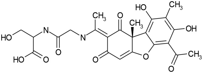
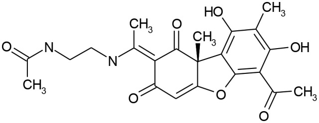
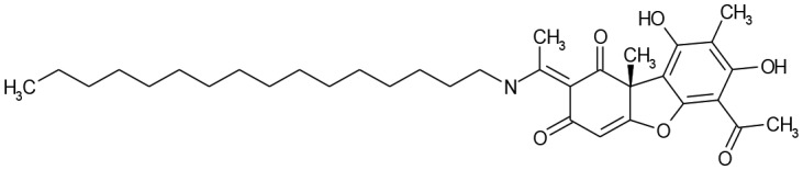
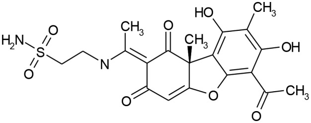
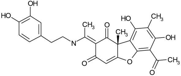
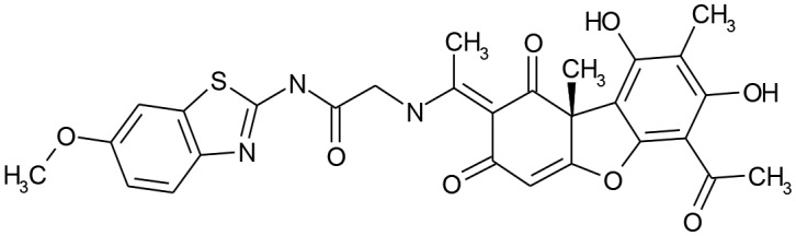
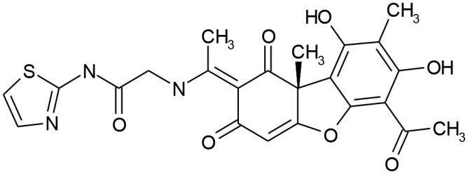
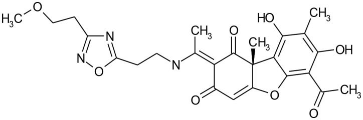
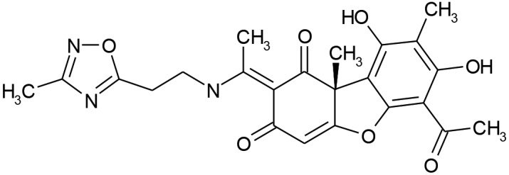
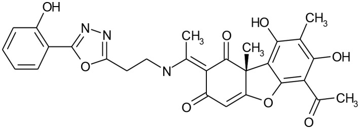
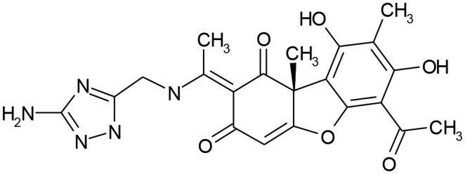
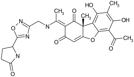
Taking all of the above into account, the aim of this work is to design and synthesize new UA derivatives and evaluate their biological activity. The prediction of biological activity spectra of the designed compounds using the computer program PASS53 and the calculation of solubility by the AlogPs program54 have been performed. The prediction of biological activity spectra of the designed compounds revealed that all of them are potent antimicrobial agents, despite the fact that, for some of them, hepatotoxicity was predicted with quite high probability. Nevertheless, hepatoprotective and antioxidant activity was also predicted. Herein, we report the design and synthesis of thirteen derivatives of usnic acid and the evaluation of their antimicrobial activity. The structures of the compounds are presented in Table 1.
Table 1. Structures of the synthesized compounds.
| Comp. | Structure | Comp. | Structure |
| 1 |
|
8 |
|
| 2 |
|
9 |
|
| 3 |
|
10 |
|
| 4 |
|
11 |
|
| 5 |
|
12 |
|
| 6 |
|
13 |
|
| 7 |
|
2. Results and discussion
2.1. Chemistry
The target products were obtained by condensation of (R)-usnic acid and appropriate amines in refluxing ethanol in the presence of triethylamine. The synthetic method is regioselective, simple and convenient and allows derivatives of usnic acid to be prepared using a wide range of starting amines. Compounds 1–13 were synthesized according to the process in Scheme 1. Their solubility was calculated using the AlogPs program54 and was found to be higher (log S from –3.13 to –6.27) than that of usnic acid (log S of –2.95). Furthermore, it was observed that the compounds are more soluble than usnic acid in polar solvents, while the opposite was observed in nonpolar solvents. All compounds were characterized spectroscopically (IR, NMR).
Scheme 1. Synthesis of usnic acid derivatives.

2.2. Biological evaluation
2.2.1. Antibacterial activity
Compounds 1–13 were tested for their antibacterial activity towards Gram-(–) and Gram-(+) bacteria, ATCC strains and clinical isolates (Table 2).
Table 2. Antibacterial activity of usnic acid derivatives (MIC/MBC in mmol ml–1 × 10⁻²).
| Compound | S. a. | MRSA | B. c. | P. a. | PaO1 | E. coli | E. coli res | En. cl. | |
| 1 | MIC | 1.02 ± 0.02c | 8.19 ± 0.03c | 4.09 ± 0.06c | 8.19 ± 0.05f | 24.57 ± 0.20d | 4.09 ± 0.08d | 12.28 ± 0.40b | 4.09 ± 0.03d |
| MBC | 2.05 ± 0.06c | 16.38 ± 0.50c | 8.19 ± 0.08c | 16.38 ± 0.6d | 32.76 ± 0.50c | 8.19 ± 0.04d | 16.38 ± 0.40b | 8.19 ± 0.08d | |
| 2 | MIC | 3.52 ± 0.03d | 56.36 ± 0.80g | 7.05 ± 0.03d | 7.05 ± 0.06f | 21.14 ± 0.20d | 14.09 ± 0.30f | 28.18 ± 0.20c | 3.52 ± 0.06d |
| MBC | 7.05 ± 0.03e | 88.06 ± 0.80de | 14.09 ± 0.30d | 14.09 ± 0.20d | 28.18 ± 0.40bc | 28.18 ± 0.40e | 56.36 ± 0.60cd | 14.09 ± 0.10e | |
| 3 | MIC | 3.13 ± 0.02d | 8.34 ± 0.03c | 1.56 ± 0.02b | 4.17 ± 0.05e | 25.03 ± 0.20d | 3.13 ± 0.02cd | 16.68 ± 0.20b | 2.09 ± 0.06c |
| MBC | 4.17 ± 0.06d | 16.68 ± 0.50c | 2.09 ± 0.03b | 16.68 ± 0.03d | 33.36 ± 0.20c | 4.17 ± 0.03b | 33.36 ± 0.20 | 4.17 ± 0.03b | |
| 4 | MIC | 33.09 ± 0.5h | 103.41 ± 1.20i | 49.64 ± 0.80g | 41.35 ± 0.60i | 24.82 ± 0.80d | 74.46 ± 0.90c | 33.09 ± 0.30c | 49.64 ± 0.20c |
| MBC | 66.18 ± 0.3h | 124.1 ± 0.80e | 66.18 ± 0.60f | 103.41 ± 0.8f | 33.09 ± 0.30c | 124.1 ± 1.20f | 66.18 ± 0.80d | 66.18 ± 0.30g | |
| 5 | MIC | 26.46 ± 0.8g | 52.92 ± 0.30g | 13.23 ± 0.20e | 17.64 ± 0.80g | 26.46 ± 0.3d | 17.64 ± 0.06f | 52.93 ± 0.30cd | 26.46 ± 0.80g |
| MBC | 70.57 ± 0.6h | 132.32 ± 1.20e | 35.28 ± 0.10e | 35.28 ± 0.08e | 35.28 ± 0.09c | 35.28 ± 0.06e | 70.57 ± 0.60d | 35.28 ± 0.30f | |
| 6 | MIC | 18.21 ± 0.08f | 136.54 ± 1.20j | 27.31 ± 0.03f | 72.82 ± 0.60j | 18.21 ± 0.30cd | 72.82 ± 0.06h | 13.65 ± 0.60b | 2.28 ± 0.08c |
| MBC | 36.42 ± 0.50g | 159.30 ± 1.80e | 72.84 ± 0.90f | 113.78 ± 1.2f | 36.42 ± 0.80c | 113.78 ± 1.20f | 18.21 ± 0.05b | 9.11 ± 0.03d | |
| 7 | MIC | 22.58 ± 0.20g | 30.10 ± 0.10f | 15.05 ± 0.30e | 15.05 ± 0.20g | 45.16 ± 0.60e | 45.16 ± 0.50g | 45.16 ± 0.30c | 15.05 ± 0.20f |
| MBC | 60.20 ± 0.50h | 60.20 ± 0.30d | 30.10 ± 0.10e | 30.10 ± 0.10e | 60.20 ± 0.20d | 60.20 ± 0.30f | 60.20 ± 0.30d | 60.20 ± 0.20g | |
| 8 | MIC | 7.00 ± 0.05e | 37.34 ± 0.20f | 7.00 ± 0.03d | 9.34 ± 0.03f | 18.67 ± 0.06cd | 9.34 ± 0.05e | 37.34 ± 0.05c | 7.00 ± 0.06e |
| MBC | 9.34 ± 0.02e | 74.68 ± 0.50d | 18.67 ± 0.20d | 18.67 ± 0.20d | 37.34 ± 0.50c | 18.67 ± 0.50de | 74.68 ± 0.60a | 9.34 ± 0.02d | |
| 9 | MIC | 1.11 ± 0.02c | 2.22 ± 0.03a | 1.11 ± 0.03b | 1.66 ± 0.02c | 26.62 ± 0.30d | 0.55 ± 0.002b | 55.50 ± 0.30cd | 4.44 ± 0.02d |
| MBC | 2.22 ± 0.03c | 8.88 ± 0.20b | 2.22 ± 0.03b | 8.88 ± 0.20cd | 35.50 ± 0.60c | 1.11 ± 0.02b | 71.04 ± 0.30d | 8.88 ± 0.03d | |
| 10 | MIC | 28.39 ± 0.30g | 88.72 ± 0.60h | 28.39 ± 0.40f | 28.39 ± 0.30b | 14.20 ± 0.10c | 14.20 ± 0.20f | 56.80 ± 0.50cd | 56.80 ± 0.80h |
| MBC | 56.80 ± 0.80h | 106.46 ± 0.90e | 56.80 ± 0.60ef | 56.80 ± 0.80f | 28.39 ± 0.60bc | 28.39 ± 0.60e | 88.72 ± 0.80de | 88.72 ± 1.20g | |
| 11 | MIC | 0.50 ± 0.003b | 32.16 ± 0.50f | 0.20 ± 0.003a | 2.01 ± 0.03d | 120.60 ± 1.20f | 2.01 ± 0.03c | 32.16 ± 0.80c | 0.50 ± 0.002b |
| MBC | 1.00 ± 0.03b | 64.32 ± 0.80d | 1.00 ± 0.03a | 4.02 ± 0.03c | 140.70 ± 2.10e | 4.02 ± 0.02c | 64.32 ± 0.60d | 1.00 ± 0.03b | |
| 12 | MIC | 7.53 ± 0.60e | 22.59 ± 0.80e | 7.53 ± 0.08d | 7.53 ± 0.06f | 15.05 ± 0.30c | 15.05 ± 0.40f | 15.05 ± 0.60b | 7.53 ± 0.60e |
| MBC | 15.05 ± 0.06f | 40.51 ± 1.00a | 15.05 ± 0.090d | 15.05 ± 0.08d | 30.10 ± 0.90c | 30.10 ± 0.80e | 30.10 ± 0.90c | 15.05 ± 0.090e | |
| 13 | MIC | 3.93 ± 0.020d | 7.86 ± 0.06c | 15.72 ± 0.08e | 0.94 ± 0.080b | 108.16 ± 1.20f | 15.73 ± 0.06f | 137.66 ± 0.9d | 1.97 ± 0.05c |
| MBC | 31.47 ± 0.08g | 62.94 ± 0.60d | 31.47 ± 0.10e | 1.87 ± 0.02b | 118.00 ± 0.90e | 31.47 ± 0.50e | 157.33 ± 1.2e | 15.73 ± 0.06e | |
| Streptomycin | MIC | 4.3 ± 0.02d | 17.20 ± 0.05d | 4.3 ± 0.02c | 17.20 ± 0.05g | 8.60 ± 0.06b | 17.20 ± 0.06f | 17.20 ± 0.05b | 4.3 ± 0.02d |
| MBC | 34.40 ± 0.20g | — | 8.60 ± 0.06c | 34.40 ± 0.20e | 17.20 ± 0.30b | 34.4 ± 0.20e | 34.40 ± 0.20c | 8.6 ± 0.02d | |
| Ampicillin | MIC | 24.8 ± 0.02g | — | 24.80 ± 0.03f | 74.4 ± 0.20j | 57.24 ± 0.20e | 37.2 ± 0.30g | 57.24 ± 0.50cd | 24.8 ± 0.03g |
| MBC | 37.2 ± 0.03g | — | 37.2 ± 0.04e | 124.0 ± 0.80f | — | 49.2 ± 0.60e | — | 37.2 ± 0.60f | |
| Usnic acid | MIC | 0.20 ± 0.002a | 4.28 ± 0.50b | 1.07 ± 0.05b | 0.15 ± 0.02a | 3.21 ± 0.10a | 0.15 ± 0.02a | 3.21 ± 0.80a | 0.03 ± 0.002a |
| MBC | 0.30 ± 0.004a | 10.68 ± 1.50b | 2.18 ± 0.08b | 0.30 ± 0.06a | 6.54 ± 0.80a | 0.30 ± 0.03a | 6.54 ± 1.00a | 0.06 ± 0.003a |
From the obtained results, it can be seen that all compounds showed antibacterial effects. The antibacterial potency could be rated in the following order: 9 > 3 > 1 > 2 > 8 > 12 > 7 > 10 > 13 > 11 > 5 > 4 > 6. The best antibacterial activity against all tested bacteria was observed for compound 9, with a minimal inhibitory concentration (MIC) of 0.55–55.50 × 10⁻² mmol mL–1 and a minimal bactericidal concentration (MBC) of 1.11–71.04 × 10⁻² mmol mL–1. Compound 6 possessed the lowest antibacterial potential with MIC of 2.28–136.54 mmol mL–1 and MBC of 9.11–159.30 × 10⁻² mmol mL–1.
It should be mentioned that bacteria in general showed different sensitivities to usnic acid derivatives. The antibacterial potency against S. aureus can be presented as follows: 11 > 1 > 9 > 3 > 2 > 8 > 12 > 13 > 6 > 7 > 10 > 5 > 4, while against B. cereus: 11 > 9 > 3 > 1 > 2 > 12 > 8 > 7 > 13 > 5 > 10 > 6 > 4.
The order of activity of usnic acid derivatives against P. aeruginosa was 13 > 11 > 9 > 7 > 3 > 2 > 12 > 1 > 8 > 5 > 10 > 4 > 6 and that against E. coli was 9 > 11 > 3 > 1 > 8 > 6 > 2 > 10 > 13 > 5 > 12 > 7 > 4, while for E. cloacae it was 11 > 12 > 3 > 1 > 6 > 9 > 8 > 2 > 13 > 4 > 5 > 10 > 7. From these results, it is obvious that Gram-positive bacteria as well as E. coli appeared to be sensitive to compounds 11, 1, 9 and 3, while compounds 11, 13, 9 and 7 were more potent against P. aeruginosa and compounds 11, 12, 3 and 1 against E. cloacae.
The most sensitive bacterial species were Staphylococcus aureus and Bacillus cereus, while Pseudomonas aeruginosa and Escherichia coli were the most resistant among the ATCC strains, and MRSA was the most resistant among all tested bacteria (ATCC and clinical isolates). These findings confirm previous studies48 that usnic acid is not active against Gram-negative bacteria.
Streptomycin showed MIC of 4.30–17.20 × 10⁻² mmol mL–1 and MBC of 8.60–34.40 × 10⁻² mmol mL–1, but no bactericidal effect was observed against MRSA. Ampicillin possessed an inhibitory concentration of 24.8–74.4 × 10⁻² mmol mL–1 and showed bactericidal activity at 37.2–124.0 × 10⁻² mmol mL–1 but neither bacteriostatic nor bactericidal effects were observed against MRSA.
Usnic acid exhibited much better antibacterial activity than all compounds tested, with the exception of compounds 3, 9 and 11 against Bacillus cereus.
A study of structure–activity relationships revealed that the activity of compounds depends on the nature of the substituent in the side chain of the 6-acetyl-7,9-dihydroxy-8-methyldibenzo[b,d]furan-1,3(2H,9bH)-dione core.
It was observed that the presence of substituents such as sulfamoylethyl (9), 3,4-dihydroxyphenethyl (3), (1-carboxy-2-hydroxyethylcarbamoyl)methyl (1) and 2-(3-(2-methoxyethyl)-1,2,4-oxadiazol-5-yl)ethyl (11) at position 2 of 6-acetyl-7,9-dihydroxy-8,9b-dimethyl-1,3-dioxo-1,9b-dihydrodibenzo[b,d]furan-2(3H)-ylidene in general is favourable for antibacterial activity. The latter group appeared to be favourable for the antibacterial activity of the compounds against all tested bacterial strains. The presence of oxopyrrolidin-2-yl as a substituent on the 1,2,4-oxadiazole moiety (13) seems to be more beneficial for antibacterial activity against P. aeruginosa than a 2-methoxyethyl group (11), while a (3-methyl-1,2,4-oxadiazol-5-yl)ethyl substituent (5) had a negative effect on antibacterial activity. Better activity was observed for the 2-(5-(2-hydroxyphenyl)-1,3,4-oxadiazol-2-yl)ethyl substituent (12) against E. cloacae. The lowest activity was observed for the (3-amino-1H-1,2,4-triazol-5-yl)methyl-substituted (6) as well as the (thiazol-2-ylcarbamoyl)methyl-substituted derivatives (4) against all tested bacteria except E. cloacae. Replacement of the (3-amino-1H-1,2,4-triazol-5-yl)methyl substituent (6) by the 2-(3-methyl-1,2,4-oxadiazol-5-yl)ethyl group (5) led to a dramatic decrease of antibacterial activity against S. aureus, E. coli and E. cloacae, while the opposite was observed in the case of B. cereus and P. aeruginosa. Replacement of the 2-acetylaminoethyl (8) substituent by 2-(3-hydroxy-2-oxoquinoxalin-1(2H)-yl)ethyl (7) resulted in a notably decreased activity of UA derivatives against S. aureus, E. coli and E. cloacae, while the opposite was observed for P. aeruginosa. On the other hand, replacement of the sulfamoylethyl moiety (9) by a (6-methoxybenzothiazol-2-ylcarbamoyl)methyl (10) group had a negative effect on the antibacterial activity of these compounds against all tested bacteria, especially in the case of E. cloacae. No correlation between the calculated clog P and antibacterial activity was observed.
2.2.2. Antifungal activity
All compounds were tested for their antifungal activity (Table 3) and all showed very good potency with MIC of 0.35–7.53 × 10⁻² mmol mL–1 and MFC of 0.7–15.05 × 10⁻² mmol mL–1, in some cases almost more than 10-fold higher than those of the reference drugs, ketoconazole and bifonazole.
Table 3. Antifungal activity of title compounds (MIC/MFC in mmol mL–1 × 10⁻²).
| Comp. | A. f. | A. v. | A. o. | A. n. | T. v. | P. f. | P. o. | P. v. c. | |
| 1 | MIC | 1.84 ± 0.02b | 1.84 ± 0.02b | 1.84 ± 0.02b | 1.84 ± 0.04b | 0.92 ± 0.03ab | 1.84 ± 0.04b | 1.84 ± 0.02b | 1.84 ± 0.04b |
| MFC | 3.69 ± 0.03b | 3.69 ± 0.03b | 3.69 ± 0.06b | 3.69 ± 0.03b | 1.84 ± 0.02b | 3.69 ± 0.04 | 3.69 ± 0.06b | 3.69 ± 0.04b | |
| 2 | MIC | 0.70 ± 0.01a | 0.35 ± 0.005a | 0.70 ± 0.01a | 0.70 ± 0.02a | 0.35 ± 0.01a | 0.70 ± 0.01a | 0.35 ± 0.005a | 0.70 ± 0.02a |
| MFC | 1.41 ± 0.05a | 0.70 ± 0.01a | 1.41 ± 0.03a | 1.41 ± 0.02a | 0.70 ± 0.01a | 1.41 ± 0.05a | 0.70 ± 0.01a | 1.41 ± 0.02a | |
| 3 | MIC | 2.09 ± 0.03b | 2.09 ± 0.03b | 1.04 ± 0.02a | 4.17 ± 0.03c | 1.04 ± 0.03ab | 2.09 ± 0.005b | 2.09 ± 0.03b | 2.09 ± 0.005b |
| MFC | 4.17 ± 0.03bc | 4.17 ± 0.03bc | 2.09 ± 0.04a | 8.34 ± 0.25c | 2.09 ± 0.06b | 4.17 ± 0.04b | 4.17 ± 0.03b | 4.17 ± 0.04b | |
| 4 | MIC | 1.86 ± 0.02b | 1.86 ± 0.03b | 0.93 ± 0.03a | 0.93 ± 0.05a | 1.86 ± 0.02b | 1.86 ± 0.05b | 1.86 ± 0.02b | 1.86 ± 0.04b |
| MFC | 3.72 ± 0.05b | 3.72 ± 0.06b | 1.86 ± 0.02a | 1.86 ± 0.04a | 3.72 ± 0.01c | 3.72 ± 0.05c | 3.72 ± 0.06b | 3.72 ± 0.05b | |
| 5 | MIC | 1.87 ± 0.02b | 1.87 ± 0.01b | 0.93 ± 0.03a | 1.87 ± 0.04b | 0.93 ± 0.02ab | 1.87 ± 0.02b | 1.87 ± 0.04b | 1.87 ± 0.04b |
| MFC | 3.73 ± 0.06b | 3.73 ± 0.05b | 1.87 ± 0.02a | 3.73 ± 0.03b | 1.87 ± 0.02b | 3.73 ± 0.05b | 3.73 ± 0.04b | 3.73 ± 0.06b | |
| 6 | MIC | 2.21 ± 0.10b | 2.21 ± 0.05b | 4.41 ± 0.04b | 2.21 ± 0.05b | 4.41 ± 0.10c | 2.21 ± 0.10b | 2.21 ± 0.10b | 4.41 ± 0.02c |
| MFC | 4.41 ± 0.10bc | 4.41 ± 0.06bc | 8.82 ± 0.20c | 4.41 ± 0.04b | 8.82 ± 0.20d | 4.41 ± 0.05b | 4.41 ± 0.10bc | 8.82 ± 0.20c | |
| 7 | MIC | 1.82 ± 0.04b | 1.82 ± 0.02b | 0.91 ± 0.005a | 1.82 ± 0.01b | 0.91 ± 0.01ab | 1.82 ± 0.02b | 1.82 ± 0.03b | 3.64 ± 0.05c |
| MFC | 3.64 ± 0.03b | 3.64 ± 0.06b | 1.82 ± 0.02a | 3.64 ± 0.10b | 1.82 ± 0.02b | 3.64 ± 0.10b | 3.64 ± 0.02b | 7.28 ± 0.20c | |
| 8 | MIC | 3.76 ± 0.05c | 3.76 ± 0.05bc | 7.53 ± 0.10c | 7.53 ± 0.20d | 7.53 ± 0.20d | 3.76 ± 0.02c | 3.76 ± 0.04c | 7.53 ± 0.20d |
| MFC | 7.53 ± 0.05c | 7.53 ± 0.20c | 15.05 ± 0.20d | 15.05 ± 0.30d | 15.05 ± 0.20e | 7.53 ± 0.05c | 7.53 ± 0.20c | 15.05 ± 0.30d | |
| 9 | MIC | 3.55 ± 0.05c | 3.55 ± 0.01bc | 1.78 ± 0.10b | 3.55 ± 0.40c | 1.78 ± 0.10b | 3.55 ± 0.20c | 3.55 ± 0.30c | 3.55 ± 0.30c |
| MFC | 7.10 ± 0.20c | 7.10 ± 0.20c | 3.55 ± 0.20b | 7.10 ± 0.30c | 3.55 ± 0.30c | 7.10 ± 0.20c | 7.10 ± 0.10c | 7.10 ± 0.30c | |
| 10 | MIC | 7.10 ± 0.30d | 3.55 ± 0.05bc | 7.10 ± 0.10c | 7.10 ± 0.20d | 0.89 ± 0.06ab | 7.10 ± 0.20d | 7.10 ± 0.30d | 7.10 ± 0.30d |
| MFC | 14.95 ± 0.20d | 7.10 ± 0.20c | 14.95 ± 0.40d | 14.95 ± 0.40d | 1.77 ± 0.03b | 14.95 ± 0.50d | 14.95 ± 0.20d | 14.95 ± 0.50d | |
| 11 | MIC | 2.01 ± 0.02b | 2.01 ± 0.04b | 1.01 ± 0.005a | 2.01 ± 0.04b | 1.01 ± 0.005ab | 2.01 ± 0.02b | 2.01 ± 0.01b | 2.01 ± 0.01b |
| MFC | 4.02 ± 0.04b | 4.02 ± 0.02bc | 2.01 ± 0.02a | 4.02 ± 0.02b | 2.01 ± 0.01b | 4.02 ± 0.05b | 4.02 ± 0.02b | 4.02 ± 0.04c | |
| 12 | MIC | 1.51 ± 0.02b | 1.51 ± 0.05b | 0.75 ± 0.002a | 3.01 ± 0.06c | 0.75 ± 0.002ab | 1.51 ± 0.02b | 1.51 ± 0.03b | 1.51 ± 0.04b |
| MFC | 3.01 ± 0.05b | 3.01 ± 0.02b | 1.51 ± 0.005a | 6.02 ± 0.10bc | 1.51 ± 0.02b | 3.01 ± 0.02b | 3.01 ± 0.05b | 3.01 ± 0.02b | |
| 13 | MIC | 1.77 ± 0.03b | 1.77 ± 0.02b | 1.77 ± 0.02b | 1.77 ± 0.03b | 0.88 ± 0.002ab | 0.88 ± 0.01a | 0.88 ± 0.002ab | 1.77 ± 0.05b |
| MFC | 3.54 ± 0.10b | 3.54 ± 0.20b | 3.54 ± 0.20b | 3.54 ± 0.10b | 1.77 ± 0.02b | 1.77 ± 0.02b | 1.77 ± 0.03ab | 3.54 ± 0.20b | |
| Ketocon. | MIC | 38.0 ± 1.15e | 285.0 ± 2.50e | 38.0 ± 1.00d | 38.0 ± 1.15e | 475.0 ± 2.00f | 38.0 ± 1.00e | 380.0 ± 1.50f | 38.0 ± 1.00e |
| MFC | 95.0 ± 1.17f | 380.0 ± 2.30e | 95.0 ± 1.50e | 95.0 ± 1.00f | 570.0 ± 2.70g | 95.0 ± 1.50e | 380.0 ± 2.50e | 94.0 ± 1.17f | |
| Bifonaz. | MIC | 48.0 ± 1.50f | 48.0 ± 1.00d | 48.0 ± 1.50e | 48.0 ± 1.20f | 64.0 ± 2.20e | 64.0 ± 2.00f | 48.0 ± 1.00e | 32.0 ± 0.85e |
| MFC | 64.0 ± 2.20e | 64.0 ± 2.20d | 80.0 ± 2.50e | 64.0 ± 2.20e | 80.0 ± 1.70f | 80.0 ± 1.50d | 64.0 ± 2.30e | 48.0 ± 1.15e | |
| Usnic acid | MIC | 34.9 ± 1.70e | 4.4 ± 0.50c | 3.20 ± 0.60b | 6.40 ± 0.60d | 3.20 ± 0.10c | 6.40 ± 0.60d | 6.40 ± 0.50d | 6.40 ± 0.60d |
| MFC | 69.8 ± 1.20e | 8.8 ± 1.00c | 4.40 ± 0.60b | 8.80 ± 1.00c | 4.40 ± 0.60c | 8.80 ± 1.00c | 8.80 ± 0.60c | 8.80 ± 1.00c |
The order of activity of compounds tested as antifungals can be presented as: 2 > 13 > 12 > 4 > 5 > 1 > 11 > 7 > 3 > 6 > 9 > 8 > 10. The best activity was observed for compound 2 with MIC ranging from 0.35–0.7 × 10⁻² mmol mL–1 and MFC of 0.7–0.41 × 10⁻² mmol mL–1 compared with ketoconazole (MIC = 38–475 × 10–2 and MFC = 95–570 × 10–2 mmol mL–1, respectively) and bifonazole (MIC = 48–64 × 10–2 and MFC = 64–80 × 10–2 mmol mL–1, respectively) while compound 10 showed the lowest activity (MIC = 3.55–7.10 × 10⁻² mmol mL–1 and MFC = 7.10–14.95 × 10⁻² mmol mL–1).
Despite the fact that all UA derivatives appeared to be active against all fungi tested, the sensitivity of the fungi was different. Nevertheless, some similarities in the activity of compounds against different fungi were observed. Thus, the order of activity against A. fumigatus and A. versicolor was similar (2 > 12 > 13 > 7 > 1 > 4 > 5 > 11 > 3 > 6 > 9 > 8 > 10) as was the activity of these compounds against P. funiculosum and P. ochrochloron (2 > 13 > 12 > 7 > 1 > 4 > 5 > 11 > 3 > 6 > 9 > 8 > 10).
The most sensitive fungal species was T. viride while P. versicolor var. cyclopium appeared to be the most resistant.
The analysis of structure–activity relationships revealed that, as in the case of antibacterial activity, antifungal activity depends on the nature of the substituent at position 2 of 6-acetyl-7,9-dihydroxy-8,9b-dimethyl-1,3-dioxo-1,9b-dihydrodibenzo[b,d]furan-2(3H)-ylidene. The presence of a hexadecyl group at position 2 of 6-acetyl-7,9-dihydroxy-8-methyldibenzo[b,d]furan-1,3(2H,9bH)-dione (2) appeared to be the most beneficial for antifungal activity of UA derivatives against all fungi tested. It was observed that oxopyrrolidin-2-yl as a substituent on the 1,2,4-oxadiazole unit (13) as well as 2-(5-(2-hydroxyphenyl)-1,3,4-oxadiazol-2-yl)ethyl (12) had a positive effect on antifungal activity while their influence on antibacterial activity was not beneficial. It was found that replacement of the (1-carboxy-2-hydroxyethylcarbamoyl)methyl substituent (1) by the 2-(3-(2-methoxyethyl)-1,2,4-oxadiazol-5-yl)ethyl substituent (11) led to a decrease of activity against all fungi except A. ochraceus. In this case, the opposite effect was observed. On the other hand, the replacement of the 2-sulfamoylethyl group of 9 by a 2-acetylaminoethyl substituent, as in 8, resulted in slightly decreased activity against almost all fungi. In general, 2-acetylaminoethyl (8) as well as (6-methoxybenzothiazol-2-ylcarbamoyl)methyl (10) and in some cases (3-amino-1H-1,2,4-triazol-5-yl)methyl (6) substitution resulted in a negative effect on antifungal activity. It was observed that, in general, the antifungal activity of these compounds was better than the antibacterial one. This is probably due to the completely different cellular make-up of bacteria and fungi: bacteria are prokaryotic organisms, without a nucleus, while fungi are eukaryotic organisms with a well-defined nucleus. Thus, the available antibacterial and antifungal agents target structures and functions most relevant to the organisms to be inhibited. For example, many antibacterial agents inhibit steps important for the formation of peptidoglycan, the essential component of the bacterial cell wall. In contrast, most antifungal compounds target either the formation or the function of ergosterol, an important component of the fungal cell membrane.
It should be mentioned that all the designed compounds appeared to be much more active than the initial compound, usnic acid, with the exception of compounds 6, 8 and 10 against some fungi (Table 3).
2.3. Docking studies
Docking studies were performed to predict the most suitable binding pose and inhibition mechanism of the synthesized usnic acid derivatives. Several targets such as Escherichia coli primase (DnaG, PDB ID: ; 1DDE), gyrase (PDB ID: ; 1KZN) and thymidylate kinase (PDB ID: ; 4QGG) for antibacterial activity and human lanosterol 14α-demethylase (CYP51, PDB ID: ; 3LD6) since half of our compounds have an azole moiety in the molecule and dihydrofolate reductase (PDB ID: ; 4HOF) for antifungal activity were chosen. Among all these enzymes, Escherichia coli primase and human lanosterol 14α-demethylase for antibacterial and antifungal activities, respectively, showed the best estimated binding energies (Table 4).
Table 4. Estimated binding energy of selected targets.
| Est. binding energy (kcal mol–1) | |||||
| Comp. | Escherichia coli primase (DnaG) PDB ID: ; 1DDE | Gyrase PDB ID: 1KZN | Thymidylate kinase PDB ID: 4QGG | Dihydrofolate reductase PDB ID: 4HOF | Human lanosterol 14 alpha-demethylase (CYP51) PDB ID: 3LD6 |
| 1 | –10.28 | –8.12 | –8.31 | –6.85 | –10.43 |
| 2 | –10.11 | –7.41 | –7.31 | –7.79 | –13.71 |
| 3 | –11.42 | –9.33 | –8.50 | –6.84 | –8.97 |
| 4 | –7.02 | –6.18 | –5.17 | –6.93 | –11.25 |
| 5 | –7.43 | –6.56 | –5.12 | –6.81 | –10.76 |
| 6 | –6.88 | –5.93 | –4.20 | –6.12 | –8.54 |
| 7 | –8.26 | –7.86 | –6.23 | –6.21 | –9.12 |
| 8 | –9.74 | –8.09 | –6.81 | –5.02 | –7.49 |
| 9 | –11.98 | –9.54 | –8.65 | –6.01 | –8.11 |
| 10 | –8.50 | –7.81 | –5.82 | –5.33 | –7.24 |
| 11 | –8.00 | –6.24 | –6.03 | –6.13 | –9.64 |
| 12 | –9.00 | –8.04 | –6.24 | –7.21 | –12.06 |
| 13 | –8.12 | –6.49 | –5.32 | –7.55 | –12.40 |
| Usnic acid | –12.85 | –9.77 | –8.21 | –6.76 | –8.62 |
2.3.1. Docking to Escherichia coli primase (DnaG)
Maciag-Dorszynska33 reported that the primary effects of usnic acid on bacteria such as B. subtilis and S. aureus are inhibition of RNA and DNA syntheses. DnaG, which is a bacterial DNA primase, is responsible for the synthesis of short strands of RNA known as oligonucleotides during DNA replication. The latter are the starting point for DNA synthesis and are known as primers. Taking into account that the antibacterial activity of usnic acid, as reported in the literature,33,55,56 is the result of inhibition of DNA and RNA syntheses, the synthesized compounds were docked against Escherichia coli primase (DnaG, PDB code: ; 1DDE), which is a flexible molecule composed of three sub-domains: the NH2-terminal, the central and the COOH-terminal sub-domains.57 The estimated binding energies and binding affinity scores of all the compounds are presented in Table 5. The most active compound (9) binds to DnaG in a way that includes four favorable H-bonding interactions.
Table 5. Escherichia coli primase (DnaG) ; 1DDE binding affinities.
| Comp. | Est. binding energy (kcal mol–1) | Binding affinity score | I–H | Residues |
| 1 | –10.28 | –35.16 | 2 | Glu265, Asp311 |
| 2 | –10.11 | –34.84 | 2 | Glu265, Asn232 |
| 3 | –11.42 | –38.12 | 3 | Glu265, Asp311, Asp345 |
| 4 | –7.02 | –21.03 | 1 | Asn232 |
| 5 | –7.43 | –22.79 | 1 | Glu265 |
| 6 | –6.88 | –20.77 | — | — |
| 7 | –8.26 | –27.46 | 2 | Leu285, Tyr267 |
| 8 | –9.74 | –33.41 | 2 | Glu265, Tyr267 |
| 9 | –11.98 | –29.32 | 4 | Glu265, Leu285, Asp311, Asn232 |
| 10 | –8.50 | –26.45 | 1 | Glu265 |
| 11 | –8.00 | –25.81 | 2 | Tyr267, Ser284 |
| 12 | –9.00 | –30.71 | 1 | Glu265 |
| 13 | –8.12 | –24.43 | 2 | Tyr267, Asp269 |
| Usnic acid | –12.85 | –47.16 | 4 | Glu265, Glu266, Thr287, Asn232 |
The first hydrogen bond is between the hydroxyl group on the benzene ring and the oxygen of the side chain of Asn232 (distance = 2.92 Å). The second and third are between the NH2 and the oxygen of the side chain of Asp311 and Glu265 (distances = 2.74 Å and 2.16 Å, respectively). Another hydrogen bond is formed between the hydroxyl group on the benzene ring and the oxygen of the side chain of Leu285 (distance = 3.06 Å). The fused rings form hydrophobic contacts with Thr287, Asp309, Pro228, Tyr230, Tyr267, Lys229, Leu231, Asp269, Gly266 and Glu265 (Fig. 2, A1 and 2).
Fig. 2. Docked conformation of the two most active compounds 9 (A1 and 2) and 3 (B1 and 2) in Escherichia coli primase.

The second most active compound (3) showed several hydrophobic interactions between the fused rings and the amino acids Asp309, Asp269, Pro228, Tyr230, Tyr267, Lys229, Leu231, Asp269, Gly266 and Leu231 (Fig. 2, B1 and 2) and three hydrogen bond interactions between the –NH of the side chain of compound 3 and the oxygen of the side chain of Glu265 (distance = 2.39 Å), between the hydroxyl substituent of the benzene ring and the oxygen of the side chain of Asp345 (distance = 2.27 Å) and between the hydroxyl substituent of the benzene ring and the oxygen of the side chain of Asp311 (distance = 2.02 Å). It is shown that the residue Glu265 plays a major role in DnaG function serving as the active site for RNA elongation in DnaG.58 This is, probably, the explanation why compounds such as 9 and 3, forming a hydrogen bond with this residue, show such a high inhibition level.
2.3.2. Docking to human lanosterol 14α-demethylase (CYP51)
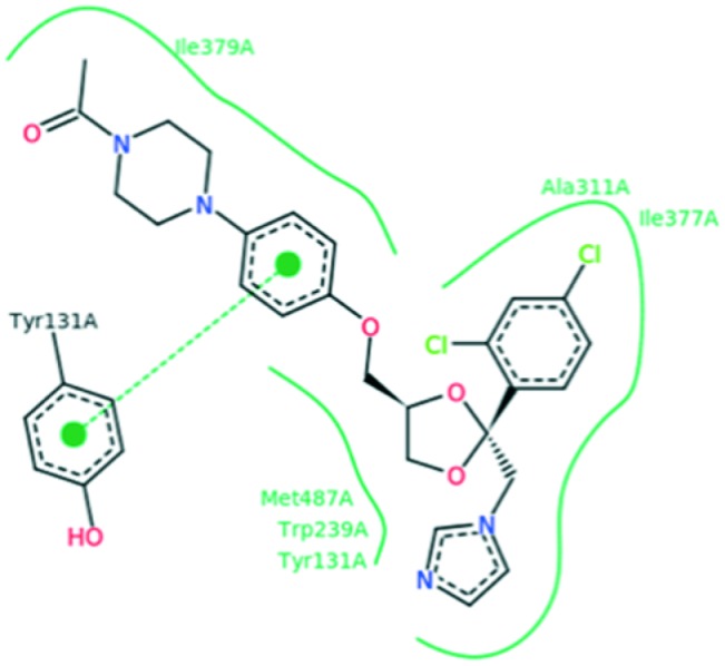
Further, in order to elucidate the mode of action of these compounds as antifungals, all the designed ligands and standards were docked against lanosterol 14α-demethylase (CYP51) in complex with ketoconazole (PDB code: 3LD6, Fig. 3) using Autodock 4.2 software.
Fig. 3. Docked conformation of ketoconazole in human lanosterol 14 alpha-demethylase (black dashed lines indicate hydrogen bonds, salt bridges, and metal interactions, green solid lines show hydrophobic interactions and green dashed lines show π–π and π–cation interactions).
The docking results of the ligands and the reference drug are given in Table 6. The interaction energy, comprised of van der Waals and electrostatic interactions, as well as intermolecular hydrogen bonding, was calculated for every minimized complex. The docking scores using Autodock 4.2® varied between –7.24 kcal mol–1 and –13.71 kcal mol–1 while the docking score for the standard ketoconazole was –7.18 kcal mol–1. This proves that usnic acid derivatives could be lead compounds for antifungal drug development. The most active compound (2) binding within CYP51 includes 3 favorable H-bonding interactions between the carboxyl group oxygen and the hydrogen of the side chain of Tyr131 (distance = 3.17 Å), the hydrogen of the alkyl chain and the oxygen of the side chain of Ile379 (distance = 2.22 Å), and the carboxyl group oxygen and the sulfur24 of the side chain of Cys449 (distance = 3.66 Å). The fused rings form hydrophobic contacts with Ile377, Met380, Tyr131, Tyr145, Met381, Met378, Phe234, Leu310, Ile488, Met387, His314, Thr315 and Ala311 (Fig. 4, A1 and 2), further stabilizing the complex.
Table 6. Human lanosterol 14 alpha-demethylase (CYP51) 3LD6 binding affinities.
| Compound | Est. binding energy (kcal mol–1) | Binding affinity score | I–H | Residues |
| 1 | –10.43 | –32.12 | 2 | Tyr145, Ile379 |
| 2 | –13.71 | –42.35 | 3 | Tyr131, Ile379, Cys449 |
| 3 | –8.97 | –28.14 | 1 | Ile379 |
| 4 | –11.25 | –37.22 | 3 | Tyr131, Ile379, Pro441 |
| 5 | –10.76 | –34.12 | 2 | Pro376, Ile379 |
| 6 | –8.54 | –26.77 | 1 | Tyr131 |
| 7 | –9.12 | –29.56 | 1 | Tyr131 |
| 8 | –7.49 | –23.52 | 1 | Tyr131 |
| 9 | –8.11 | –23.79 | 1 | Ile379 |
| 10 | –7.24 | –21.75 | — | — |
| 11 | –9.64 | –31.32 | 2 | Tyr131, Tyr145 |
| 12 | –12.06 | –38.72 | 3 | Tyr145, Pro376, Pro441 |
| 13 | –12.40 | –39.13 | 4 | Tyr145, Ile379, Met487 |
| Usnic acid | –8.62 | –24.98 | 1 | Met487 |
| Ketoconazole | –7.18 | –21.47 | — | — |
Fig. 4. (A1 and 2) Docked conformation of the most active compound 2 in human lanosterol 14 alpha-demethylase (CYP51).

The same hydrophobic interactions were observed for ketoconazole. However, compound 2 entered deeper than ketoconazole into the active center of the enzyme, forming a strong hydrogen bond with Cys449 (Fig. 5). These results suggest that this compound docks well in the binding pocket and explain its high inhibitory effect.
Fig. 5. Docked conformation of the most active compound 2 in human lanosterol 14 alpha-demethylase (CYP51).

The second most active compound (13) showed several hydrophobic interactions between the benzene rings and the amino acids Ile377, Met380, Tyr131, Met381, Met378, Phe234, Leu134, Ile488, Met378, His314, Thr135 and Ala311 (Fig. 6, A1 and 2) and four hydrogen bonds were formed: the first one between the carboxylic group oxygen and the backbone OH group of the side chain of Tyr145 (distance = 2.64 Å), the second one between the hydrogen of the side chain and the oxygen of the side chain of Tyr145 (distance = 2.71 Å), another hydrogen bond is formed between the OH group of the aromatic ring and the oxygen of the side chain of Ile379 (distance = 2.49 Å) and the last one between the other OH group of the aromatic ring and the oxygen of the side chain of Met287 (distance = 2.99 Å, Fig. 6, A1 and 2).
Fig. 6. (A1 and 2) Docked conformation of compound 13 in human lanosterol 14 alpha-demethylase (CYP51).

Regarding the docking of usnic acid in the 14α-demethylase active site, hydrophobic interactions were found with Tyr131, Tyr145, His314, Asp231, Ile377, Ile379 and Phe234 and only one hydrogen bond between the OH group of the aromatic ring and the oxygen of the carbonyl group of the side chain of Met487 was observed. This probably explains why usnic acid was less active than its synthetic derivatives (Fig. 7).
Fig. 7. (A1 and 2) Docked conformation of usnic acid in human lanosterol 14 alpha-demethylase (CYP51).

3. Experimental
3.1. Materials and methods
Solvents, unless otherwise specified, were of analytical reagent grade or of the highest quality commercially available. Synthetic starting materials, reagents and solvents were purchased from InterBioscreen (Chernogolovka, Russia, https://www.ibscreen.com/) and Aldrich Chemie (Steinheim, Germany). Melting points (°C) were determined with a Boetius apparatus without correction. 1H NMR spectra of the novel synthesized compounds in DMSO-d6 solutions were recorded on a Bruker AC 300 instrument at 298 K. Chemical shift (δ) values for the 1H NMR spectra are reported in parts per million (ppm) with the solvent resonance as the internal standard. TLC analyses were performed with Merck silica gel 60 F254 precoated plates, and each of the synthesized compounds showed a single spot. Specific rotations [α]20D were measured with a Jasco DIP-360 instrument at 589 nm.
3.2. Synthesis of usnic acid derivatives (general procedure)
To a mixture of (R)-usnic acid (0.52 g, 0.0015 mol) and the corresponding amine (0.0017 mol), triethylamine (0.2 g, 0.002 mol) was added (in the case when amine hydrochloride was used) and the mixture was heated to reflux in EtOH (7 mL) for 1 h. Then, the reaction mixture was diluted with 1% hydrochloric acid (50 mL), stirred for 2 h at room temperature and left overnight. The resulting precipitate was filtered, washed with 1% hydrochloric acid (3 × 20 mL) and water (3 × 20 mL), and dried to afford the pure title compounds. The yields of synthesized compounds were calculated after recrystallization from aqueous EtOH.
3.2.1. 2-(2-((E)-1-((R)-6-Acetyl-7,9-dihydroxy-8,9b-dimethyl-1,3-dioxo-1,9b-dihydrodibenzo[b,d]furan-2(3H)-ylidene)ethylamino)acetamido)-3-hydroxypropanoic acid (1)
Yield 62%, pale yellow solid, m.p. 188–191 °C. [α]20D = +319.46 (c = 0.43, CHCl3). 1H NMR (300 MHz, DMSO-d6) δ 1.71 (s, 3H, CH3), 1.99 (s, 3H, CH3), 2.65 (s, 3H, CH3), 2.69 (s, 3H, CH3), 3.75 (d, J = 15.6 Hz, 2H, CH2), 4.39 (m, 3H, CH2 + CH), 4.83 (br. s, 1H, OH), 5.77 (s, 1H, CH), 8.36 (s, 1H, NH), 12.12 (s, 1H, OH), 13.16 (s, 1H, NH), 13.28 (s, 1H, OH). 13C NMR (75 MHz, DMSO-d6) δ 7.41, 18.98, 30.92, 31.63, 46.22, 54.78, 56.17, 61.09, 100.79, 101.85, 102.39, 105.06, 106.23, 155.67, 157.59, 162.44, 166.33, 171.45, 172.71, 174.58, 188.40, 197.27, 200.81.
3.2.2. (R,E)-6-Acetyl-2-(1-(hexadecylamino)ethylidene)-7,9-dihydroxy-8,9b-dimethyldibenzo[b,d]furan-1,3(2H,9bH)-dione (2)
Yield 76%, pale yellow solid, m.p. 68–70 °C. [α]20D = +524.57 (c = 0.07, CHCl3). 1H NMR (300 MHz, DMSO-d6) δ 0.88 (t, J = 6.6 Hz, 3H, CH3), 1.25–1.42 (m, 26H, CH2), 1.69 (m, 5H, CH3 + CH2), 1.99 (s, 3H, CH3), 2.60 (s, 3H, CH3), 2.66 (s, 3H, CH3), 3.54 (m, 2H, CH2), 5.72 (s, 1H, CH), 12.09 (s, 1H, OH), 13.16 (s, 1H, NH), 13.27 (s, 1H, OH).
3.2.3. (R,E)-6-Acetyl-2-(1-(3,4-dihydroxyphenethylamino)ethylidene)-7,9-dihydroxy-8,9b-dimethyldibenzo[b,d]furan-1,3(2H,9bH)-dione (3)
Yield 57%, pale yellow solid, m.p. 145–148 °C. [α]20D = +244.85 (c = 0.34, CHCl3). 1H NMR (300 MHz, DMSO-d6) δ 1.67 (s, 3H, CH3), 1.99 (s, 3H, CH3), 2.61 (s, 3H, CH3), 2.68 (s, 3H, CH3), 2.81 (t, J = 7.1 Hz, 2H, CH2), 3.74 (m, 2H, CH2), 5.75 (s, 1H, CH), 6.51 (d, J = 8.1 Hz, 1H, CH), 6.65 (m, 2H, 2CH), 8.44 (s, 2H, 2OH), 12.10 (s, 1H, OH), 13.11 (s, 1H, NH), 13.28 (s, 1H, OH). 13C NMR (75 MHz, DMSO-d6) δ 7.45, 17.99, 30.95, 31.65, 33.72, 45.16, 56.24, 100.80, 101.45, 102.25, 105.10, 106.26, 115.54, 116.14, 119.49, 128.47, 143.94, 145.23, 155.68, 157.63, 162.46, 172.80, 174.74, 188.76, 197.21, 200.80.
3.2.4. (R,E)-2-(1-(6-Acetyl-7,9-dihydroxy-8,9b-dimethyl-1,3-dioxo-1,9b-dihydrodibenzo[b,d]furan-2(3H)-ylidene)ethylamino)-N-(thiazol-2-yl)acetamide (4)
Yield 81%, pale yellow solid, m.p. >250 °C. [α]20D = +363.89 (c = 0.29, CHCl3). 1H NMR (300 MHz, DMSO-d6) δ 1.71 (s, 3H, CH3), 2.00 (s, 3H, CH3), 2.62 (s, 3H, CH3), 2.68 (s, 3H, CH3), 4.63 (d, J = 5.2 Hz, 2H, CH2), 5.80 (s, 1H, CH), 7.14 (d, J = 3.6 Hz, 1H, CH), 7.43 (d, J = 3.6 Hz, 1H, CH), 12.04 (s, 1H, OH), 12.35 (s, 1H, NH), 13.28 (s, 1H, OH), 13.31 (s, 1H, NH). 13C NMR (75 MHz, DMSO-d6) δ 7.39, 18.92, 30.90, 31.62, 46.39, 56.30, 100.85, 101.97, 102.34, 105.01, 105.79, 106.28, 113.87, 137.57, 155.84, 157.50, 162.47, 165.49, 172.82, 174.99, 188.59, 197.47, 200.78.
3.2.5. (R,E)-6-Acetyl-7,9-dihydroxy-8,9b-dimethyl-2-(1-(2-(3-methyl-1,2,4-oxadiazol-5-yl)ethylamino)ethylidene)dibenzo[b,d]furan-1,3(2H,9bH)-dione (5)
Yield 56%, pale yellow solid, m.p. 106–109 °C. [α]20D = +372.97 (c = 0.40, CHCl3). 1H NMR (300 MHz, DMSO-d6) δ 1.68 (s, 3H, CH3), 1.99 (s, 3H, CH3), 2.36 (s, 3H, CH3), 2.65 (s, 6H, 2CH3), 3.33 (t, J = 6.7 Hz, 2H, CH2), 4.03 (m, 2H, CH2), 5.76 (s, 1H, CH), 12.03 (s, 1H, OH), 13.27 (s, 2H, OH + NH). 13C NMR (75 MHz, DMSO-d6) δ 7.41, 11.04, 17.92, 25.66, 30.91, 31.61, 45.88, 56.29, 100.79, 101.75, 102.23, 105.00, 106.29, 155.65, 157.55, 162.47, 166.80, 172.85, 175.28, 176.77, 188.72, 197.36, 200.76.
3.2.6. (R,E)-6-Acetyl-2-(1-((3-amino-1H-1,2,4-triazol-5-yl)methylamino)ethylidene)-7,9-dihydroxy-8,9b-dimethyldibenzo[b,d]furan-1,3(2H,9bH)-dione (6)
Yield 67%, pale yellow solid, m.p. >250 °C. [α]20D = +328.17 (c = 0.36, CHCl3). 1H NMR (300 MHz, DMSO-d6) δ 1.68 (s, 3H, CH3), 1.99 (s, 3H, CH3), 2.64 (s, 3H, CH3), 2.71 (s, 3H, CH3), 4.60 (d, J = 5.3 Hz, 2H, CH2), 5.77 (s, 1H, CH), 5.85 (s, 2H, NH2), 11.77 (s, 1H, NH), 12.10 (s, 1H, OH), 13.28 (s, 1H, OH), 13.34 (s, 1H, NH). 13C NMR (75 MHz, DMSO-d6) δ 7.43, 18.40, 30.93, 31.64, 41.85, 56.27, 100.78, 101.73, 102.33, 102.94, 105.11, 106.26, 155.70, 157.02, 157.61, 162.45, 172.83, 175.09, 188.63, 197.34, 200.83.
3.2.7. (R,E)-6-Acetyl-7,9-dihydroxy-2-(1-(2-(3-hydroxy-2-oxoquinoxalin-1(2H)-yl)ethylamino)ethylidene)-8,9b-dimethyldibenzo[b,d]furan-1,3(2H,9bH)-dione (7)
Yield 71%, pale yellow solid, m.p. >250 °C. [α]20D = +391.52 (c = 0.45, CHCl3). 1H NMR (300 MHz, DMSO-d6) δ 1.65 (s, 3H, CH3), 2.00 (s, 3H, CH3), 2.65 (s, 6H, 2CH3), 3.91 (m, 2H, CH2), 4.44 (t, J = 6.8 Hz, 2H, CH2), 5.72 (s, 1H, CH), 7.15 (m, 2H, 2CH), 7.23 (m, 1H, CH), 7.57 (m, 1H, CH), 12.00 (br. s, 2H, 2OH), 13.02 (s, 1H, NH), 13.27 (s, 1H, OH). 13C NMR (75 MHz, DMSO-d6) δ 7.50, 17.94, 31.00, 31.61, 38.34, 41.03, 56.26, 100.88, 102.11, 102.36, 105.12, 106.33, 114.97, 115.81, 123.24, 123.75, 125.78, 126.16, 153.31, 155.55, 155.74, 157.66, 162.52, 172.80, 175.50, 188.37, 197.29, 200.89.
3.2.8. (R,E)-N-(2-(1-(6-Acetyl-7,9-dihydroxy-8,9b-dimethyl-1,3-dioxo-1,9b-dihydrodibenzo[b,d]furan-2(3H)-ylidene)ethylamino)ethyl)acetamide (8)
Yield 83%, pale yellow solid, m.p. 133–135 °C. [α]20D = +357.63 (c = 0.49, CHCl3). 1H NMR (300 MHz, DMSO-d6) δ 1.68 (s, 3H, CH3), 1.85 (s, 3H, CH3), 2.00 (s, 3H, CH3), 2.60 (s, 3H, CH3), 2.66 (s, 3H, CH3), 3.34 (m, 2H, CH2), 3.65 (m, 2H, CH2), 5.77 (s, 1H, CH), 7.96 (s, 1H, NH), 12.10 (s, 1H, OH), 13.08 (s, 1H, NH), 13.27 (s, 1H, OH). 13C NMR (75 MHz, DMSO-d6) δ 7.42, 18.07, 22.46, 30.92, 31.64, 37.74, 42.87, 56.22, 100.79, 101.75, 102.27, 105.05, 106.26, 155.66, 157.61, 162.46, 169.82, 172.82, 175.19, 188.67, 197.16, 200.77.
3.2.9. (R,E)-2-(1-(6-Acetyl-7,9-dihydroxy-8,9b-dimethyl-1,3-dioxo-1,9b-dihydrodibenzo[b,d]furan-2(3H)-ylidene)ethylamino)ethanesulfonamide (9)
Yield 48%, pale yellow solid, m.p. 159–162 °C. [α]20D = +285.04 (c = 0.33, CHCl3). 1H NMR (300 MHz, DMSO-d6) δ 1.68 (s, 3H, CH3), 1.99 (s, 3H, CH3), 2.65 (s, 6H, 2CH3), 3.38 (t, J = 6.6 Hz, 2H, CH2), 3.97 (m, 2H, CH2), 5.75 (s, 1H, CH), 6.96 (s, 2H, NH2), 12.10 (s, 1H, OH), 13.12 (s, 1H, NH), 13.29 (s, 1H, OH). 13C NMR (75 MHz, DMSO-d6) δ 8.65, 19.28, 32.15, 32.86, 46.97, 54.14, 57.45, 102.00, 103.06, 103.51, 106.34, 107.49, 156.87, 158.79, 163.68, 173.98, 176.31, 189.71, 198.48, 202.00.
3.2.10. (R,E)-2-(1-(6-Acetyl-7,9-dihydroxy-8,9b-dimethyl-1,3-dioxo-1,9b-dihydrodibenzo[b,d]furan-2(3H)-ylidene))-N-(6-methoxybenzo[d]thiazol-2-yl)acetamide (10)
Yield 87%, pale yellow solid, m.p. >250 °C. [α]20D = +309.81 (c = 0.28, CHCl3). 1H NMR (300 MHz, DMSO-d6) δ 1.71 (s, 3H, CH3), 2.01 (s, 3H, CH3), 2.63 (s, 3H, CH3), 2.66 (s, 3H, CH3), 3.84 (s, 3H, OCH3), 4.67 (d, J = 5.2 Hz, 2H, CH2), 5.81 (s, 1H, CH), 6.94 (d, J = 8.9 Hz, 1H, CH), 7.45 (s, 1H, CH), 7.61 (d, J = 8.9 Hz, 1H, CH), 12.02 (s, 1H, OH), 12.45 (br. s, 1H, NH), 13.25 (s, 1H, OH), 13.33 (s, 1H, NH). 13C NMR (75 MHz, DMSO-d6) δ 7.42, 18.97, 30.94, 31.65, 46.58, 55.57, 56.32, 100.82, 101.96, 102.34, 104.65, 105.02, 106.28, 115.00, 121.18, 132.72, 136.55, 154.82, 155.66, 156.20, 157.53, 162.47, 166.30, 172.86, 174.99, 188.65, 197.51, 200.81.
3.2.11. (R,E)-6-Acetyl-7,9-dihydroxy-2-(1-(2-(3-(2-methoxyethyl)-1,2,4-oxadiazol-5-yl)ethylamino)ethylidene)-8,9b-dimethyldibenzo[b,d]furan-1,3(2H,9bH)-dione (11)
Yield 61%, pale yellow solid, m.p. 92–95 °C. [α]20D = +370.58 (c = 0.25, CHCl3). 1H NMR (300 MHz, DMSO-d6) δ 1.67 (s, 3H, CH3), 2.01 (s, 3H, CH3), 2.65 (s, 6H, 2CH3), 2.93 (t, J = 6.5 Hz, 2H, CH2), 3.28 (s, 3H, OCH3), 3.36 (m, 2H, CH2), 3.72 (t, J = 6.5 Hz, 2H, CH2), 4.04 (m, 2H, CH2), 5.75 (s, 1H, CH), 12.03 (s, 1H, OH), 13.28 (s, 2H, OH + NH). 13C NMR (75 MHz, DMSO-d6) δ 8.65, 19.13, 26.96, 27.21, 32.17, 32.82, 41.22, 57.50, 58.99, 69.57, 102.03, 102.98, 103.50, 106.25, 107.50, 156.89, 158.78, 163.68, 169.33, 174.09, 176.52, 178.08, 189.90, 198.56, 202.05.
3.2.12. (R,E)-6-Acetyl-7,9-dihydroxy-2-(1-(2-(5-(2-hydroxyphenyl)-1,3,4-oxadiazol-2-yl)ethylamino)ethylidene)-8,9b-dimethyldibenzo[b,d]furan-1,3(2H,9bH)-dione (12)
Yield 78%, pale yellow solid, m.p. 130–133 °C. [α]20D = +431.70 (c = 0.27, CHCl3). 1H NMR (300 MHz, DMSO-d6) δ 1.67 (s, 3H, CH3), 1.99 (s, 3H, CH3), 2.65 (s, 3H, CH3), 2.69 (s, 3H, CH3), 3.42 (t, J = 6.6 Hz, 2H, CH2), 4.10 (m, 2H, CH2), 5.77 (s, 1H, CH), 6.93 (m, 1H, CH), 7.11 (m, 1H, CH), 7.43 (m, 1H, CH), 7.79 (m, 1H, CH), 10.00 (s, 1H, OH), 12.03 (s, 1H, OH), 13.26 (s, 1H, OH), 13.34 (s, 1H, NH). 13C NMR (75 MHz, DMSO-d6) δ 8.66, 19.22, 25.97, 32.18, 32.82, 46.95, 57.52, 102.05, 103.01, 103.51, 106.28, 107.50, 110.54, 118.23, 120.88, 129.68, 134.55, 156.90, 157.39, 158.80, 163.68, 164.83, 165.73, 174.14, 176.52, 189.98, 198.56, 202.07.
3.2.13. (9bR,E)-6-Acetyl-7,9-dihydroxy-8,9b-dimethyl-2-(1-((5-(5-oxopyrrolidin-2-yl)-1,2,4-oxadiazol-3-yl)methylamino)ethylidene)dibenzo[b,d]furan-1,3(2H,9bH)-dione (13)
Yield 54%, pale yellow solid, m.p. 172–174 °C. [α]20D = +302.09 (c = 0.56, CHCl3). 1H NMR (300 MHz, DMSO-d6) δ 1.70 (s, 3H, CH3), 2.00 (s, 3H, CH3), 2.18–2.45 (m, 2H, CH2), 2.52–2.60 (m, 2H, CH2), 2.62 (s, 3H, CH3), 2.68 (s, 3H, CH3), 5.04 (m, 3H, CH2 + CH), 5.81 (s, 1H, CH), 8.24 (s, 1H, NH), 11.92 (s, 1H, OH), 13.27 (s, 1H, OH), 13.51 (s, 1H, NH). 13C NMR (75 MHz, DMSO-d6) δ 8.66, 19.66, 26.99, 29.94, 32.19, 32.70, 41.12, 50.60, 57.71, 102.07, 103.34, 103.48, 106.20, 107.57, 156.86, 158.68, 163.69, 167.67, 174.33, 177.38, 178.00, 182.16, 190.18, 198.99, 202.06.
3.3. Biological evaluation
3.3.1. Tested microorganisms
Laboratory and clinical strains of bacteria (two gram-positive species, Staphylococcus aureus (ATCC 6538) and Bacillus cereus (clinical isolate), three gram-negative species, Escherichia coli (ATCC 35210), Pseudomonas aeruginosa (ATCC 27853) and Enterobacter cloacae (clinical isolate), and three resistant bacteria, methicillin-resistant Staphylococcus aureus (MRSA), Escherichia coli and Pseudomonas aeruginosa PAO1) were used for testing the antibacterial activity of the usnic acid derivatives. For the antifungal bioassays, eight fungi were used: Aspergillus niger (ATCC 6275), Aspergillus ochraceus (ATCC 12066), Aspergillus fumigatus (ATCC 1022), Aspergillus versicolor (ATCC 11730), Penicillium funiculosum (ATCC 36839), Penicillium ochrochloron (ATCC 9112), Penicillium verrucosum var. cyclopium (food isolate) and Trichoderma viride (IAM 5061). The microorganisms are deposited in the Mycological Laboratory, Department of Plant Physiology, Institute for Biological Research “Siniša Stanković”, University of Belgrade, Belgrade, Serbia. The bacterial suspensions were adjusted with sterile saline to a concentration of 1.0 × 10⁵ CFU mL–1. The inocula were prepared daily and stored at +4 ° C until use. Dilutions of the inocula were cultured on solid medium to verify the absence of contamination and to check the validity of the inoculum. All experiments were performed in duplicate and repeated three times.
3.3.2. Isolation and determination of clinical bacteria
3.3.2.1. Methicillin-resistant Staphylococcus aureus (MRSA)
This strain is isolated from cows with subclinical mastitis. Milk samples were streaked onto Columbia agar plates (Torlak, Serbia) containing 5% sheep blood, Baird-Parker agar plates (HiMedia, India) and chromogenic culture media (chromID MRSA, bioMerieux). After incubation at 37 °C for 24 h, the colonies were presumptively identified according to morphological features, pigment production, Gram stain results, catalase and oxidase test results, type of haemolysis and characteristic growth on BP agar and chromID MRSA.
Supposed colonies of S. aureus on the blood agar and green colonies on chromogenic media were transferred to individual plates to obtain pure cultures. The identification was confirmed using a BBL Crystal G/P ID kit (Becton Dickinson). Antimicrobial susceptibility testing was performed by the disk diffusion method with 30 μg cefoxitin discs (Rosco, Denmark) in accordance with the Clinical and Laboratory Standard Institute recommendations. All isolated strains of S. aureus were tested for the presence of penicillin-binding protein (PBP2) with latex agglutination tests (Slidex MRSA detection, bioMerieux). Staphylococcus aureus ATCC 25923 was used as the control strain. All isolates were tested for the presence of the mecA gene by PCR.59
3.3.2.2. E. coli
Samples of rectal swabs, feces and intestines from diseased pigs were taken. In order to isolate E. coli strains, the following nutrition media were used: MacConkey agar (Torlak), Columbia agar (Torlak) with 5% defibrinated sheep blood and Brilliant Green agar (Torlak). For the identification of the isolated strains, laboratory tests with the following nutritious media and reagents were performed: Simmons citrate agar (Torlak), MR/VP broth (Torlak), Christensen urea agar (Torlak), peptone water for the indole test (Torlak), catalase and oxidase, triple sugar agar (Torlak), as well as identification systems BBL Crystal Entero/nonfermenter ID kit (Becton Dickinson).
Sensitivity studies on the isolated bacteria were completed by the disc diffusion method on Mueller Hinton agar with the use of antibiogram discs (Bioanalyse) and tablets (Torlak) for the following antibiotics: penicillin, ampicillin, amoxicillin, tetracycline, neomycin, gentamicin, colistin, ceftriaxone, sulphamethaxasole with trimethoprim, enrofloxacin and florfenicol. All isolated E. coli strains were resistant to all tested antibiotics with the exception of enrofloxacin, colistin and florfenicol.60
3.3.2.3. Pseudomonas aeruginosa
The strains were isolated from cats and dogs. Samples were inoculated on Columbia agar plates (Torlak, Serbia) containing 5% sheep blood, nutrition agar (HiMedia) and MacConkey agar (Torlak) and incubated under aerobic conditions at temperatures of 37 °C and 42 °C for 24 hours. Pure cultures were identified on the basis of morphological and biochemical characteristics. For identification of pigment production, sub-cultivation on the corresponding medium was carried out. Identification was confirmed using a BBL Crystal Entero/nonfermenter ID kit (Becton Dickinson). Sensitivity studies were completed by the disc diffusion method on Mueller Hinton agar with the use of antibiogram discs (Bioanalyse) and tablets (Torlak) for the following antibiotics: penicillin, ampicillin, amoxicillin, tetracycline, neomycin, gentamicin, ceftriaxone, sulphamethaxasole with trimethoprim, enrofloxacin and florfenicol.
All isolated Pseudomonas aeruginosa strains were resistant to all tested antibiotics with the exception of enrofloxacin and florfenicol.60
3.4. In vitro assays for determination of antimicrobial activity
3.4.1. Microdilution method
The compounds were tested for antibacterial activity using a microdilution method.61–63 Minimal inhibitory (MIC), minimal bactericidal (MBC) and minimal fungicidal (MFC) concentrations of the tested samples for each species of the studied microorganisms were determined by serial dilutions of compounds dissolved in a 5% DMSO–water solution in 96-well microtitre plates. Bacterial inoculum, 1.0 × 10⁴ CFU mL–1, was added to LB medium and the compounds were dissolved in 5% DMSO solution containing 0.1% Tween 80 (v/v) (1 mg mL–1). Minimal inhibitory concentration (MIC) was defined as the lowest concentration that inhibited bacterial growth, without visible growth under a binocular microscope. The lowest concentration with no visible growth was defined as the MBC, indicating 99.5% death of the original inoculum. All wells were measured at a wavelength of 655 nm using Microplate Manager 4.0® (Bio-Rad Laboratories) and compared with a blank and the positive control. Antibiotics used as positive controls were streptomycin (Sigma P 7794) and ampicillin (Panfarma, Belgrade, Serbia) (1 mg mL–1 in sterile physiological saline), while 5% DMSO was used as a negative control. All experiments were performed in duplicate and repeated three times.
3.4.2. Statistical analysis
All the assays were carried out in triplicate and the results are expressed as mean values and standard deviation (SD). The results were analyzed using one-way analysis of variance (ANOVA) followed by Tukey's HSD test with α = 0.05. This treatment was carried out using the SPSS v. 18.0 program.
3.5. Docking studies
All docking calculations were carried out using the software AutoDock 4.2® which is one of the most appropriate tools for performing molecular docking of ligands to their macromolecular receptors. The free energies of binding (ΔG) of DnaG, gyrase, thymidylate kinase, human lanosterol 14 alpha-demethylase (CYP51) and dihydrofolate reductase complexes with inhibitors 1–13 were generated using this molecular docking program. The X-ray crystal structure data of all proteins used were obtained from the Protein Data Bank (PDB ID: IDDE, IKZN, ; 4QGG, ; 3LD6 and ; 4HOF, respectively).
For preparation of all proteins mentioned above, polar hydrogens were added, and then Kollman united atom charges and atomic solvation parameters were assigned. For ligand preparation, Gasteiger partial charges were added, non-polar hydrogen atoms were merged, and rotatable bonds were defined. The grid maps of docking studies were computed using the Autogrid® program. The grid must surround the region of interest in the macromolecule. Three-dimensional structures of the aforementioned compounds were constructed using Chem 3D Ultra 12.0® software (Chemical Structure Drawing Standard; Perkin Elmer Informatics, Waltham, MA, USA). The compounds were subjected to flexible docking using the pre-computed grid files. All parameters used in docking were default values. A preliminary blind docking study was performed in order to discriminate the preferential binding sites of the ligand to the receptor. The translation, quaternion, and torsions steps were taken from default values in Autodock®. The Lamarckian genetic algorithm and pseudo-Solis and Wets methods were applied for minimization using default parameters. The number of docking runs was 100. After docking, the 100 solutions were clustered into groups with RMS lower than 1.0 E. The clusters were ranked by the lowest energy representative of each cluster. In order to describe the ligand-binding pocket interactions, the top ranked binding mode was found by Autodock® in a complex with the binding pocket of the enzyme. The resulting positions and potential interactions were visualized in Discovery Studio 2017 R2 Silent®.
4. Conclusion
Thirteen new usnic acid derivatives were synthesized and evaluated for their antimicrobial activity. The newly synthesized derivatives of usnic acid exhibited remarkable inhibition of the growth of a wide spectrum of Gram-positive and Gram-negative bacteria and fungi. Some of our compounds exhibited comparable or better antibacterial activity than the reference drugs, with the most potent among them being compound 9 followed by 3. Nevertheless, none of the compounds exhibited antibacterial activity better than usnic acid. The most sensitive bacterial species for these compounds was S. aureus, while P. aeruginosa and E. coli were the more resistant ones.
As far as fungi are concerned, the tested compounds possessed excellent activity against all the fungal species tested, being 220–230 times more active than ketoconazole and 8–89 times more active than bifonazole. The most promising is compound 2 followed by 13 and 12. It should be mentioned that all compounds, in general, exhibited much better activity than usnic acid, especially against Aspergillus fumigatus. The most sensitive fungal species was Trichoderma viride, while P. versicolor var. cyclopium was the most resistant.
The results of the docking studies coincided well with the antifungal activity of the four most active compounds (2, 13, 12 and 4) with binding energies of –13.71, –12.40, –12.06 and –11.25, respectively.
Conflicts of interest
The authors declare no competing interest.
References
- Stocker-Worgotter E. Nat. Prod. Rep. 2008;25:188–200. doi: 10.1039/b606983p. [DOI] [PubMed] [Google Scholar]
- Cocchietto M., Skert N., Nimis P. L., Sava G. Naturwissenschaften. 2002;89:137–146. doi: 10.1007/s00114-002-0305-3. [DOI] [PubMed] [Google Scholar]
- Kinosha Y., Yamamoto Y., Yoshimura I., Kurokawa T., Huneck S. J. Hattori Bot. Lab. 1997;83:173–178. [Google Scholar]
- Shtro A. A., Zarubaev V. V., Luzina O. A., Sokolov D. N., Salakhutdinov N. F. Antiviral Chem. Chemother. 2015;24(3–4):92–98. doi: 10.1177/2040206616636992. [DOI] [PMC free article] [PubMed] [Google Scholar]
- Shtro A. A., Zarubaev V. V., Luzina O. A., Sokolov D. N., Kiselev O. I., Salakhutdinov N. F. Bioorg. Med. Chem. 2014;22(24):6826–6836. doi: 10.1016/j.bmc.2014.10.033. [DOI] [PubMed] [Google Scholar]
- Vanga N. R., Kota A., Sistla R., Uppuluri M. Mol. Diversity. 2017;21(2):273–282. doi: 10.1007/s11030-016-9716-5. [DOI] [PubMed] [Google Scholar]
- Khan M. F., Nabila S. A., Rashid R. B., Rahman M. S., Chowdhary A. A., Rashid M. A. Bangladesh Pharm. J. 2015;18(2):90–96. [Google Scholar]
- Ananthi R., Tinabaye A., Ganesan T., Selvaraj G., Arulmozhi S., Kumar S. Int. J. Sci. Eng. Res. 2016;7(8):39–44. [Google Scholar]
- Oran S., Sahin S., Sahinturk P., Ozturk S., Demir C. Iran. J. Pharm. Res. 2016;15(2):527–535. [PMC free article] [PubMed] [Google Scholar]
- Luzina O. A., Salakhutdinov N. F. Russ. J. Bioorg. Chem. 2016;42(3):249–268. [Google Scholar]
- White A. S., Oliveira R. C., Oliveira A. P., Serafini M. R., Araujo A. A. S., Gelain D. P., Moreira J. C. F., Almeida J. R. G. S., Quintas J. S. S., Quintans-Junior L. J., Santos M. R. V. Molecules. 2014;19:14496–14527. doi: 10.3390/molecules190914496. [DOI] [PMC free article] [PubMed] [Google Scholar]
- Jiang C.-S., Liang L.-F., Guo Y.-W. Acta Pharmacol. Sin. 2012;33(10):1217–1245. doi: 10.1038/aps.2012.90. [DOI] [PMC free article] [PubMed] [Google Scholar]
- Zakharenko A., Sokolov D., Luzina O., Sukhanova M., Khodyreva S., Zakharova O., Salakhutdinov N., Lavrik O. Med. Chem. 2012;8(5):883–893. doi: 10.2174/157340612802084225. [DOI] [PubMed] [Google Scholar]
- Paudel B., Bhattarai H. D., Lee H. K., Oh H., Shin H. W., Yim J. H. Z. Naturforsch., C: J. Biosci. 2010;65(1–2):34–38. doi: 10.1515/znc-2010-1-206. [DOI] [PubMed] [Google Scholar]
- Ramos D. F., Almeida da Silva P. E. Pharm. Biol. 2010;48(3):260–263. doi: 10.3109/13880200903085490. [DOI] [PubMed] [Google Scholar]
- Luzina O., Salakhutdinov N. F. Russ. J. Bioorg. Chem. 2016;42(2):115–132. [Google Scholar]
- Pires R. H., Lucorini R., Mendes-Giannini M. J. S. Antimicrob. Agents Chemother. 2011;56(1):595–597. doi: 10.1128/AAC.05348-11. [DOI] [PMC free article] [PubMed] [Google Scholar]
- De Carvalho E. A., Andrade P. P., Silva N. H., Pereira E. C., Fiqueiredo R. C. Micron. 2005;36(2):155–161. doi: 10.1016/j.micron.2004.09.003. [DOI] [PubMed] [Google Scholar]
- Koparal A. T. Z. Naturforsch., C: J. Biosci. 2015;70(5–6):159–164. doi: 10.1515/znc-2014-4178. [DOI] [PubMed] [Google Scholar]
- Varol M. J. J. Appl. Pharm. 2015;8:1. [Google Scholar]
- Song Y., Dai F., Zhai D., Dong Y., Zhang J., Lai L., Zhang T., Li D., Pang X., Liu M., Yi Z. Angiogenesis. 2012;15:421–432. doi: 10.1007/s10456-012-9270-4. [DOI] [PubMed] [Google Scholar]
- O'Neill M. A., Mayer M., Murray K. E., Rolim-Santos H. M., Santos-Magalhães N. S., Thompson A. M., Appleyard V. C. Braz. J. Microbiol. 2010;70(3):659–664. doi: 10.1590/s1519-69842010005000013. [DOI] [PubMed] [Google Scholar]
- Takai M., Uehara Y., Beisler J. A. J. Med. Chem. 1979;22:1380–1384. doi: 10.1021/jm00197a019. [DOI] [PubMed] [Google Scholar]
- Mayer M., O'Neill M. A., Murray K. E., Santos-Magalhães N. S., Carneiro-Leão A. M., Thompson A. M., Appleyard V. C. Anti-Cancer Drugs. 2005;16(8):805–809. doi: 10.1097/01.cad.0000175588.09070.77. [DOI] [PubMed] [Google Scholar]
- Kupchan S. M., Kopperman H. L. Experientia. 1975;31(6):625. doi: 10.1007/BF01944592. [DOI] [PubMed] [Google Scholar]
- Singh N., Nambiar D., Kale R. K., Singh R. P. Nutr. Cancer. 2013;65(sup1):36–43. doi: 10.1080/01635581.2013.785007. [DOI] [PubMed] [Google Scholar]
- Bačkorová M., JendŽelovský R., Kello M., Bačkor M., Mikeš J., Fedoročko P. Toxicol. In Vitro. 2012;26:462–468. doi: 10.1016/j.tiv.2012.01.017. [DOI] [PubMed] [Google Scholar]
- Brisdelli F., Perilli M., Sellitri D., Piovano M., Garbarino J. A., Nicoletti M., Bozzi A., Amicosante G., Celenza G. Phytother. Res. 2012;25:431–437. doi: 10.1002/ptr.4739. [DOI] [PubMed] [Google Scholar]
- Yang Y., Nguyen T. T., Jeong M.-H., Crişan F., Yu Y. H., Ha H.-H., Choi K. H., Jeong H. G., Jeong T. C., Lee K. Y., Kim K. K., Hur J.-S., Kim H. PLoS One. 2016;11(1):e0146575. doi: 10.1371/journal.pone.0146575. [DOI] [PMC free article] [PubMed] [Google Scholar]
- Koparal A. T., Tüylü B. A., Türk H. Nat. Prod. Res. 2006;20:1300–1307. doi: 10.1080/14786410601101910. [DOI] [PubMed] [Google Scholar]
- Molnar K., Farkas E. Z. Naturforsch., C: J. Biosci. 2010;65:153–173. doi: 10.1515/znc-2010-3-401. [DOI] [PubMed] [Google Scholar]
- Lucarini R., Tozatti M. G., De Oliveira Salloum A. I., Crotti A. E. M., Silva M. L. A., Gimenez V. M. M., Groppo M., Januario A. H., Martins C. H. G., Cunha W. R. Afr. J. Biotechnol. 2012;11:4636–4639. [Google Scholar]
- Maciag-Dorszynska M., Wegryn G., Guzow-Krzeminska B. FEMS Microbiol. Lett. 2014;353:57–62. doi: 10.1111/1574-6968.12409. [DOI] [PubMed] [Google Scholar]
- Gupta V. K., Verma S., Singh A., Darokar P. Eur. J. Clin. Microbiol. Infect. Dis. 2012;31:3375–3383. doi: 10.1007/s10096-012-1706-7. [DOI] [PubMed] [Google Scholar]
- Kekuda T. R. P., Syed J., Dileep N., Ralesh K. N., Vinayaka K. S. Sci., Technol. Arts Res. J. 2013:87–90. [Google Scholar]
- Paudel B., Bhattarai H. D., Lee H. K., Oh H., Shin H. W., Yim J. H. Z. Naturforsch., C: J. Biosci. 2010;65(1–2):34–38. doi: 10.1515/znc-2010-1-206. [DOI] [PubMed] [Google Scholar]
- Moreira A. S. N., Braz-Filho R., Mussi-Dias V., Vieira I. J. C. Molecules. 2015;20:8952–8987. doi: 10.3390/molecules20058952. [DOI] [PMC free article] [PubMed] [Google Scholar]
- Yu X., Guo Q., Su G., Yang A., Hu Z., Qu C., Wan Z., Li R., Tu P., Chai X. J. Nat. Prod. 2016;79(5):1373–1380. doi: 10.1021/acs.jnatprod.6b00109. [DOI] [PubMed] [Google Scholar]
- Taresco V., Francolini I., Padella F., Bellusci M., Boni A., Innocenti C., Martinelli A., D'Ilario L., Piozzi A. Mater. Sci. Eng., C. 2015;52:72–81. doi: 10.1016/j.msec.2015.03.044. [DOI] [PubMed] [Google Scholar]
- Nithianand P., Shareen R. M. B., Muthamil S., Pandian S. K. Microbiol. Res. 2015;179:20–28. doi: 10.1016/j.micres.2015.06.009. [DOI] [PubMed] [Google Scholar]
- Francolini I., Norris P., Piozzi A., Donelli G., Stoodley P. Antimicrob. Agents Chemother. 2004;48:4360–4365. doi: 10.1128/AAC.48.11.4360-4365.2004. [DOI] [PMC free article] [PubMed] [Google Scholar]
- Pompilio A., Pomponio S., Di Vincenzo V., Crocetta V., Nicoletti M., Piovano M., Garbarino J. A., Di Bonaventura G. Future Microbiol. 2013;8(2):281–292. doi: 10.2217/fmb.12.142. [DOI] [PubMed] [Google Scholar]
- Gupta V. K., Verma S., Gupta S., Singh A., Pal A., Srivastava S. K., Srivastava P. K., Singh S. C., Darokar M. P. Eur. J. Clin. Microbiol. Infect. Dis. 2012;31(12):3375–3383. doi: 10.1007/s10096-012-1706-7. [DOI] [PubMed] [Google Scholar]
- Grumezescu V., Holban A. M., Grumezescu A. M., Socol G., Ficai A., Vasile B. S., Truscă R., Bleotu C., Lazar V., Chifiriuc C. M. Biofabrication. 2014;6(035002):1–12. doi: 10.1088/1758-5082/6/3/035002. [DOI] [PubMed] [Google Scholar]
- Weckesser S., Engel K., Simon Haarhaus B., Wittmer A., Pelz K., Schempp C. M. Phytomedicine. 2007;14(7–8):508–516. doi: 10.1016/j.phymed.2006.12.013. [DOI] [PubMed] [Google Scholar]
- Vijayakumar C. S., Viswanathan S., Reddy M. K., Parvathavarthini S., Kundu A. B., Sukumar E. Fitoterapia. 2000;71:564–566. doi: 10.1016/s0367-326x(00)00209-4. [DOI] [PubMed] [Google Scholar]
- Ingolfsdottir K. Phytochemistry. 2002;61:729–731. doi: 10.1016/s0031-9422(02)00383-7. [DOI] [PubMed] [Google Scholar]
- Leandro L. F., Munari C. C., Sato V. L., Alves J. M., de Oliveira P. F., Mastrocola D. F., da Silva Morales M. T., Oliveira A. I., Tozatti M. G., Cunha W. R., Tavare D. C. Mutat. Res., Genet. Toxicol. Environ. Mutagen. 2013;753(2):101–106. doi: 10.1016/j.mrgentox.2013.03.006. [DOI] [PubMed] [Google Scholar]
- Fournet A., Ferreira M. E., De Rojas A., de Torres O. S., Inchausti A., Yaluff G., Quilhot W., Fernandez E., Hidalgo M. E. Comp. Biochem. Physiol. 1997;111:69–74. doi: 10.1016/s0742-8413(96)00127-2. [DOI] [PubMed] [Google Scholar]
- Pramyothin P., Janthasoot W., Pongnimitprasert N., Phrukudom S., Ruangrungsi N. J. Ethnopharmacol. 2004;90:381–387. doi: 10.1016/j.jep.2003.10.019. [DOI] [PubMed] [Google Scholar]
- Odabasoglu F., Cakir A., Suleyman H., Aslan A., Bayir Y., Halici M., Kazaz C. J. Ethnopharmacol. 2006;103:59–65. doi: 10.1016/j.jep.2005.06.043. [DOI] [PubMed] [Google Scholar]
- Halici M., Odabasoglu F., Suleyman H., Cakir A., Aslan A., Bayir Y. Phytomedicine. 2005;12:656–662. doi: 10.1016/j.phymed.2004.06.021. [DOI] [PubMed] [Google Scholar]
- Murtazalieva K. A., Druzhilovskii D. S., Goel R. K., Sastry G. N., Poroikov V. V. SAR QSAR Environ. Res. 2017;28(10):843–862. doi: 10.1080/1062936X.2017.1399448. [DOI] [PubMed] [Google Scholar]
- www.vclab.org .
- Wang J. D., Sanders G. M., Grossman A. D. Cell. 2007;128:865–875. doi: 10.1016/j.cell.2006.12.043. [DOI] [PMC free article] [PubMed] [Google Scholar]
- Maciag M., Kochanowska M., Lyzeń R., Wegrzyn G., Szalewska-Pałasz A. Plasmid. 2010;63:61–67. doi: 10.1016/j.plasmid.2009.11.002. [DOI] [PubMed] [Google Scholar]
- Aravind I., Leipe D. D., Koonin E. Y. Nucleic Acids Res. 1998;26:4205–4213. doi: 10.1093/nar/26.18.4205. [DOI] [PMC free article] [PubMed] [Google Scholar]
- Ziegelin G., Linderoth N. A., Calendar R., Lanka E. J. Bacteriol. 1995;177:4333–4341. doi: 10.1128/jb.177.15.4333-4341.1995. [DOI] [PMC free article] [PubMed] [Google Scholar]
- Stegger P. S., Andersen A., Kearns B., Pichon B., Holmes M. A., Edwards G., Laurent F., Teale C., Skov R., Larsen A. R. Clin. Microbiol. Infect. 2012;18(4):395–400. doi: 10.1111/j.1469-0691.2011.03715.x. [DOI] [PubMed] [Google Scholar]
- CLSI. (2009): Approved standard, 8th ed. CLSI publication M07-A8. Clinical and Laboratory Standards Institute, Wayne, PA.
- Hanel H., Raether W. Mycoses. 1988;31:148–154. doi: 10.1111/j.1439-0507.1988.tb03718.x. [DOI] [PubMed] [Google Scholar]
- Daouk K. D., Dagher M. S., Sattout J. E. J. J. Food Prot. 1995;58:1147. doi: 10.4315/0362-028X-58.10.1147. [DOI] [PubMed] [Google Scholar]
- Espinel-Ingroff A. J. J. Clin. Microbiol. 2001;39:1360–1367. doi: 10.1128/JCM.39.4.1360-1367.2001. [DOI] [PMC free article] [PubMed] [Google Scholar]