Abstract
Bouveret’s syndrome is a rare presentation of duodenal obstruction or gastric outlet obstruction caused by a large gallstone migrating through a cholecystoduodenal or choledochoduodenal fistula. Most patients are elderly and often have underlying comorbidities, complicating surgery. Endoscopic therapy should be used as first-line treatment for these patients who are not good surgical candidates. We report a case of a 98-year-old Chinese female who presented with vomiting for three days. Esophagogastroduodenoscopy and computed tomography confirmed the diagnosis of Bouveret’s syndrome. The patient successfully underwent endoscopic lithotripsy with the Holmium: Yttrium–Aluminum–Garnet (Ho: YAG) laser. Ho: YAG laser lithotripsy has been used to treat Bouveret’s syndrome in four case reports. It can be recommended in patients with Bouveret’s syndrome who are poor candidates for surgery.
Bouveret’s syndrome is a rare presentation of duodenal obstruction or gastric outlet obstruction caused by a large gallstone migrating through a cholecystoduodenal or choledochoduodenal fistula. In the majority of reported cases, surgical intervention was performed. However, the major complication and mortality rates for surgical treatment are approximately 13%–22.7%.1–3,9 Moreover, most patients are elderly and often have underlying comorbidities, complicating surgery. Previous reports have shown the effectiveness of minimal invasive treatment modalities such as extracorporeal shock wave lithotripsy, mechanical lithotripsy, electrohydraulic lithotripsy, and laser lithotripsy for treating Bouveret’s syndrome. Although the success rates of these non-surgical therapies are unsatisfactory, endoscopic treatment with laser lithotripsy seems to be more promising than other non-surgical methods.3 In recent years, there have been some reports on using endoscopy alone for successfully treating Bouveret’s syndrome. We describe an extremely elderly patient with high surgical risk treated using the Ho: YAG laser for endoscopic lithotripsy.
CASE
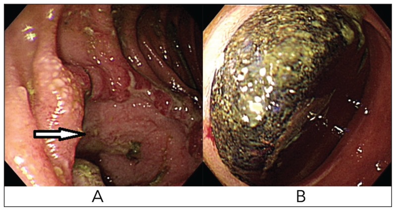
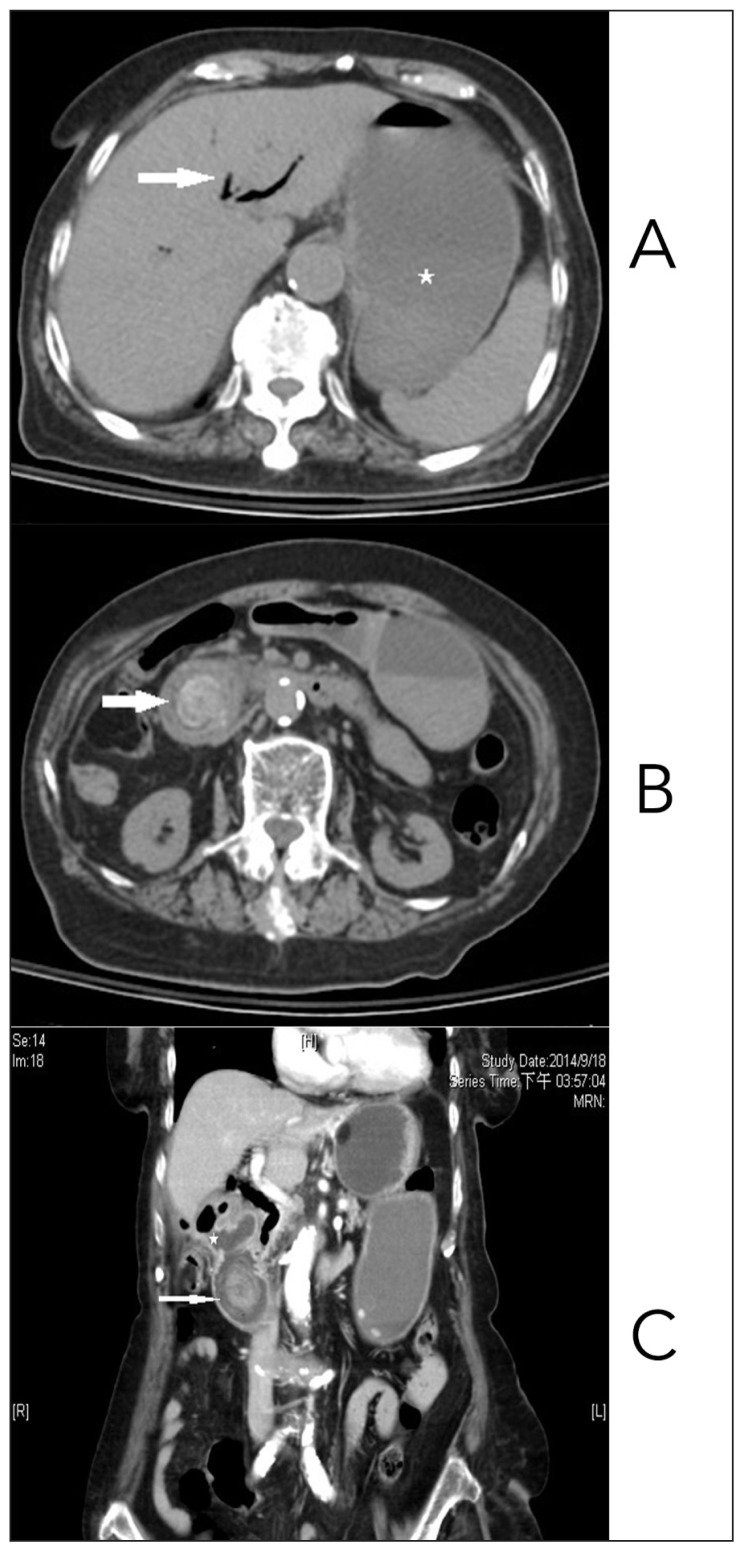
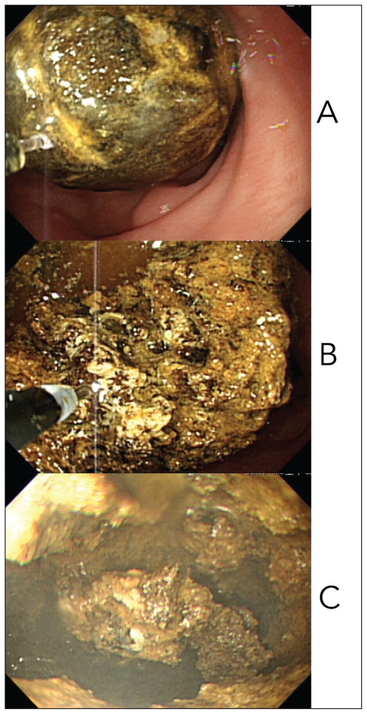
A 98-year-old Chinese female with cryptogenic liver cirrhosis, Child-Pugh A, presented with complaints of nausea, vomiting, abdominal pain, abdominal fullness, and anorexia for three days. She denied constipation, fever, or chills. Her vital signs were stable, and no peritoneal signs were noted on physical examination. Laboratory findings of the patient were as follows: white blood cell count, 13000/μL (80.7% segmented neutrophils); total bilirubin, 2.1 mg/dL; albumin, 3.4 g/dL; and normal creatinine level. Esophagogastroduodenoscopy showed a lot of fluid in the stomach, fistulous orifice on the proximal second portion of the duodenum near the bulb, and a large gallstone obstructing the second portion of the duodenum (Figure 1A and 1B). Abdominal computed tomography (CT) revealed an obstructed duodenum with a distended stomach, containing a lot of fluid, air in the biliary tree and gallbladder, a cholecystoduodenal fistula, and a 3.2×3.0-cm gallstone within the second portion of the duodenum (Figure 2A, 2B, 2C). Because of old age and underlying liver cirrhosis, the patient was a poor candidate for surgical intervention. Endoscopic mechanical lithotripsy was attempted to fragment and remove the stone. However, the large and firm stone broke two sets of our mechanical lithotripters, and the first therapeutic endoscopy failed. Therefore, the AURIGA Ho: YAG laser system (Lynton Surgical, UK) was used during the second attempt of therapeutic endoscopy. A 365-μ flexible quartz laser fiber was passed through the Glo-Tip catheter first, and the Glo-tip catheter was inserted through the working channel of the upper gastrointestinal endoscope to the duodenum. The force and frequency of laser pulses were 1.8 J and 12 Hz, respectively. Adequate water irrigation was performed during the procedure, and the stone was fragmented gradually using Ho: YAG laser lithotripsy under direct endoscopic view (Figure 3A, 3B, 3C). After complete fragmentation, the large piece of stone was removed readily with a Dormia basket. The patient’s condition improved rapidly without further discomfort associated with cholelithiasis. Cholecystoduodenal fistula was not repaired, and the patient presented with no complaints due to the gallstone at the six-month follow up.
Figure 1.
A) Cholecystoduodenal fistula B) Gallstone in duodenum.
Figure 2.
A) Pneumobilia and distended stomach B) Gallstone obstructs the duodenum C) CT scan confirmed Bouveret’s syndrome.
Figure 3.
A) Start of laser lithotripsy B) Partial fragmentation of the gallstone C) Complete fragmentation of the gallstone.
DISCUSSION
Gallstone ileus is an uncommon but serious complication of cholelithiasis following migration of a large gallstone to the intestine through a cholecystoduodenal fistula, and it comprises only 1%–4% cases of intestinal obstruction.1 The stone can obstruct any site in the gastrointestinal tract, but the most common site of obstruction is the distal small intestine where the lumen is the narrowest. Most investigators believe that stones must be more than 2–2.5 cm in diameter to obstruct the intestine. 4,5 In a review of 1001 reported cases by Reisner and Cohen,6 impaction of the stone occurs in the ileum (60.5%), jejunum (16.1%), stomach (14.2%), colon (4.1%), and duodenum (3.5%). A gallstone impacting the duodenum and resulting in gastric outlet obstruction or duodenal obstruction is known as Bouveret’s syndrome and was first described in 1896 by Leon Bouveret (1850–1929), a French physician who played an important role in studying gastric diseases.7 Base on the above description and the review of 128 cases by Mitchell et al,3 Bouveret’s syndrome is categorized as a rare disease. The mean patient age was 74.1 years, and the female-to-male sex ratio was 1.86. Common symptoms included nausea/vomiting (86%) and abdominal pain or discomfort (71%), and common signs included abdominal tenderness (44%), dehydration (31%), and abdominal distention (27%). All cases had gastroduodenal obstruction on esophagogastroduodenoscopy, but the obstructing stone could be detected in only 69% cases during endoscopic examination. Fistulous stoma was seen in only 13% esophagogastroduodenoscopies. Plain abdominal radiography may show pneumobilia, calcified right upper quadrant mass or gallstone, gastric distention, and dilated small bowel loops in some cases, but the sensitivity is low. Abdominal CT is the preferred imaging modality to evaluate biliary complications and to reveal the classical Rigler’s triad of pneumobilia, ectopic gallstone, and gastroduodenal obstruction.8 Most patients with Bouveret’s syndrome have been treated by surgery, but the major complications and mortality rates are high despite modern surgical techniques.1–3,9 Thus endoscopic treatments have been popular because of the advanced age and comorbidities seen in this patient group. Endoscopic laser lithotripsy seems to be more promising than other endoscopic treatment.3 The Ho: YAG laser lithotripsy was reported only in four cases.10–13 One of these cases was complicated with distal gallstone ileus after Ho: YAG laser lithotripsy; hence, surgical enterotomy was performed.11 Another case was treated using Ho: YAG laser lithotripsy with electrohydraulic lithotripter,13 and the remaining two cases were successfully treated using Ho: YAG laser lithotripsy.10,12
Our case was a characteristic Bouveret’s syndrome patient who was extremely old, showing characteristic features and poor risk factors for surgery. Initial endoscopic mechanical lithotripsy failed because the stone was too large and firm. We chose the Ho: YAG laser lithotripsy because of its wide use in the surgical management of urinary lithiasis and prior success rates in treating Bouveret’s syndrome.
The Ho: YAG laser is a solid-state pulsed-wave laser with a wavelength of 2140 nm and a pulse duration of 350–700 μs.14 It is capable of disintegrating urinary stones of all compositions while maintaining a wide margin of safety.15 A previous study also demonstrated the safety and efficacy of the Ho: YAG laser lithotripsy in the management of complex biliary tract stones.16 The laser deploys via a flexible optical fiber, and we can work with the laser through the working channel of the endoscope. The laser fiber can be placed under endoscopic view and makes the treatment technically easy and safe. The mechanism of stone fragmentation with the Ho: YAG laser mainly includes superheating the surrounding water and forming vaporization bubbles, which create a thermal effect in the localized area within 3 mm of the probe.17 Some investigators have commented that Ho: YAG laser lithotripsy occurs through a “drilling effect”, whereby small bits of stone are vaporized, emitting a fine stone dust, which obscures visualization. 18 For endoscopic use, an effective irrigation system would be essential.
In our case, the large and firm gallstone was fixed in the duodenum and it broke two sets of our mechanical lithotripters. Hence, the fact that it could be fragmented in one session using Ho: YAG laser lithotripsy shows the superior fragmentation power of this device. The rhodamine-6 G dye laser system also has been used successfully for endoscopic treatment of Bouveret’s syndrome, but multiple sessions are needed to achieve complete fragmentation.19,20
A potential complication of endoscopic treatment is that partial fragmentation or dislocation of the stone can cause distal gallstone ileus, often requiring surgical intervention.11,21 Although surgery ultimately is necessary in these cases, endoscopic lithotripsy helps to avoid the potentially high-risk duodenotomy and change to easier distal enterotomy.22
Direct endoscopic lithotomy of the complete gallstone and mechanical lithotripsy or electrohydraulic lithotripsy in cases of Bouveret’s syndrome with high failure rates have been demonstrated in previous reports. 3 Ho: YAG laser lithotripsy has been used successfully and safely in patients with Bouveret’s syndrome, so it should be considered as first-line treatment for patients with poor surgical risk. There are no procedure-related complications or long-term adverse outcomes in these four case reports.10–13
The limitation of the Ho: YAG laser lithotripsy is the inability to close the fistula or do further cholecystectomy as surgery. Therefore, it has potential for recurrence, if there are residual stones in gallbladder. Further systematic study might reveal more experience and make it possible to make better recommendations, but this may be unavailable because of the rarity of Bouveret’s syndrome. A special endoscopic suite of Ho: YAG laser for a rare disease is not cost-effective. However, the Ho: YAG laser is very popular in urology, and is used for urolithiasis. In our hospital, instead of possessing a special Ho: YAG laser system in gastroenterology division, we borrow it from the urology division when needed.
In conclusion, we believe that Ho: YAG laser lithotripsy can be recommended in patients with Bouveret’s syndrome who are poor candidates for surgery. Thus, endoscopists should be familiar with Ho: YAG laser lithotripsy which is available in many hospitals for efficient treatment of these difficult cases.
Footnotes
SIMILAR CASES PUBLISHED: 4
Conflicts of interest
The authors declare that they have no conflicts of interest related to the subject matter or materials discussed in this article.
REFERENCES
- 1.Clavien PA, Richon J, Burgan S, Rohner A. Gallstone ileus. Br J Surg. 1990 Jul;77(7):737–42. doi: 10.1002/bjs.1800770707. [DOI] [PubMed] [Google Scholar]
- 2.Ayantunde AA, Agrawal A. Gallstone Ileus: Diagnosis and Management. World J Surg. 2007;31:1292–1297. doi: 10.1007/s00268-007-9011-9. [DOI] [PubMed] [Google Scholar]
- 3.Cappell MS, Davis M. Characterization of Bouveret’s syndrome: a comprehensive review of 128 cases. Am J Gastroenterol. 2006;101(9):2139–2146. doi: 10.1111/j.1572-0241.2006.00645.x. [DOI] [PubMed] [Google Scholar]
- 4.Abou-Saif A, Al-Kawas FH. Complications of gallstone disease: Mirizzi syndrome, cholecystocholedochal fistula and gallstone ileus (clinical reviews) Am J Gastroenetrol. 2002;97:249–254. doi: 10.1111/j.1572-0241.2002.05451.x. [DOI] [PubMed] [Google Scholar]
- 5.Dai Xin-Zheng, Li Guo-Qiang, Zhang Feng, Wang Xue-Hao, Zhang Chuan-Yong. Gallstone ileus: Case report and literature review. World J Gastroenterol. 2013 Sep 7;19(33):5586–5589. doi: 10.3748/wjg.v19.i33.5586. [DOI] [PMC free article] [PubMed] [Google Scholar]
- 6.Reisner RM, Cohen JR. Gallstone ileus: a review of 1001 reported cases. Am Surg. 1994;60:441–446. [PubMed] [Google Scholar]
- 7.Bouveret L. Sténose du pylore adhérent à la vésicule. Rev Med (Paris) 1896;16:1–16. [Google Scholar]
- 8.Gan S, et al. More than meets the eye: subtle but important CT findings in Bouveret’s syndrome. AJR Am J Roentgenol. 2008;191(1):182–185. doi: 10.2214/AJR.07.3418. [DOI] [PubMed] [Google Scholar]
- 9.Lowe AS, Stephenson S, Kay CL, May J. Duodenal obstruction by gallstone (Bouveret’s syndrome): a review of the literature. Endoscopy. 2005;37(1):82–87. doi: 10.1055/s-2004-826100. [DOI] [PubMed] [Google Scholar]
- 10.Sinha Anubha, Nazareth Michelle, Shah AN, Eurtuk Etal, Uma Sundaram MD. Successful endoscopic therapy of Bouveret’s syndrome using holmium laser lithotripsy: A case report. Am J Gastroeterol. 2002;97:S146. [Google Scholar]
- 11.Alsolaiman MM, Reitz C, Nawras AT, et al. Bouveret’s syndrome complicated by distal gallstone ileus after laser lithotripsy using Holmium: YAG laser. BMC Gastroenterol. 2002;2:15. doi: 10.1186/1471-230X-2-15. [DOI] [PMC free article] [PubMed] [Google Scholar]
- 12.Goldstein EB, Savel RH, Pachter HL, Cohen J, Shamamian P. Successful treatment of Bouveret syndrome using holmium: YAG laser lithotripsy. Am Surg. 2005 Oct;71(10):882–5. [PubMed] [Google Scholar]
- 13.Rogart JN, Perkal M, Nagar A. Successful multimodality endoscopic treatment of gastric outlet obstruction caused by an impacted gallstone (Bouveret’s Syndrome) Diagn Ther Endosc. 2008;47:1512. doi: 10.1155/2008/471512. [DOI] [PMC free article] [PubMed] [Google Scholar]
- 14.Marks AJ, Teichman JM. Lasers in clinical urology: state of the art and new horizons. World J Urol. 2007;25(3):227–33. doi: 10.1007/s00345-007-0163-x. [DOI] [PubMed] [Google Scholar]
- 15.Patel Abhishek P, Knudsen Bodo E. Optimizing Use of the Holmium:YAG Laser for Surgical Management of Urinary Lithiasis. Curr Urol Rep. 2014;15:397. doi: 10.1007/s11934-014-0397-2. [DOI] [PubMed] [Google Scholar]
- 16.Shamamiam P, Grasso M. Management of complex biliary tract caculi with a holmium laser. J Gastrointest Surg. 2004;8:191–9. doi: 10.1016/j.gassur.2003.10.007. [DOI] [PubMed] [Google Scholar]
- 17.Blomley MJK, Nicholson DA, Bartal G, Foster C, Bradley A, Myers M, Man W, Li S, Banks LM. Holmium-YAG laser for gall stone fragmentation: an endoscopic tool. Gut. 1995;36:442–445. doi: 10.1136/gut.36.3.442. [DOI] [PMC free article] [PubMed] [Google Scholar]
- 18.Razvi HA, Denstedt JD, Chun SS, Sales JL. Intracarporeal lithotripsy with the holmium: YAG laser. J Urol. 1996;156:912–4. [PubMed] [Google Scholar]
- 19.Maiss J. Successful treatment of Bouveret’s syndrome by endoscopic laserlithotripsy. Endoscopy. 1999;31:S4–5. [PubMed] [Google Scholar]
- 20.Langhorst J, Schumacher B, Deselaers T, Neuhaus H. Successful endoscopic therapy of a gastric outlet obstruction due to gallstone with intracorporal laser lithotripsy: a case of Bouveret’s syndrome. Gastrointest Endosc. 2000;5:209–213. doi: 10.1016/s0016-5107(00)70421-4. [DOI] [PubMed] [Google Scholar]
- 21.Reinhardt SW, et al. Bouveret’s syndrome complicated by classic gallstone ileus: progression of disease or iatrogenic? J Gastrointest Surg. 2013;17(11):2020–2024. doi: 10.1007/s11605-013-2301-7. [DOI] [PubMed] [Google Scholar]
- 22.George Justin, Aufhauser David D, Jr, Raper Steven E. Bouveret’s Syndrome Resulting in Gallstone Ileus. J Gastrointest Surg. 2015;19:1189–1191. doi: 10.1007/s11605-015-2778-3. [DOI] [PubMed] [Google Scholar]


