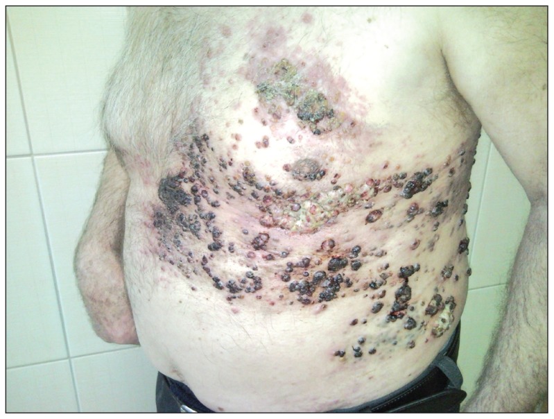
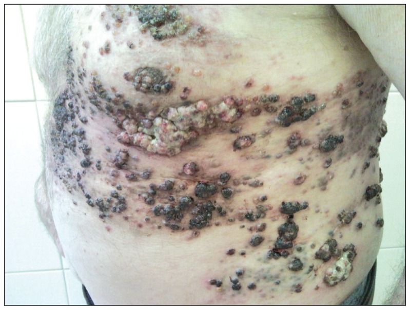
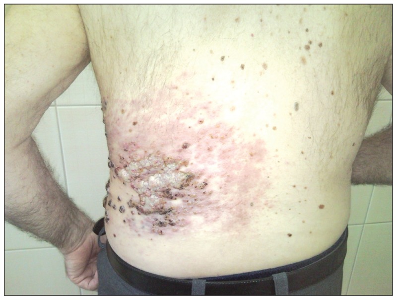
A 77-year-old male patient presented with a dark-brown nodular lesion, which grew within an oval melanocytic nevus of 6×4 cm2 in size and located on the skin of the lower abdomen. The nodule appeared 6 months earlier and reached the size of 2 cm in diameter. The growing cutaneous finding manifested with the bleeding sore and crust. The nevus was excised entirely together with the nodule. The histopathologic examination revealed the nodular type of amelanotic melanoma, which was ulcerated and classified as Clark II and Breslow IV (5 mm of infiltration thickness). Essentially, no clear margins were noted both vertically and horizontally, and the infiltration of the tumor encompassed the border and subcutaneous tissue. Additionally, some satellite metastases were detected subcutaneously. The most of laboratory tests stayed within the normal range and only erythrocyte sedimentation rate was elevated (70–90 mm/h). No increase was observed in concentrations of tumor markers (CEA, Ca125, total-PSA), and diagnostic imaging (chest x-ray, abdominal ultrasound, head CT [computed tomography]) detected no abnormalities. One month after the surgical excision of the primary lesion, a fine needle aspiration of inguinal lymph nodes was performed bilaterally. Subsequently, the metastases of melanoma were detected in the left groin. A palliative radiotherapy of the skin of abdomen and chest was initiated on an outpatient basis. Nevertheless, the therapy was ceased at a dose of 14 Gy, before the target dose of 20 Gy, because of the sudden worsening of the general health condition of the patient. Simultaneously, multiple, non-painful, brown papules started to occur on the skin of the left side of the chest that were interpreted as herpes zoster infection. Hence, the patient was administered with 800 mg acyclovir orally 4 times daily for the following 2 months. As no clinical improvement of cutaneous lesions was observed and the new ones started to spread over the left side of the chest, the patient was again admitted to the Dermatology Clinic. The skin biopsy of these spreading lesions revealed melanoma metastases. The patient was given an external treatment, and, additionally, was referred to the CT scan of the chest and abdomen. Unfortunately, there was no further possibility to realize the whole diagnostic plan since the patient died 2 weeks later due to the cardiorespiratory failure.
Figure 1.
A 77-year-old male patient, with metastases of malignant melanoma located on the skin of abdomen.
Figure 2.
A 77-year-old male patient, with multiple scattered black and dark-brown nodules.
Figure 3.
A 77-year-old male patient, with skin nodules mostly covered by bleeding sores and crusts.


