Abstract
BACKGROUND AND OBJECTIVES
This study aims to compare the findings of the shoulder ultrasonography (US) of patients with a supraspinatus (SS) tendon rupture with those of the shoulder arthroscopy, to determine the reliability and diagnostic performance of the shoulder US in the algorithm of the SS tendon pathologic lesions and their secondary ultrasonographic findings.
DESIGN AND SETTINGS
A prospective study conducted with patients scheduled for arthroscopy of the shoulder due to an SS tendon rupture in Yildirım Beyazit Education and Research Center and Gazi University, Ankara, Turkey.
MATERIALS AND METHODS
Fifty patients scheduled for an arthroscopy of the shoulder due to an SS tendon rupture were evaluated by shoulder US 1 week before the surgery. SS tendon pathologic lesions (tendinosis, partial tears, and full-thickness tears) and humeral degeneration were recorded, and the results of shoulder US were compared with those of arthroscopy.
RESULTS
With reference to the arthroscopic data, the sensitivity of the ultrasonographic evaluation for the diagnosis of a full-thickness SS tendon rupture was 91%, with a specificity of 88%; the sensitivity for the diagnosis of a partial-thickness rupture was 86%, with a specificity of 82%; and the sensitivity for the diagnosis of a tendinosis was 98%, with a specificity of 71%. With reference to the arthroscopic data, the sensitivity of US for the diagnosis of humeral degeneration was 93%, with a specificity of 91%.
CONCLUSION
The high sensitivity and specificity rates of US in detecting SS tendon rupture and its secondary imaging findings make it an efficient and reliable diagnostic modality, which should be preferred to other more expensive and more invasive methods in the algorithm.
In patients with shoulder pain, the clinical and the physical examination findings are not entirely reliable for the diagnosis. Imaging methods are needed for differential diagnosis.1 Instead of the expensive and invasive methods such as magnetic resonance imaging (MRI) and arthrography, the diagnostic imaging of the shoulder can be done using a rapid, inexpensive, and easily available method in many cases.2–4
Shoulder ultrasonography (US) is a cost-effective and non-invasive method for the evaluation of the rotator cuff pathologic lesion.5,6 In previous studies carried out by orthopedic surgeons and radiologists, remarkable results in the detection of full-thickness rotator cuff ruptures have been achieved.7–9 Due to difficulties in the detection of rotator cuff tears with US, a variety of secondary findings as humeral degeneration and joint space effusion have been described.10,11
The purpose of this study was to determine the diagnostic performance and accuracy of US in the diagnosis of rotator cuff pathologic lesions and their secondary imaging findings with reference to arthroscopic findings.
MATERIALS AND METHODS
Our final population including 50 patients (37 women, 13 men; age range: 36–66 years; mean age 51.0 [8.3]) scheduled for an arthroscopy due to a supraspinatus (SS) tendon rupture were included in this study. All patients were admitted with persistent pain or limitation of motion in the shoulder, not responding to rehabilitation.
The study was designed as a prospective study. Patients were recruited for this project during a period of 16 months from August 2009 to December 2010. Our institutional review board approved the study protocol, and informed consent was obtained from all patients. The gender, age, and the side of the arthroscopy were recorded. Patients with complaints in both shoulders were initially excluded. Patients who were scheduled for arthroscopy of the shoulder were referred to our clinic 1 week before the procedure for a US of the shoulder.
The US examinations were performed by 2 certified musculoskeletal radiologists, each with 4-year experience, who evaluated each patient and reached a consensus diagnosis. Both radiologists were not aware of the clinical examination findings, nor were they aware which patients were enrolled in the study and which patients were not. A GE Logiq S6 (GE Health Care, Milwaukee, WI) 7 to 12 MHz high-frequency linear transducer was used in the examination. In addition, a wide field of view was also used in the examination. Sonographic findings were classified as SS pathologic lesions (tendinosis, partial tears, and full-thickness tears) and humeral degeneration.9 Tendinosis was diagnosed when a partial increase in thickness of SS tendon and focal—generally nodular—hypoechoic area were seen in this thick segment. Partial-thickness tears were diagnosed when a focal, well-defined hypoechoic or anechoic defects in the tendon was seen, but this involved only the bursal or articular surface. Full-thickness tears extend from the bursal surface to the articular surface. Full-thickness rotator cuff tears were diagnosed when hypoechoic or anechoic defects in which fluid had replaced the area of the torn tendon were seen. Humeral degeneration is imaged as an irregularity in the humeral head and absence of regular hyperechoic cartilage line, and in cases of chronic SS tear, crash hypoechoic areas consistent with fracture. The patient was examined in the sitting position on a stool. The evaluating physician examined the symptomatic shoulder sitting on a higher stool. The findings of the SS tendon were classified as full-thickness tears of the SS tendon, partial tears of the SS tendon, tendinosis, and cases with no pathologic lesion. In addition, the findings of the humeral degeneration and the acromioclavicular joint degeneration were recorded.
Following those procedures, the patients underwent shoulder arthroscopy with a diagnosis of a rotator cuff tear by the surgeon who had 10 years of experience in shoulder arthroscopy and was blinded to the US results. The images obtained during arthroscopy and the arthroscopic findings were recorded, and the recorded data were analyzed in detail. The ultrasonographic findings were compared with those found on arthroscopy.
A cross-tabulation was prepared to compare the findings of US and arthroscopy. The diagnostic performance (sensitivity, specificity, positive predictive value, and negative predictive value) were calculated for US results. Chi-square test was used to evaluate the significance of results. The data were analyzed using SPSS, version 15.0 (SPSS Inc., Chicago, IL USA), and P value less than .05 was considered significant.
RESULTS
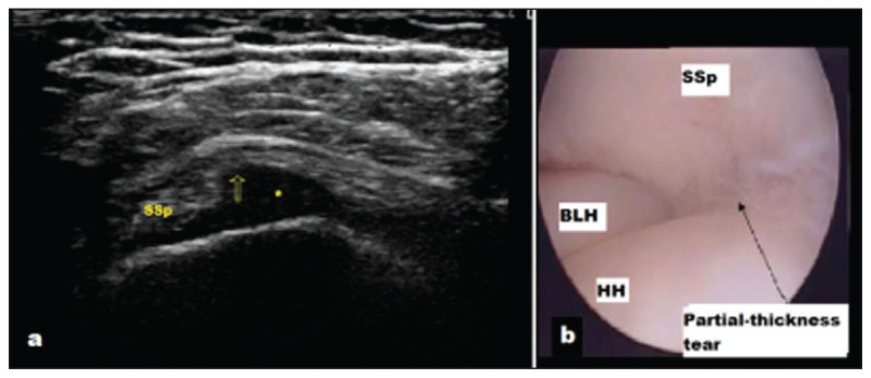
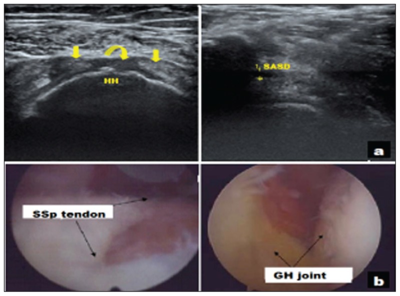
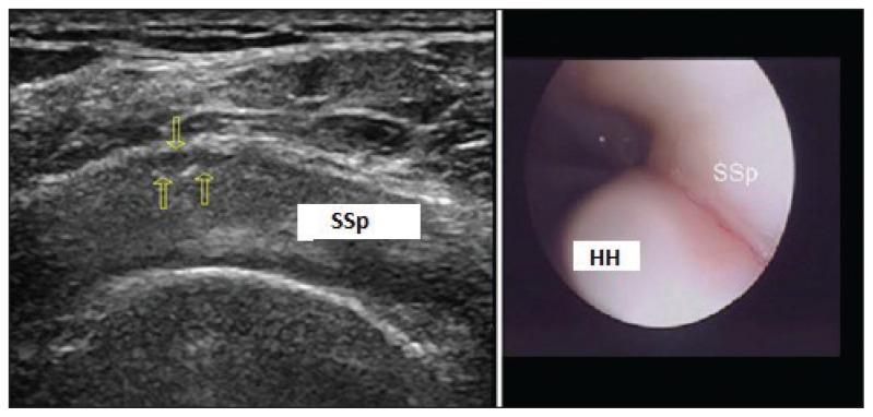
There was a significant correlation between the US findings and the arthroscopic findings in the examination of the SS tendon (P<.05) (Figures 1–3). The results from US and arthroscopy with regard to the involved SS tendon are summarized in Table 1.
Figure 1.
Ultrasonographic image showing full-thickness supraspinatus tear.
Figure 2.
Ultrasonographic image of rotator cuff tear.
Figure 3.
Supraspinatus tendon at the rotator cuff interval.
Table 1.
The comparison of the ultrasonographic findings of the SS tendon on the operated side with the arthroscopic findings.
| US examination, SS tendon findings | Arthroscopic evaluation, SS tendon findings | Total | Chi square | SD | P | |||
|---|---|---|---|---|---|---|---|---|
| None | Partial tear | Tendinosis | Full-thickness tear | |||||
|
| ||||||||
| None | 3 | 1 | 0 | 0 | 4 | 81.399 (a) | 9 | .0a |
| Partial tear | 1 | 18 | 1 | 2 | 22 | |||
| Tendinosis | 0 | 1 | 5 | 0 | 6 | |||
| Full-thickness tear | 0 | 2 | 1 | 15 | 18 | |||
| Total | 4 | 22 | 7 | 17 | 50 | |||
|
| ||||||||
| In 12 cells (75.0%), a frequency less than 5 was expected. The smallest expected frequency was 0.32. | ||||||||
P<.05.
SS: Supraspinatus; US: ultrasonography.
Humeral head degeneration results for both US and arthroscopy are summarized in Table 2. There was a significant correlation between the ultrasonographic and arhroscopic findings in the detection of the humeral degeneration (P<.05), with ultrasonographic sensitivity and specificity of 93% and 91%, respectively.
Table 2.
The comparison of the ultrasonographic findings with that of the arthroscopy in the detection of the humeral degeneration.
| US examination, humeral degeneration | Arthroscopic evaluation, humeral degeneration | Total | Fisher exact test P value | |
|---|---|---|---|---|
| Yes | No | |||
|
| ||||
| Yes | 25 | 2 | 27 | .000a |
| No | 2 | 21 | 23 | |
| Total | 27 | 23 | 50 | |
P<.05.
US: Ultrasonography.
DISCUSSION
Our study compared the findings of the shoulder US of 50 patients with an SS tendon rupture with those of the shoulder arthroscopy. Studies reported that there is an increased risk of rotator cuff tendon tears with patient’s age.12,13 In the current study, the mean age of the patients was 51.4 (8.3) (range: 36–66)
With reference to the arthroscopic data, the high rates of detection of full-thickness and partial-thickness tears, the high sensitivity and specificity rates in detecting the pathologic lesions of the SS tendon, and the secondary pathologic lesions indicate that the US examination is an important diagnostic method.
Halma and colleagues14 concluded that radiologist and orthopedic surgeons differed in predicting not all but some types (osteoarthritis, impingement, Hill-Sachs lesions) of pathologic lesions of the glenohumeral joint on MRI. However, there are some limitations of the US examination of the shoulder. The most important issue is the dependence on the capability of the radiologist performing the examination, and, in particular, the lack of experience on the examination of the shoulder. In addition, obesity, pain, and reduced range of motion are the causes that make the evaluation difficult and reduce the diagnostic sensitivity.15,16
Yamaguchi et al17 have found full-thickness tear in 35% and a partial tear in 21% of the patients in their study, which included 588 symptomatic patients. Our study was in concordance with the published studies in terms of full-thickness tears; however, we obtained a higher rate of partial-thickness tears. Nonetheless, the number of patients in our study was small to obtain a significant result.
In our study, the ultrasonographic whereas the arthroscopic surgery revealed a full-thickness tear in 15 patients (84%). With reference to the arthroscopic data, the sensitivity of the ultrasonographic evaluation for the diagnosis of full-thickness tendon tears was 91%, with a specificity of 88%. This study’s findings are in concordance with the published studies.18,19 The reasons for the high rate of diagnosis of the full-thickness tears on US examination are the objective ultrasonographic findings and pronounced clinical symptoms, which may provide clues to the radiologist.
In our study, the ultrasonographic evaluation revealed a partial-thickness tear in 22 patients, whereas the arthroscopic surgery revealed a partial-thickness tear in 18 patients (82%). With reference to the arthroscopic data, the sensitivity of the ultrasonographic evaluation for the diagnosis of partial-thickness tendon tears was 86%, with a specificity of 82%. Our study’s results are in concordance with the published studies.20,21 However, the varying sensitivity and specificity rates for the diagnosis of the partial-thickness tears in US examination can be explained by the size of the tear, the necessity to evaluate the partial tear separately on the articular and the bursal surface, and the technical specifications of the US device.
In their published studies review, Ottenheijm et al22 have reported that the sensitivity and specificity of US for detecting tendinosis were 67% to 93% and 88% to 100%, respectively. In our study, out of the 6 patients with a sonographic diagnosis of tendinosis, 5 (83%) were positive on arthroscopy, resulting in a sensitivity of 98% and a specificity of 71%.
Our study had some limitations. Our sample number was small; therefore, our study can be called as a small-scale prospective study. We did not compare the US results with MRI or arthrography. Comparing the US results with any other diagnostic methods such as MRI or magnetic resonance arthrography, in addition to comparing these with arthroscopy, could be better. A randomized prospective study with larger groups and multi-center studies could be better to discuss the place of shoulder US in the algorithm of shoulder-related pathologic lesions.
In conclusion, the high accuracy and diagnostic performance of shoulder US in detecting SS tendon pathologic lesions make it an efficient, fast, less-expensive, non-invasive, and reliable evaluation method to be used by experienced musculoskeletal radiologists, and, therefore, it should be the first modality of evaluation in patients with rotator cuff symptoms.
REFERENCES
- 1.Dinnes J, Loveman E, McIntyre L, Waugh N. The effectiveness of diagnostic tests for the assessment of shoulder pain due to soft tissue disorders: a systematic review. Health Technol Assess. 2003;7(29):iii, 1–166. doi: 10.3310/hta7290. [DOI] [PubMed] [Google Scholar]
- 2.Frei R, Chládek P, Trc T, Kopecný Z, Kautzner J. Arthroscopic evaluation of ultrasonography and magnetic resonance imaging for diagnosis of rotator cuff tear. Ortop Traumatol Rehabil. 2008;10(2):111–4. [PubMed] [Google Scholar]
- 3.Schneider TL, Schmidt R, Drescher W, Fink B, Appel JC. The significance of subacromial arthrography to verify partial bursal-side rotator cuff ruptures. Arch Orthop Trauma Surg. 2003;123:481–4. doi: 10.1007/s00402-003-0492-1. [DOI] [PubMed] [Google Scholar]
- 4.Küllmer K, Sievers KW, Rompe JD, Nagele M, Harland U. Sonography and MRI of experimental muscle injuries. Arch Orthop Trauma Surg. 1997;116:357–1. doi: 10.1007/BF00433990. [DOI] [PubMed] [Google Scholar]
- 5.Al-Shawi A, Badge R, Bunker T. The detection of full thickness rotator cuff tears using ultrasound. J Bone Joint Surg Br. 2008;90:889–92. doi: 10.1302/0301-620X.90B7.20481. [DOI] [PubMed] [Google Scholar]
- 6.Burk DL, Jr, Karasick D, Kurtz AB, Mitchell DG, Rifkin MD, Miller CL, Levy DW, Fenlin JM, Bartolozzi AR. Rotator cuff tears: prospective comparison of MR imaging with arthrography, sonography, and surgery. AJR Am J Roentgenol. 1989;153:87–92. doi: 10.2214/ajr.153.1.87. [DOI] [PubMed] [Google Scholar]
- 7.Ziegler DW. The use of in-office, orthopaedist-performed ultrasound of the shoulder to evaluate and manage rotator cuff disorders. J Shoulder Elbow Surg. 2004;13:291. doi: 10.1016/j.jse.2004.01.017. [DOI] [PubMed] [Google Scholar]
- 8.Farin PU, Kaukanen E, Jaroma H, Vaatainen U, Miettinen H, Soimokallio S. Findings at ultrasound, double-contrast arthrography, and computed tomography arthrography with surgical correlation. Invest Radiol. 1996;31:387. doi: 10.1097/00004424-199607000-00001. [DOI] [PubMed] [Google Scholar]
- 9.Teefey SA, Hasan SA, Middleton WD, Patel M, Wright R, Yamaguchi K. Ultrasonography of the rotator cuff: a comparison of ultrasonographic and arthroscopic findings in one hundred consecutive cases. J Bone Joint Surg Am. 2000;82:498–504. [PubMed] [Google Scholar]
- 10.Wohlwend JR, Van Holsbeeck M, Craig J, Shirazi K, Habra G, Jacobsen G, Bouffard JA. The association between irregular greater tuberosities and rotator cuff tears: a sonographic study. AJR Am J Roentgenol. 1998;171:229–33. doi: 10.2214/ajr.171.1.9648794. [DOI] [PubMed] [Google Scholar]
- 11.Hollister MS, Mack LA, Patten RM, Winter TC, Matsen FA, Veith RR. Association of sonographically detected subacromial/subdeltoid bursal effusion and intraarticular fluid with rotator cuff tear. Am J Roentgenol. 1995;165:605–8. doi: 10.2214/ajr.165.3.7645478. [DOI] [PubMed] [Google Scholar]
- 12.Worland RL, Lee D, Orozco CG, SozaRex F, Keenan J. Correlation of age, acromial morphology, and rotator cuff tear pathology diagnosed by ultrasound in asymptomatic patients. J South Orthop Assoc. 2003;12(1):23–6. [PubMed] [Google Scholar]
- 13.Lehman C, Cuomo F, Kummer FJ, Zuckerman JD. The incidence of full thickness rotator cuff tears in a large cadaveric population. Bull Hosp Jt Dis. 1995;54(1):30–1. [PubMed] [Google Scholar]
- 14.Halma JJ, Eshuis R, Krebbers MJ, Weits T, Gast A. Interdisciplinary inter-observer agreement and accuracy of MR imaging of the shoulder with arthroscopic correlation. Arch Orthop Trauma Surg. 2012;132:311–20. doi: 10.1007/s00402-011-1370-x. [DOI] [PMC free article] [PubMed] [Google Scholar]
- 15.Teefey SA, Rubin DA, Middleton WD, Hildebolt CF, Leibold RA, Yamaguchi K. Detection and quantification of rotator cuff tears. Comparison of ultrasonographic, magnetic resonance imaging, and arthroscopic findings in seventy-one consecutive cases. J Bone Joint Surg Am. 2004;86-A(4):708–16. [PubMed] [Google Scholar]
- 16.Brenneke SL, Morgan CJ. Evaluation of ultrasonography as a diagnostic technique in the assessment of rotator cuff tendon tears. Am J Sports Med. 1992;20(3):287–9. doi: 10.1177/036354659202000309. [DOI] [PubMed] [Google Scholar]
- 17.Yamaguchi K, Ditsios K, Middleton WA, Hildebolt CF, Galatz LM, Teefey SA. The Demographic and Morphological Features of Rotator Cuff Disease: A Comparison of Asymptomatic and Symptomatic Shoulders. J Bone Joint Surg Am. 2006;88:1699–704. doi: 10.2106/JBJS.E.00835. [DOI] [PubMed] [Google Scholar]
- 18.Kluger R, Mayrhofer R, Kröner A, Pabinger C, Pärtan G, Hruby W, Engel A. Sonographic versus magnetic resonance arthrographic evaluation of full-thickness rotator cuff tears in millimeters. J Shoulder Elbow Surg. 2003;12(2):110–6. doi: 10.1067/mse.2003.10. [DOI] [PubMed] [Google Scholar]
- 19.Martín-Hervás C, Romero J, Navas-Acién A, Reboiras JJ, Munuera L. Ultrasonographic and magnetic resonance images of rotator cuff lesions compared with arthroscopy or open surgery findings. J Shoulder Elbow Surg. 2001;10(5):410–5. doi: 10.1067/mse.2001.116515. [DOI] [PubMed] [Google Scholar]
- 20.Van Holsbeeck MT, Kolowich PA, Eyler WR, Craig JG, Shirazi KK, Habra GK, Vanderschueren GM, Bouffard JA. US depiction of partial-thickness tear of the rotator cuff. Radiology. 1995;197(2):443–6. doi: 10.1148/radiology.197.2.7480690. [DOI] [PubMed] [Google Scholar]
- 21.Labanauskaite G. Usefulness of ultrasonography in the diagnosis of rotator cuff tears. Medicina (Kaunas) 2002;38(2):131–4. [PubMed] [Google Scholar]
- 22.Ottenheijm RP, Jansen MJ, Staal JB, van den Bruel A, Weijers RE, de Bie RA, Dinant GJ. Accuracy of diagnostic ultrasound in patients with suspected subacromial disorders: a systematic review and meta-analysis. Arch Phys Med Rehabil. 2010;91(10):1616–25. doi: 10.1016/j.apmr.2010.07.017. [DOI] [PubMed] [Google Scholar]


