Abstract
Here, two methods are described for efficient genetic modification of Saccharomyces cerevisiae using CRISPR/Cas9. The first method enables the modification of a single genetic locus using in vivo assembly of a guide RNA (gRNA) expression plasmid without the need for prior cloning. A second method using in vitro assembled plasmids that could contain up to two gRNAs was used to simultaneously introduce up to six genetic modifications (e.g. six gene deletions) in a single transformation step by transforming up to three gRNA expression plasmids simultaneously. The method is not only suitable for gene deletion but is also applicable for in vivo site-directed mutagenesis and integration of multiple DNA fragments in a single locus. In all cases, the strain transformed with the gRNA expression plasmids was equipped with a genomic integration of Spcas9, leading to strong and constitutive expression of SpCas9. The protocols detailed here have been streamlined to be executed by virtually any yeast molecular geneticist.
Keywords: CRISPR/Cas9, S. cerevisiae, gRNA, genetic modification, webtool, plasmid
Streamlined CRISPR-Cas9 protocols for genome editing in Saccharomyces cerevisiae executable by virtually any yeast molecular geneticist.
INTRODUCTION
Protocols for highly efficient transformation of Saccharomyces cerevisiae have been well established. However, prior to the emergence of CRISPR/Cas9 as a genetic engineering tool, most methods for gene deletion or integration relied on the simultaneous integration of a selection marker gene and thus required a subsequent marker-recycling step (Baudin et al.1993; Güldener et al.1996; Gietz and Woods 2002; Carter and Delneri 2010; Solis-Escalante et al. 2013, 2014). Furthermore, introduction of multiple genetic modifications has so far remained a time-consuming and labour-intensive process, as each individual alteration requires a cycle of transformation, selection and confirmation. This work describes the steps involved in two methods for CRISPR/Cas9-mediated transformation of S. cerevisiae (Mans et al.2015). The first method (based on pMEL plasmids) makes use of in vivo assembly of plasmids encoding a single guide RNA (gRNA) via homologous recombination. This method enables fast modification of a single genetic locus without the need for prior cloning. The second method (based on pROS plasmids) is based on in vitro pre-assembled plasmids containing two gRNA coding sequences, which enable the simultaneous modification of two genetic loci per transformed plasmid.
MATERIALS
This protocol offers a series of pMEL and pROS plasmids with a broad range of selection markers. The materials listed below are for application of these selection markers. When transforming with a subset of plasmid markers, not all listed materials might be required.
Saccharomyces cerevisiae and Escherichia coli strains
Chemical- or electrocompetent E. coli XL1-Blue cells for plasmid transformation and propagation. Chemically competent cells can be prepared and transformed according to the protocol of Zymo Research (T3001, http://www.biocenter.hu/pdf/T3001.PDF). Electrocompetent cells can be prepared and transformed according to the protocol of Bio-Rad (165–2100, http://www.bio-rad.com/webroot/web/pdf/lsr/literature/4006174B.pdf).
Saccharomyces cerevisiae strain(s) with a genomic integration of cas9. The yeast strains in Table 1 can be obtained via EUROSCARF (https://tinyurl.com/y8xxzpr9). cas9 is flanked by the TEF1 promoter and the CYC1 terminator and is equipped with the SV40 nuclear localisation signal for targeting to the nucleus (DiCarlo et al.2013; Mans et al.2015).
Note: The transfer of Streptococcus pyogenes cas9 in a S. cerevisiae strain can be achieved via assembly and simultaneous integration of a cassette carrying cas9 and another containing the natNT2 marker into the CAN1 locus. The cas9 cassette was PCR amplified from p414-TEF1p-Cas9-CYC1t (Addgene plasmid #43802) (DiCarlo et al.2013) (Table 2), using primers 2873 & 4653 (Table 3). The natNT2 cassette was PCR amplified from pUG-natNT2 (Addgene plasmid #110922) (de Kok et al.2012) with primers 3093 & 5542. 2.5 μg cas9 and 800 ng natNT2 cassette were pooled and used for each transformation as described in (Mans et al.2015).
Plasmids for gRNA expression. The singe gRNA (pMEL) and double gRNA (pROS) plasmids can be obtained via EUROSCARF (Table 2).
Table 1.
Saccharomyces cerevisiae strains deposited at EUROSCARF with a genomic integration of cas9 (Mans et al.2015).
| Name (Accession no.) | Relevant genotype | Parental strain |
|---|---|---|
| IMX585 (Y40592) | MATa can1Δ::Spcas9-natNT2 URA3 TRP1 LEU2 HIS3 | CEN.PK113–7D |
| IMX581 (Y40593) | MATa ura3–52 can1Δ::Spcas9-natNT2 TRP1 LEU2 HIS3 | CEN.PK113–5D |
| IMX664 (Y40594) | MATa/MATα CAN1/can1Δ::Spcas9-natNT2 URA3/URA3 TRP1/TRP1 LEU2/LEU2 HIS3/HIS3 | CEN.PK122 |
| IMX672 (Y40595) | MATa ura3–52 trp1–289 leu2–3112 his3Δ can1Δ::Spcas9-natNT2 | CEN.PK2–1C |
| IMX673 (Y40596) | MATa/MATα ura3–52/ura3–52 CAN1/can1Δ::Spcas9-natNT2 TRP1/TRP1 LEU2/LEU2 HIS3/HIS3 | CEN.PK115 |
Table 2.
E. coli strains deposited at EUROSCARF (https://tinyurl.com/y8xxzpr9) and at Addgene (https://www.addgene.org) containing the single gRNA (pMEL) and double gRNA (pROS) plasmid series (Mans et al.2015).
| Name | Accession no Euroscarf | Accession no Addgene | Relevant characteristics |
|---|---|---|---|
| p414-TEF1p-cas9-CYC1t | 43802 | ARS4 CEN6 bla TRP1 TEF1 p::Spcas9::CYC1t | |
| pUG-natNT2 | 110922 | bla AgTEF1p::nat::AgTEF1t | |
| pMEL10 | P30779 | 107916 | 2μm bla KlURA3 gRNA-CAN1.Y |
| pMEL11 | P30780 | 107917 | 2μm bla amdSYM gRNA-CAN1.Y |
| pMEL12 | P30781 | 107918 | 2μm bla hphNT1 gRNA-CAN1.Y |
| pMEL13 | P30782 | 107919 | 2μm bla kanMX gRNA-CAN1.Y |
| pMEL14 | P30783 | 107920 | 2μm bla KlLEU2 gRNA-CAN1.Y |
| pMEL15 | P30784 | 107921 | 2μm bla natNT2 gRNA-CAN1.Y |
| pMEL16 | P30785 | 107922 | 2μm bla HIS3 gRNA-CAN1.Y |
| pMEL17 | P30786 | 107923 | 2μm bla TRP1 gRNA-CAN1.Y |
| pROS10 | P30787 | 107924 | 2μm bla URA3 gRNA-CAN1.Y gRNA-ADE2.Y |
| pROS11 | P30788 | 107925 | 2μm bla amdSYM gRNA-CAN1.Y gRNA-ADE2.Y |
| pROS12 | P30789 | 107926 | 2μm bla hphNT1 gRNA-CAN1.Y gRNA-ADE2.Y |
| pROS13 | P30790 | 107927 | 2μm bla kanMX gRNA-CAN1.Y gRNA-ADE2.Y |
| pROS14 | P30791 | 107928 | 2μm bla KlLEU2 gRNA-CAN1.Y gRNA-ADE2.Y |
| pROS15 | P30792 | 107929 | 2μm bla natNT2 gRNA-CAN1.Y gRNA-ADE2.Y |
| pROS16 | P30793 | 107930 | 2μm bla HIS3 gRNA-CAN1.Y gRNA-ADE2.Y |
| pROS17 | P30794 | 107931 | 2μm bla TRP1 gRNA-CAN1.Y gRNA-ADE2.Y |
Table 3.
Primers used to construct and verify insertion of CRISPR-Cas9 gRNA in pMEL and pROS plasmid series.
| Name | Sequence 5΄ → 3΄ | Purpose |
|---|---|---|
| 6005 | GATCATTTATCTTTCACTGCGGAGAAG | pMEL + pROS plasmid construction |
| 6006 | GTTTTAGAGCTAGAAATAGCAAGTTAAAATAAGGCTAGTC | pMEL plasmid construction |
| 2873 | TCAGACTTCTTAACTCCTGTAAAAACAAAAAAAAAAAAAGGCATAGCAATAAGCTGGAGCTCATAGCTTC | Construction Spcas9 integration cassette |
| 4653 | GTGCCTATTGATGATCTGGCGGAATGTCTGCCGTGCCATAGCCATGCCTTCACATATAGTCCGCAAATTA AAGCCTTCGAG | Construction Spcas9 integration cassette |
| 3093 | ACTATATGTGAAGGCATGGCTATGGCACGGCAGACATTCCGCCAGATCATCAATAGGCACCTTCGTACGC TGCAGGTCGAC | Construction of natNT2 integration cassette |
| 5542 | CTATGCTACAACATTCCAAAATTTGTCCCAAAAAGTCTTTGGTTCATGATCTTCCCATACGCATAGGCCA CTAGTGGATCTG | Construction of natNT2 integration cassette |
| Primer α | CACCTTTCGAGAGGACGATG | Confirmation pROS plasmids |
| Primer β | GCTGGCCTTTTGCTCACATG | Confirmation pROS plasmids |
| Ptarget FWa | TGCGCATGTTTCGGCGTTCGAAACTTCTCCGCAGTGAAAGATAAATGATCN 20 GTTTTAGAGCTAGAAATAGC AAGTTAAAATAAGGCTAGTCCGTTATCAAC | pMEL construction |
| Ptarget RVb | GTTGATAACGGACTAGCCTTATTTTAACTTGCTATTTCTAGCTCTAAAACN 20c GATCATTTATCTTTCACTG CGGAGAAGTTTCGAACGCCGAAACATGCGCA | pMEL construction |
| Ptarget ROS | TGCGCATGTTTCGGCGTTCGAAACTTCTCCGCAGTGAAAGATAAATGATCN 20 GTTTTAGAGCTAGAAATAGC AAGTTAAAATAAG | pROS construction |
| Ptargetcas9 ROSc | TGCGCATGTTTCGGCGTTCGAAACTTCTCCGCAGTGAAAGATAAATGATCCTAGGCTGTCCAAATCCCGGGTT TTAGAGCTAGAAATAGCAAGTTAAAATAAG | Construction of a pROS targeting cas9 |
| PrepairD FW | TTTCAGAGTTCTTCAGACTTCTTAACTCCTGTAAAAACAAAAAAAAAAAAAGGCATAGCATATGAGGGTGAGA ATGCGAAATGGCGTGGAAATGTGATCAAAGGTAATAAAACGTCATAT | Repair DNA for cas9 deletion leaving a can1Δ locus |
| PrepairD RV | ATATGACGTTTTATTACCTTTGATCACATTTCCACGCCATTTCGCATTCTCACCCTCATATGCTATGCCTTTT TTTTTTTTTGTTTTTACAGGAGTTAAGAAGTCTGAAGAACTCTGAAA | Repair DNA for cas9 deletion leaving a can1Δ locus |
| PrepairCAN1 FWd | TTTCAGAGTTCTTCAGACTTCTTAACTCCTGTAAAAACAAAAAAAAAAAAAGGCATAGCAATGACAAATTCAA AAGAAGACGCC | Repair DNA for cas9 deletion restoring a CAN1 locus |
| PrepairCAN1 RVd | ATATGACGTTTTATTACCTTTGATCACATTTCCACGCCATTTCGCATTCTCACCCTCATATCTATGCTACAAC ATTCCAAAATTTG | Repair DNA for cas9 deletion restoring a CAN1 locus |
N20 corresponds to the chosen target sequence. This nucleotide sequence does not include the PAM sequence (NGG).
N20c corresponds to the reverse complement of the chosen target sequence found in Ptarget FW.
Primer for the construction of pROS plasmid carrying a spacer targeting cas9, which can be used to remove chromosomal integration of the endonuclease expression cassette.
Primer suitable to PCR amplify the CAN1 ORF.
Equipment
Thermocycler
Gel electrophoresis equipment for DNA separation
UV transilluminator
Safe Imager 2.0 Blue-Light Transilluminator
Incubators with a temperature range of at least 30°C and 42°C
Water bath or heat block with a temperature range of at least 30°C to 95°C
Nanodrop or Qubit fluorometer with Qubit dsDNA BR Assay Kit (Thermo Fisher Scientific (Waltham, MA), Q32853) for DNA quantification
Computer with internet access and DNA manager software (e.g. Clone Manager (Scientific & Educational Software, Denver, CO) Snapgene (Chicago, IL, (http://www.snapgene.com/))
Optional: 2 mm cuvette (Bio-Rad, 165–2086) and MicroPulser Electroporator (Bio-Rad, 165–2100)
Chemicals
Bacto tryptone (Difco-Thermo Fisher Scientific; 211705)
Bacto yeast extract (Difco-Thermo Fisher Scientific; 212750)
Sodium chloride (J.T.Baker (Center Valley, PA); 0278)
Bacto agar (Difco-Thermo Fisher Scientific; 214010)
Bacto peptone (Difco-Thermo Fisher Scientific; 211677)
Ammonium sulphate (Merck (Kenilworth, NJ); 101211)
Monopotassium phosphate (Merck; 104877)
Magnesium sulphate heptahydrate (J.T.Baker; 2500)
Potassium hydroxide (J.T.Baker; 0222)
L-glutamic acid monosodium salt (Sigma Aldrich (Saint-Louis, MO); G1626)
100% Glycerol (Merck; 8.18709)
100% Ethanol (Merck; 1.00983)
Sodium dodecyl sulphate (Sigma Aldrich; L4390)
Polyethylene glycol 3350 (Sigma Aldrich; P4338)
Deoxyribonucleic acid sodium salt (Sigma Aldrich; D1626)
Trizma base (Sigma Aldrich; T6066)
Hydrochloric acid (Sigma Aldrich; 30721)
Ethylene diamine tetra-acetic acid (EDTA) solution (Sigma Aldrich; E7889)
Lithium acetate dehydrate (Sigma Aldrich; L6883)
Acetamide (Sigma Aldrich; A0500)
Potassium sulphate (Merck; 105153)
Molecular biology reagents
-
DNA purification kits:
GenElute Plasmid Miniprep Kit (Sigma Aldrich; PLN350)
ZymoClean Gel DNA Recovery Kit (Zymo Research; D4002)
Optional: YeaStar Genomic DNA Kit (Zymo Research: D2002)
-
PCR reagents:
DreamTaq PCR Master Mix (2x) (Thermo Fisher Scientific; K1071)
Phusion High Fidelity DNA polymerase (2 U μL−1) and 5x Phusion HF buffer (Thermo Fisher Scientific; F530)
dNTPs (10 mM) (Thermo Fisher Scientific; R0181)
MilliQ or RNase-free water.
Primers (Table 3):
FastDigest DpnI (Thermo Fisher Scientific; FD1704)
Optional: FastDigest enzymes and FastDigest Green Buffer (10x) for confirmation of correct assembly pROS plasmids.
GeneRuler DNA Ladder Mix (Thermo Fisher Scientific; SM0331)
SERVA DNA stain G (SERVA (Heidelberg, Germany); 39803.02)
50X TAE Buffer (Thermo Fisher Scientific; B49)
TopVision Agarose (Thermo Fisher Scientific; R0492)
NEBuilder HiFi DNA Assembly Master Mix (New England Biolabs (Ipswich, MA); E2621)
0.2M lithium acetate 1% SDS solution; dissolve 5 g sodium dodecyl sulphate in 50 mL distilled water, add 10.2 g lithium acetate dehydrate and fill up to 500 mL with distilled water.
TE buffer (pH 8.0); prepare 1.0M Tris by dissolving 60.57 g Trizma base in 500 mL distilled deionised water and set pH to 8.0 using HCl. Mix 5 mL 1M Tris pH 8.0 with 1 mL 0.5M EDTA and fill up to 500 mL with deionised water. When necessary, heat sterilise for 20 min at 121°C.
-
Transformation reagents:
50% PEG. Prepare a 50% w/v solution of polyethylene glycol 3350 in distilled deionised water and autoclave at 121°C for 20 min.
1.0M Lithium acetate. Prepare in distilled deionised water and filter sterilise. Final pH should be between 8.4 and 8.9.
ssDNA. Dissolve deoxyribonucleic acid sodium salt in TE buffer (pH 8.0) to a concentration of 2 mg L−1. Mix vigorously on a magnetic stirrer for 2–3 h or until fully dissolved, aliquot and store at –20°C. Prior to use, boil for 5 min at 100°C and cool down on ice.
Growth medium
LB medium: For preparation of 2 L LB medium, mix 20 g Bacto tryptone, 10 g Bacto yeast extract and 20 g sodium chloride and fill up to 2 L with deionised water. For solid medium also add 2% (w/v) Bacto agar. Heat sterilise for 20 min at 121°C. After sterilisation, antibiotics are added separately.
YP (yeast peptone) medium: For 2 L YP medium, mix 10 g Bacto yeast extract, 20 g Bacto peptone and fill up to 2 L with demineralised water. Split the volume over five Schott bottles and, for solid medium, add 2% (w/v) Bacto agar to each bottle and heat sterilise for 20 min at 121°C. After sterilisation, the carbon source and antibiotics of choice are sterilised and added separately.
SM (synthetic medium): For 2 L synthetic medium, start with 1.5 L of demineralised water and add 10 g ammonium sulphate [(NH4)2SO4], 6 g monopotassium phosphate [KH2PO4] and 1 g magnesium sulphate heptahydrate [MgSO4.7·H2O] and add 2 mL trace element solution (Verduyn et al.1992). When these salts are dissolved, set the pH to 6.0 with 2M potassium hydroxide [KOH] and add demineralised water to reach a final volume of 2 L. For solid medium, add 2% (w/v) Bacto agar to each bottle and heat sterilise for 20 min at 121°C. The carbon source, auxotrophic growth requirements and antibiotics of choice are sterilised and added separately after sterilisation.
SMglut (synthetic medium with glutamate as nitrogen source): SMglut is prepared similar to SM with the exception that the ammonium sulphate is replaced by 1 g L−1 L-glutamic acid monosodium salt and 6.6 g L−1 potassium sulphate to maintain a stable pH during cell growth, which improves selection when using antibiotic resistance selection markers (kanMX, natNT2, hphNT1).
SMace (synthetic medium with acetamide as nitrogen source): SMace is prepared similar to SM, with the exception that ammonium sulphate is replaced by 0.6 g L−1 acetamide and 6.6 g L−1 potassium sulphate. SMace is used when using the amdSYM selection marker (Solis-Escalante et al.2013).
Supplements for selection
Depending on marker usage, the following compounds are supplemented to the growth medium (Pronk 2002).
100 mg L−1 Ampicillin (Sigma Aldrich; A9518)
150 mg L−1 Uracil (Sigma Aldrich; U0750)
125 mg L−1 L-Histidine (Sigma Aldrich; H8000)
150 mg L−1 L-Leucine (Sigma Aldrich; 61819)
750 mg L−1 L-Tryptophan (Sigma Aldrich; T0254)
100 mg L−1 Nourseothricin (Jena Bioscience (Jena, Germany); AB-101)
200 mg L−1 G418 (Invivogen, Toulouse, France; ant-gn-1)
200 mg L−1 Hygromycin B (Thermo Fisher Scientific; 10687010)
Protocol
Method (1): In vivo assembly of CRISPR-Cas9 gRNA plasmid using the pMEL series. Method (1) can be used to introduce marker-free and scarless genetic modifications. This method is highly efficient for editing a single locus and has the advantage of a very simple workflow prior to yeast transformation (steps 1–4). When aiming for multiple simultaneous genetic modifications, Method (2), In vitro assembly of single and double gRNA plasmids using the pROS, is recommended. The pROS method can be used to introduce multiple marker‐free and scarless genetic modifications in a single transformation step. This method makes use of gRNA plasmids containing two gRNA coding sequences, facilitating restriction at two loci (steps 1–6).
-
Design of the guideRNA (gRNA) primer(s)
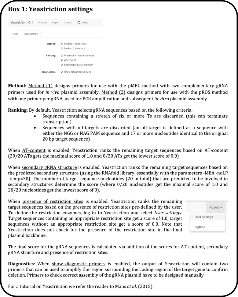
Use the Yeastriction tool at http://yeastriction.tnw.tudelft.nl (Fig. 1)
-
Alternatively, design the primer(s) manually (Box S4):
-
For the pMEL method (1)
Ptarget FW (5΄ → 3΄, N20 is the target sequence without the PAM) (Table 3): TGCGCATGTTTCGGCGTTCGAAACTTCTCCGCAGTGAAAGATAAATGATCN20GTTTTAGAGCTAGAAATAGCAAGTTAAAATAAGGCTAGTCCGTTATCAAC
Ptarget RV (5΄ → 3΄, N20c is the complementary target sequence without the PAM):
GTTGATAACGGACTAGCCTTATTTTAACTTGCTATTTCTAGCTCTAAAACN20cGATCATTTATCTTTCACTGCGGAGAAGTTTCGAACGCCGAAACATGCGCA
-
For the pROS method (Fig. 2) (2)
Forward & Reverse primer (Ptarget ROS (Table 3)) (5΄ → 3΄, where N20 is the target sequence without the PAM sequence, choosing a target site that contains a restriction site facilitates confirmation by restriction enzymes later on):
TGCGCATGTTTCGGCGTTCGAAACTTCTCCGCAGTGAAAGATAAATGATCN20GTTTTAGAGCTAGAAATAGCAAGTTAAAATAAG
NOTE: When aiming to construct a pROS plasmid targeting two distinct loci, two Ptarget ROS primers with different target sequences should be designed.
-
Order the primers PAGE-purified.
-
Construction of the gRNA insert fragment
-
For the pMEL method (1)
Dissolve the primers designed in 1.a or 1.b.i in deionised water to a final concentration of 10 μM
Mix the two complementary primers in a 1:1 molar ratio
Heat the mixture to 95°C for 5 min and subsequently cool down the mixture to room temperature on the bench to anneal both primers
Optional: Confirm efficient primer dimerisation using the Qubit fluorometer with Qubit dsDNA BR Assay Kit, repeat the process (2.a) when a concentration of over 200 ng μL−1 is observed.
-
For the pROS method (2)
-
Use the primer(s) designed in 1.a or 1.b.ii to set up the following PCR mixture:
Component Amount (μL) DreamTaq Master Mix (2x) 50 pROS template* 1–100 ng gRNA (Ptarget ROS1)(10 μM) 2 gRNA (Ptarget ROS2) (10 μM)** 2 MilliQ X Total volume 100 *This can be any pROS plasmid**When aiming to construct a pROS plasmid targeting a single distinct locus, only gRNA primer 1 is used.Note: The primer Ptargetcas9 ROS (Table 3) can be used to construct a pROS plasmid carrying a gRNA targeting cas9 gene.
Divide the reaction mixture over two PCR tubes (50 μL each).
- Run the PCR with the following conditions:
Step Temperature (°C) Time (s) 1 95 240 2 95 30 3 55 30 40x* 4 66 120 5 66 600 *Steps 2–4 are repeated 40 times sequentially (2→3→4→2→3→4 etc.) After the PCR is finished, combine the PCR reactions and add 1 μL FastDigest DpnI and incubate for 30 min at 37°C.
Load the complete digested PCR reaction mixtures on a 1% agarose 1x TAE gel with SERVA staining (10 μL L−1) and run the gel at 100 V for 30 min.
Excise the 1589 bp PCR product using a transilluminator and purify using the ZymoClean Gel DNA Recovery Kit according to manufacturer's instructions.
-
-
-
Construction of the linearised gRNA plasmid backbone
-
Amplify the plasmid backbone via PCR
-
For the pMEL method (1)
- Set up the following PCR mixture:
Component Amount (μL) 5x Phusion HF buffer 10 dNTPs (10 mM) 1 pMEL template* 1–5 ng Primer 6005 (10 μM) 1 Primer 6006 (10 μM) 1 Phusion polymerase 0.75 MilliQ X Total volume 50 *This can be any pMEL plasmid - Run the PCR with the following conditions:
Step Temperature (°C) Time (s) 1 98 30 2 98 10 3 67 20 40x* 4 68 180 5 68 300 *Steps 2–4 are repeated 40 times sequentially (2→3→4→2→3→4 etc.)
-
For the pROS method (2)
- Set up the following PCR mixture:
Component Amount (μL) 5x Phusion HF buffer 10 dNTPs (10 mM) 1 pROS template* 1–100 ng Primer 6005 (10 μM) 2 Phusion polymerase 0.75 MilliQ X Total volume 50 *This can be any pROS plasmid - Run the PCR with the following conditions:
Step Temperature (°C) Time (s) 1 98 300 2 98 30 3 63 30 40x* 4 68 120 5 68 600 *Steps 2–4 are repeated 40 times sequentially (2→3→4→2→3→4 etc.)
-
Digest and purify the PCR product as described in 2.b.iv–vi.
-
-
Construction of the double-stranded repair fragment(s)
-
When aiming for deletions:
Use the Yeastriction tool at http://yeastriction.tnw.tudelft.nl to design complementary primers which, when used as repair fragment, will delete the gene ORF.
Alternatively, manually design two complementary 120 bp primers. These primers are comprised of two adjacent 60 bp sequences, homologous to the up- and downstream region of the chromosomal locus containing the target site. The sequence between the selected up- and downstream regions will be removed from the chromosome during transformation (Box S5).
Order the primers DST- or PAGE-purified.
Anneal both primers as described in 2.a.i–iii.
-
When aiming for mutations:
Design two complementary 120 bp primers. These primers are comprised of two adjacent 60 bp sequences, homologous to the up- and downstream region of the chromosomal target site. The desired mutation needs to prevent restriction by Cas9 and can therefore be introduced in the 20 bp gRNA recognition sequence or the adjacent 3 bp PAM sequence (changing the sequence from GG to any other sequence besides AG) (Box S6).
Order the primers PAGE-purified.
Anneal both primers as described in 2.a.i–iii.
-
When aiming for DNA integration
-
Design primers for amplification of the desired DNA fragment for integration and add 60 bp 5΄ tails in such a way that the resulting PCR product is flanked by 60 bp sequences homologous to the up- and downstream regions of the chromosomal target site (Box S7).
When aiming for deletion of the integration locus, design the 60 bp homologous sequences as described in 4.a.ii.
To minimise alterations in the genomic DNA, the 60 bp homologous sequences can be designed in such a way that the integration event occurs in the 20 bp gRNA recognition sequence or the adjacent 3 bp PAM sequence, leaving the surrounding DNA intact.
Order the primers PAGE-purified.
PCR amplify the insert fragment using Phusion DNA polymerase according to manufacturer's instructions.
Purify the PCR product as described in 2.b.v–vi.
-
-
-
Assembly of the gRNA expression plasmid (pROS method (2) only)
- Prepare the following NEBuilder reaction mixture:
Component Amount (μL) NEBuilder HiFi DNA Assembly Master Mix (2x) 2.5 gRNA insert fragment (2.b) 50 ng Plasmid backbone (3.a.ii & 3.b) 50 ng MilliQ X Total volume 5 Incubate the reaction mixture at 50°C for 1 h.
Transform 2 μL of reaction mixture to 50 μL chemically competent E. coli cells according to the protocol of Zymo Research (T3001). Alternatively, transform 1 μL of the reaction mixture to 40 μL electro competent E. coli cells according to the protocol of Bio-Rad (165–2100).
Plate the transformed E. coli cells on a pre-warmed (37°C) LB plate containing 100 mg L−1 ampicillin.
Incubate the plate overnight at 37°C. The next day colonies are present on the plate.
-
Confirmation and storage of the constructed plasmid (pROS (2) method only)
-
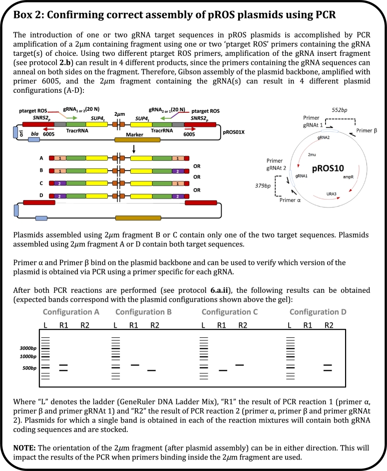
Via colony PCR (Fig. 2)
Design primers that are complementary to the N20 introduced target sequences (gRNAt 1 and 2).
- Set up the following two PCR mixtures (for each plasmid, we recommend testing 4 to 8 colonies):
Mixture 1 Component Amount (μL) DreamTaq Master Mix (2x) 5 Template * Primer gRNAt 1 (10 μM) 0.5 Primer α (10 μM) 0.25 Primer β (10 μM) 0.25 MilliQ 4 Total volume 10 Mixture 2 Component Amount (μL) DreamTaq Master Mix (2x) 5 Template * Primer gRNAt 2 (10 μM) 0.5 Primer α (10 μM) 0.25 Primer β (10 μM) 0.25 MilliQ 4 Total volume 10 *Pick a small portion of the same colony (5.e) into both reaction mixtures using for example a sterile loop or sterile pipette tip. - Run the PCR with the following conditions:
Step Temperature (°C) Time (s) 1 95 240 2 95 30 3 50–55** 30 35x* 4 72 30 5 72 600 *Steps 2–4 are repeated 35 times sequentially (2→3→4→2→3→4 etc.)**When using AT-rich (>50%) gRNA sequences, a lower (50°C) annealing temperature could provide better results. Load 5–10 μL of the reaction mixture on a 1% agarose 1x TAE gel with SERVA staining (10 μL L−1) and run the gel at 100 V for 30 min.
Confirm correct plasmid assembly by visualising bands using UV transillumination equipment. A double gRNA plasmid results in a single band (379 or 552 bp) for each reaction mixture.
Pick one single E. coli colony (5.e) corresponding to a correct plasmid into a 15 mL Greiner tube containing 5 mL of liquid LB with 100 mg L−1 ampicillin, restreak the colony first if necessary.
Incubate overnight at 37°C. Mix 3 mL of the grown culture with 1.5 mL 100% glycerol and divide over three 1.5 mL tubes and store at –80°C, the remainder or the culture can be used for isolation of the plasmid.
-
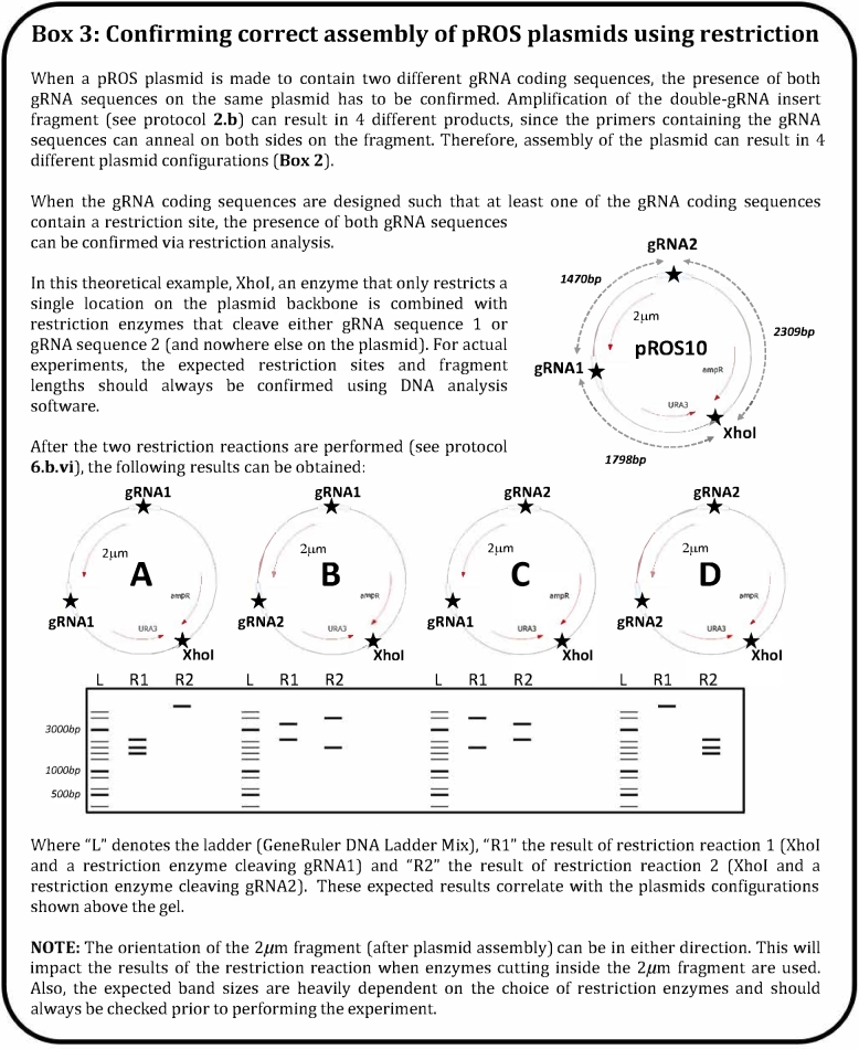
Via restriction analysis (when at least one of the gRNA primers designed in 1.b.ii contains a restriction site) (Fig. 3)
Pick 4–8 colonies (5.e) with a sterile loop and transfer to 15 mL Greiner tubes containing 5 mL of liquid LB with 100 mg L−1 ampicillin.
Incubate overnight at 37°C.
Transfer 2 mL of the E. coli cultures to 2 mL Eppendorf tubes.
Store the Greiner tubes with the remainder of the cultures at 4°C.
Extract the plasmids from the 2 mL of E. coli culture (6.b.iii) using the GenElute Plasmid Miniprep kit according to the supplier's manual, but elute the plasmid in 50 μLbuffer or water instead of the recommended 100 μL.
- Set up the following digestion reaction:
Component Amount (μL) FastDigest Green Buffer (10x) 1 Plasmid (6.b.v) 250 ng FastDigest Enzyme(s)** 0.5* MilliQ X Total volume 10 *When multiple restriction enzymes are used, add 0.5 μL of each enzyme.**When each gRNA coding sequence contains a restriction site, two separate reactions should be performed where only one of the restriction enzymes corresponding to the gRNA restriction sites is added. Incubate for 30–60 min at the temperature specified by the manufacturer.
Load 5–10 μL of the reaction mixture on a 1% agarose 1x TAE gel with SERVA staining (10 μL L−1) and run the gel at 100 V for 30 min.
Confirm correct plasmid assembly by visualising bands using UV transillumination equipment.
Mix the culture containing the correct plasmid (6.a.vii or one of the cultures stored at 6.b.iv corresponding to a correct plasmid) with 1.5 mL 100% glycerol.
Aliquot the culture in three 1.5 mL tubes and store at –80°C.
-
-
High concentration plasmid preparation using the GenElute Plasmid Miniprep kit. (NOTE: for most transformations, the protocol can be used as described under 6.b.v)
Inoculate a correct E. coli clone in 20 mL of liquid LB with 100 mg L−1 ampicillin in a 50 mL Erlenmeyer flask.
Incubate overnight at 37°C.
Transfer the whole E. coli culture to a 50 mL Greiner tube.
Spin down the culture by centrifugation.
Resuspend the cell pellet with 800 μL of resuspension solution and divide over four 1.5 mL Eppendorf tubes.
-
Follow the rest of the GenElute Plasmid Miniprep Kit protocol, with the following modifications:
Per E. coli culture, prepare four lysis reactions (see 7.e).
When loading the DNA onto the column: load the cleared lysate of two reaction tubes, in two steps of 750 μL, onto a single column.
Elute the plasmid with 50 μL distilled water at 55°C. To obtain very high plasmid concentrations, use the eluate from the first column to elute the second column.
Load the eluate back onto the columns and elute again to increase the plasmid concentration.
-
Transformation to S. cerevisiae.
Saccharomyces cerevisiae cells are prepared for transformation according to Gietz and Woods 2002.
-
Prepare the following transformation mix (we recommend to include a negative control, which consists of the same mixture of which the double-stranded repair DNA is omitted). Comparing the number of transformants obtained with and without repair DNA should provide an indication on the targeting efficiency. In case of efficient editing, the number of transformants obtained in presence of the repair DNA should be significantly (>10-fold) higher than in its absence. You should also be aware that editing efficiency might be affected by strain ploidy (haploid, polyploid, aneuploid).
- For the pMEL method (1)
Component Amount pMEL plasmid backbone (3.a.i.1–2 & 3b) 100 ng gRNA insert fragment (2.a.3) 500 ng Double stranded repair DNA (4) * 50% PEG 240 μL Lithium acetate (1M) 36 μL SSDNA 25 μL Distilled water (sterile) X μL Total volume 351 μL *When aiming for gene deletions or the introduction of SNPs, we recommend using 1 μg of repair DNA. When aiming for (multiple) gene integration, we recommend using at least 200 ng/kb for each fragment. - For the pROS method (2)
Component Amount Double gRNA plasmid (7f) 1 μg# Double stranded repair DNA (4) * 50% PEG 240 μL Lithium acetate (1M) 36 μL SSDNA 25 μL Distilled water (sterile) X μL Total volume 351 *When aiming for gene deletions or the introduction of SNPs, we recommend using 1 μg of repair DNA per target locus. When aiming for (multiple) gene integration, we recommend using at least 200 ng/kb for each fragment.#When transforming multiple plasmids, we recommend using 2–5 μg of DNA per plasmid to increase the number of transformants.
Saccharomyces cerevisiae cells are transformed according to Gietz and Schiestl (2007).
- Select for successful transformants by plating the transformation mixture on agar plates of the appropriate medium. For dominant markers, rich (YP) medium can be used.
Plasmid background Marker gene Selection medium pMEL/pROS10 URA3 SM without uracil pMEL/pROS11 amdSYM SMAce pMEL/pROS12 hphNT1 SMglut/YP + 200 mg L−1 hygromycin B pMEL/pROS13 kanMX SMglut/YP + 200 mg L−1 G418 pMEL/pROS14 LEU2 SM without leucine pMEL/pROS15 natNT2 SMglut/YP + 100 mg L−1 nourseothricin pMEL/pROS16 HIS3 SM without histidine pMEL/pROS17 TRP1 SM without tryptophan Incubate the plates at 30°C until colonies are clearly visible.
-
Selection of successful transformants.
-
Design and order diagnostic primers
Use the Yeastriction tool at http://yeastriction.tnw.tudelft.nl.
Alternatively, design forward and reverse primers, of which the resulting PCR products cover relevant homologous recombination sites to confirm correct DNA integration events during transformation (Box S8).
Order the primers DST-purified.
-
Isolate genomic DNA.
-
According to Lõoke, Kristjuhan and Kristjuhan (2011)
Inoculate a single colony in 200 μL YP medium in a sterilised Eppendorf tube and incubate overnight at 30°C. Spin down 200 μL of the grown yeast culture (this can be done directly when starting from a liquid culture (9f)), remove the supernatant and resuspend the pellet in 100 μL 0.2 M lithium acetate and 1% SDS.
Alternatively (faster but less efficient), pick a large portion of a yeast colony into an Eppendorf tube containing 100 μL 0.2M lithium acetate and 1% SDS.
Heat to 75°C for 10 min.
Add 300 μL 100% ethanol and vortex.
In a benchtop centrifuge, spin down at ≥13 000 g for 1 min and remove the supernatant.
Resuspend the pellet in 150 μL 70% ethanol.
Spin down at ≥13 000 g for 1 min and remove the supernatant.
Dry the pellet at 37°C for 15–60 min by leaving the tube open until the pellet is completely dry.
Add 10–50 μL distilled water and vortex thoroughly.
Spin down at ≥13 000 g for 1 min.
Use the supernatant directly as template for PCR or transfer to a new Eppendorf tube and store at –20°C for later use.
Alternatively, use the YeaStar Genomic DNA kit following the manufacturer's protocol.
-
- Prepare the colony PCR reactions using the following PCR mix.
Component Amount (μL) DreamTaq Master Mix (2x) 5 Template DNA (9b) 0.25 Forward primer (10 μM) 0.25 Reverse primer (10 μM) 0.25 MilliQ 4.25 Total volume 10 - Run the PCR with the following conditions:
Step Temperature (°C) Time (s) 1 95 240 2 95 30 3 55 30 40x* 4 72 60/kb 5 72 600 *Steps 2–4 are repeated 40 times sequentially (2→3→4→2→3→4 etc.) Isolate one correct colony by steaking to a new plate and incubate at 30°C until single colonies are obtained.
Inoculate a single colony in liquid medium, when the culture is grown, repeat the confirmation PCR (9b–d).
Mix 10 mL of the grown culture with 5 mL 100% glycerol, aliquot in 1.5 mL tubes and store at –80°C.
-
-
Plasmid removal (up to four plasmids simultaneously in a single round) (Box S9)
-
Inoculate the plasmid bearing yeast strain (9f or 9g) in 25 mL of non-selective liquid medium.
When using auxotrophic markers this can be achieved by growing the cells on rich medium such as YP medium or via addition of the appropriate nutrients to SM.
When using dominant markers this can be achieved by omitting or replacing the components associated with the dominant marker (8d) from the medium.
Incubate the culture at 30°C until the exponential growth phase is finished. The time required to achieve depletion of the carbon source heavily varies based on the strain background and medium composition.
Transfer 1 μL of the grown culture to 100 mL non-selective medium and incubate at 30°C until depletion of the carbon source. NOTE: This additional incubation step increases the fraction of cells that have lost the plasmid and can be repeated several times to increase the fraction of cells without plasmid(s).
Streak part of the grown culture on a non-selective agar plate and incubate at 30°C until single colonies are clearly visible.
Re-steak the obtained single colonies on non-selective plates and selective plates to confirm removal of the gRNA plasmid.
Transfer colonies (from the non-selective plate 10e) that grow on non-selective, but not on selective medium agar plates to 20 mL non-selective liquid medium.
After sufficient cell growth, the culture is stored at –80°C as described in 9e - g and can be used for subsequent round(s) of transformation.
-
- Troubleshooting
Step Solutions 2.a/4.ab: Low efficiency annealing of primers (measured by Qubit) When low primer annealing efficiencies are obtained, repeating this step usually results in a higher efficiency. 2.b: Low PCR efficiency for amplification of the gRNA insert fragment for pROS plasmids The gRNA insert fragment used in the construction scheme of the pROS plasmids (expression of two identical or different gRNA) might be difficult to PCR amplify. Empirically, the DreamTaq polymerase (Thermo Fischer Scientific) showed the best performance, and is routinely used for this purpose. Alternatively, the 2μm fragment might be split in two fragments that might be assembled with the yeast replication origin through a 25 to 60 bp overlap included in the primers sequences. This option would facilitate the construction of plasmid expressing two different gRNAs as each gRNA would be inserted in an independent PCR reaction, avoiding the need for screening for the correct plasmid (Figs 2 and 3). As a trade-off the subsequent Gibson assembly reaction will include three parts instead of two that might lower the assembly efficiency. 3.a: Low PCR efficiency for amplification of the plasmid backbone DNA fragments containing a selection marker such as nourseothricin resistance marker can be difficult to PCR, probably due to higher GC content. When low PCR yields are observed, we recommend increasing the denaturation time (step 2) to 1–2 min. 5: Low transformation efficiency of Gibson assembly mix If you observe a high rate of false-positive E. coli transformants after transformation of the Gibson assembled gRNA expression pMEL or pROS plasmids, we recommend to: 1) lower the concentration of template DNA for amplification of the plasmid backbone and gRNA insert fragment to 0.1–1 ng per 50 μL; 2) increase the duration of the DpnI digestion step; 3) if possible increase the gel separation of the PCR product by increasing the run time. 8c: Low yeast transformation efficiency If transformation is performed with the LiAc protocol, we recommend to check the troubleshooting section described in Gietz and Schiestl (2007). In addition to issues directly related to the transformation itself, causes for low efficiency might have diverse origins such as: 1) Misassembled gRNA plasmid, resulting in no active gRNA expression. This can be confirmed via Sanger sequencing of the transformed plasmid and solved by picking a different E. coli transformant or repeating plasmid construction. 2) A mutation in cas9. This can be confirmed by transforming another gRNA plasmid which is known to be highly active. This can be solved by picking a different yeast transformant from the previous transformation. 3) A mutation in the gRNA sequence. Especially for yeast strains of which no whole genome sequence is available, SNPs compared to the used reference genome can reduce efficiency of Cas9 editing leading to imperfect annealing of the gRNA. This can be confirmed by Sanger sequencing of a PCR fragment which amplified the region of interest. This can be solved by picking a new gRNA targeting sequence or modifying the gRNA design to account for the mutation identified after sequencing. 4) A non-functional gRNA. For reasons not well understood, some gRNA target sequences exhibit low restriction activity. This is solved by designing a new gRNA. 5) A notoriously difficult loci to modify. Depending on the target locus, different transformation efficiencies are obtained. This is solved by increasing the gRNA expression plasmid concentration in the transformation step, resulting in more colonies. 9c and 9d: Low colony PCR efficiency Some genetic loci are notoriously difficult to amplify via PCR. When low efficiencies are obtained in colony PCR of yeast colonies, we suggest to try the following: 1) Repeat the PCR, varying the concentration of template DNA (0,1x and 10x). 2) Repeat the DNA extraction from an exponentially growing liquid culture, preferably using a commercial high-quality DNA extraction kit. 3) Design new primers for amplification of the region of interest, paying attention to design the primers such that the length of the resulting PCR fragment is short (∼250 bp) to increase PCR efficiency. 10: Low efficiency plasmid removal When experiencing difficulties in plasmid removal, we suggest to increase the number of generations in non-selective medium. When working with counterselectable markers such as URA3 or amdS, counterselection can help improve the efficiency of plasmid loss.
Figure 1.
Yeastriction settings.
Figure 2.
Confirming correct assembly of pROS plasmids using PCR.
Figure 3.
Confirming correct assembly of pROS plasmids using restriction.
Supplementary Material
SUPPLEMENTARY DATA
Supplementary data are available at FEMSYR online.
FUNDING
This work was supported by the BE-Basic R&D Program, which was granted an FES subsidy from the Dutch Ministry of Economic Affairs, Agriculture and Innovation (EL&I).
Conflicts of interest. None declared.
REFERENCES
- Baudin A, Ozier-Kalogeropoulos O, Denouel A et al. A simple and efficient method for direct gene deletion in Saccharomyces cerevisiae. Nucleic Acids Res 1993;21:3329–30. [DOI] [PMC free article] [PubMed] [Google Scholar]
- Carter Z, Delneri D. New generation of loxP-mutated deletion cassettes for the genetic manipulation of yeast natural isolates. Yeast 2010;27:765–75. [DOI] [PubMed] [Google Scholar]
- de Kok S, Nijkamp JF, Oud B et al. Laboratory evolution of new lactate transporter genes in a jen1 Delta mutant of Saccharomyces cerevisiae and their identification as ADY2 alleles by whole-genome resequencing and transcriptome analysis. FEMS Yeast Res 2012;12:359–74. [DOI] [PubMed] [Google Scholar]
- DiCarlo JE, Norville JE, Mali P et al. Genome engineering in Saccharomyces cerevisiae using CRISPR-Cas systems. Nucleic Acids Res 2013;41:4336–43. [DOI] [PMC free article] [PubMed] [Google Scholar]
- Gietz DR, Woods RA. Transformation of yeast by lithium acetate/single-stranded carrier DNA/polyethylene glycol method. Methods Enzymol 2002;350:87–96. [DOI] [PubMed] [Google Scholar]
- Gietz RD, Schiestl RH. High-efficiency yeast transformation using the LiAc/SS carrier DNA/PEG method. Nat Protoc 2007;2:31–34. [DOI] [PubMed] [Google Scholar]
- Güldener U, Heck S, Fielder T et al. A new efficient gene disruption cassette for repeated use in budding yeast. Nucleic Acids Res 1996;24:2519–24. [DOI] [PMC free article] [PubMed] [Google Scholar]
- Lõoke M, Kristjuhan K, Kristjuhan A. Extraction of genomic DNA from yeasts for PCR-based applications. BioTechniques 2011;50:325–8. [DOI] [PMC free article] [PubMed] [Google Scholar]
- Mans R, van Rossum HM, Wijsman M et al. CRISPR/Cas9: a molecular Swiss army knife for simultaneous introduction of multiple genetic modifications in Saccharomyces cerevisiae. FEMS Yeast Res 2015;15, DOI: 10.1093/femsyr/fov004. [DOI] [PMC free article] [PubMed] [Google Scholar]
- Pronk JT. Auxotrophic yeast strains in fundamental and applied research. Appl Environ Microb 2002;68:2095–100. [DOI] [PMC free article] [PubMed] [Google Scholar]
- Solis-Escalante D, Kuijpers NGA, Bongaerts N. et al. amdSYM, a new dominant recyclable marker cassette for Saccharomyces cerevisiae. FEMS Yeast Res 2013;13:126–39. [DOI] [PMC free article] [PubMed] [Google Scholar]
- Solis-Escalante D, Kuijpers NGA, Van der Linden FH et al. Efficient simultaneous excision of multiple selectable marker cassettes using I-SceI-induced double-strand DNA breaks in Saccharomyces cerevisiae. FEMS Yeast Res 2014;14:741–54. [DOI] [PubMed] [Google Scholar]
- Verduyn C, Postma E, Scheffers WA et al. Effect of benzoic acid on metabolic fluxes in yeasts: A continuous-culture study on the regulation of respiration and alcoholic fermentation. Yeast 1992;8:501–17. [DOI] [PubMed] [Google Scholar]
Associated Data
This section collects any data citations, data availability statements, or supplementary materials included in this article.


