SUMMARY
Clinical management of chronic diseases requires periodic evaluations. Subjects transition between various levels of severity of a disease over time, one of which may trigger an intervention that requires treatment. For example, in diabetic retinopathy, patients with type 1 diabetes are evaluated yearly for either the onset of proliferative diabetic retinopathy (PDR) or clinically significant macular edema (CSME) that would require immediate treatment to preserve vision. Herein, we investigate methods for the selection of personalized cost-effective screening schedules and compare them with a fixed visit schedule (e.g., annually) in terms of both cost and performance. The approach is illustrated using the progression of retinopathy in the DCCT/EDIC study.
Keywords: Optimal screening schedule, Markov models, Undetected time, Diabetic retinopathy
1. Introduction
Periodic evaluations are common in the management of chronic diseases, and informed evaluation of their frequency is of interest. For example, the current guidelines for screening for retinopathy in type 1 diabetes (T1D) recommend yearly visits. The Diabetes Control and Complications Trial (DCCT) and its follow-up Epidemiology of Diabetes Interventions and Complications (EDIC) provide a unique opportunity to re-evaluate this recommendation in a well characterized cohort with available fundus photography for over thirty years (Aiello and others, 2015; DCCT, 2015).
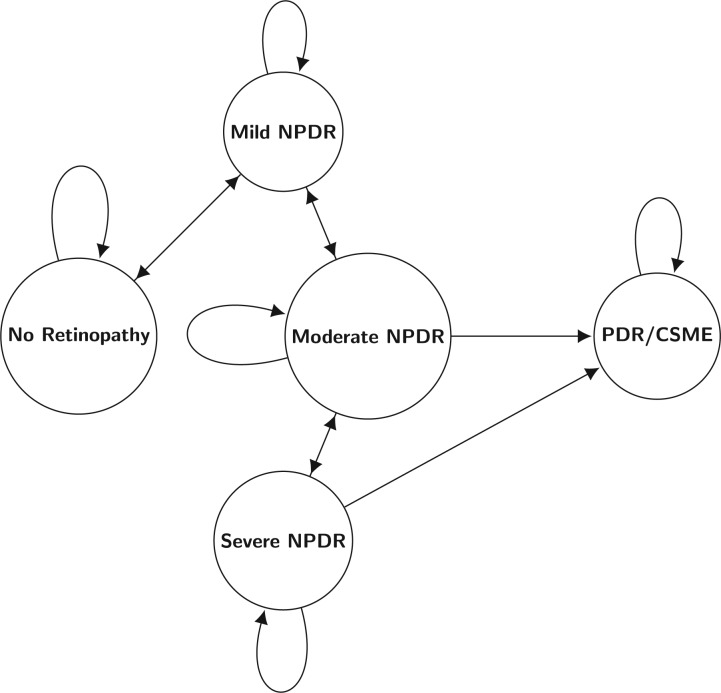
The health status at each visit is typically categorized into one of the several possible states. For example, diabetic retinopathy (DR) status is assessed on the ETDRS scale (ETDRS, 1991), which is an ordered scale. Based on this, five clinically meaningful retinopathy states can be defined: (i) no DR and no clinically significant macular edema (CSME); (ii) mild non-proliferative diabetic retinopathy (Mild NPDR) and no CSME; (iii) moderate non-proliferative diabetic retinopathy (Moderate NPDR) and no CSME; (iv) severe non-proliferative diabetic retinopathy (Severe NPDR) and no CSME; and (v) proliferative diabetic retinopathy (PDR) or CSME, either of which requires treatment to preserve vision. Thus, State 5 (PDR/CSME) is the state that triggers clinical intervention, and is considered as an absorbing state in the Markov models employed herein. Also note that a subject cannot jump from state 1 (no DR) to state 3 (Moderate NPDR) without first going through state 2 (Mild NPDR). It is important to note that the health status is observed only at the scheduled visits, and therefore the exact transition times are unknown. Moreover, a subject can either progress from state 2 to state 3 or regress to state 1. To account for these possible transitions, a Markov model in continuous time (Cox and Miller, 1965; Iosifescu, 1980) can be employed to estimate the cumulative incidence functions for each state, and to then focus on transitions to the absorbing state, at which point interventions are required.
Multi-state Markov models have been employed to identify risk factors for diabetic retinopathy (Liu and others, 2013; Marshall and Jones, 1995). Fixed schedules (e.g., annual visits) have also been considered (Dasbach and others, 1991). However, our goal is to take this one step further and to provide rational examination schedules based on the patient’s risk profile.
Herein screening schedules are compared in terms of two cost components of particular interest in this context. The first is the undetected time, defined as the elapsed time from the actual onset of progression (assumed to occur in continuous time) and the next visit at which it is detected. The less frequent the visits, the longer progression will go undetected and the harder or more costly it may be to treat when finally detected. The second component is the number of visits or screening examinations up to and including when the progression is detected. The more frequent the visit schedule, the greater the number of negative visits before progression is detected.
The goal is to introduce a screening schedule that minimizes both the undetected time and the number or frequency of examinations, and the associated costs, and at the same time is practical and easy to explain. Both fixed screening schedules (e.g., annual visits) and personalized screening schedules based on the current health state are investigated and illustrated using the progression of retinopathy in the DCCT/EDIC study. The more clinically relevant findings are presented in detail in a companion article (Nathan and others, 2017). The Supplementary Material to that paper includes an assessment of the fit of the Markov models to the DCCT/EDIC data. Based on the model results, a Shiny application within R has been developed to apply the results of the models to individual patients. A description is also provided in the Supplementary Materials to the companion paper.
2. The model
Let  be the set of all health states, where
  is the number of states (
 ). A subject is followed over time, and at each time point can be in any of the
  health states. Denote by
  the state of the subject at time
  ,
,  . The transition probability matrix
  with entries
can be defined in terms of the transition intensity matrix  with elements
Then  and
and  are related by the Kolmogorov differential equations (forward/backward) (Cox and Miller, 1965). A homogeneous Markov process (i.e.,
  or constant intensities
  ) is employed,
| (2.1) |
with elements  ,
,  .
.
The state requiring the intervention is denoted by  and it is assumed absorbing (i.e.,
  , for all
  ).
).
The focus is on the time to the absorbing state, in other words to estimate the cumulative distribution function for time from state  to state
  , denoted by
  (
( ). A closed form expression for
  is available for homogeneous Markov processes. Briefly, using the spectral decomposition of the intensity matrix
  ,
,
where  and
and  is a diagonal matrix with elements
  , then one has
| (2.2) |
where  .
.
The intensity matrix of the Markov model can incorporate covariates, and their effect is expressed as hazard ratios,
| (2.3) |
where  denotes the covariates, and
  the corresponding hazard ratios. This can be taken into account for developing personalized screening schedules.
All parameters are estimated by maximizing the likelihood (Kalbleisch and Lawless, 1985).
Let  denote the time horizon, say
  years. The total cost over this follow-up period is based on the number of visits
  , and the undetected time
  . With
  denoting the cost of each visit, and
  the cost of one unit (e.g., year) of undetected time, the expected total cost becomes:
When comparing two screening schedules, one is preferable if it performs better (i.e. lower  ) and is less costly (i.e. lower
  ).
).
3. Fixed visit schedules
Consider the case of a single sub-clinical state not requiring treatment that can lead to progression to a clinical state requiring treatment. Let  denote the cdf of
  , the time from the sub-clinical state to the clinical state. A screening schedule, denoted by
  is a partition of the interval
  , so that
  , where
  is the time horizon and
  .
.
The undetected time, denoted by  , is defined as the length of time between the onset of the clinical state requiring treatment and the next visit when it is detected. Using our notations, if
  then
then  . If
. If  , then, with
  (
( ) such that
  , then
  .
.
The expected value of the undetected time is
| (3.1) |
One can see that, for a different partition  of the interval
  (
( ), the difference in undetected time between the two visit schedules is
| (3.2) |
where  and
and  (and similar notations for
  and
and  ).
).
The expected number of visits before the onset  is
is
| (3.3) |
and therefore,
The difference in expected costs between two visit schedules  and
and  is
is
Assuming  , the visit schedule
  is more effective, i.e.,
  . It is also less costly iff
| (3.4) |
4. Personalized screening schedules
Two personalized screening approaches are presented. First, given the unit costs  and
and  , a screening schedule can be obtained as the solution of an optimization problem, illustrated by minimizing the expected cost. The second approach is to select the time to the next visit such that the risk of reaching the absorbing state is below an acceptable risk (say 5%).
4.1 Minimizing expected cost
Assume that at visit  (
( ), a subject is in state
  (
( ), denoted by
  . The decision regarding the time to the next visit is based on the retinopathy level at the current visit, namely the state
  . The action at visit
  is to follow up the patient in
  years, denoted by
  . The probability of the state at the next visit depends on the current state and the action
  ,
,
where  is defined in (2.1).
The total cost associated with the action  for a subject in state
  is computed first conditionally on the state
  reached after the
  years. There are two costs associated with transitioning from
  to
to  under action
  . The first one is the cost per year associated with the timing of the next visit, namely
which does not depend of the state  . The second type of cost
  is due to the (expected) undetected time. Notice that this cost is zero unless the patient is in the absorbing state at the next visit (i.e.,
  for
for  ), while
where  denotes the cdf of the time to the absorbing state starting from state
  .
.
The total cost of action  when in state
  conditional on reaching the state
  is
is
Then, unconditionally over the set of possible states reached, the expected total cost is
| (4.1) |
Since the time to the next visit is bounded (i.e.  is bounded), there is a value
  that minimizes the total cost
  , and the values so obtained define the optimal screening schedule
  . Notice that, for example, one could also define a screening schedule by employing a weighted sum of
  and
and  . However, this would lead to a similar objective function but with different values for
  and
and  .
.
4.2 Limiting risk of undetected time
One difficulty that arises when using the previous approach is the need to specify values for  and
and  . While the visit cost
  can be readily obtained (e.g., the cost of the ophtalmoscopy and fundus photography in the retinopathy example), it is rather difficult to elicit values for the undetected time cost
  . More importantly, while intuitive from a payer’s perspective, it is less clear how meaningful the previous cost-benefit analysis is to a particular patient who is more likely interested in a screening schedule that minimizes the undetected time. Thus, a different approach is to choose the time to the visit such that the probability of reaching the absorbing state (which requires an intervention) is below a certain cutoff value, deemed as an acceptable risk (e.g., 5% or 10%). In applications, other considerations (such as practical constraints) may play a role as well in choosing a certain screening policy.
For each state  , the time to the next visit
  is specified, so that the time to the next visit vector
  defines a screening schedule. This leads to a Markov chain (in discrete time)
  with the same states and with transition matrix
  obtained as follows. Transition probabilities from state
  are given by the state probabilities of the Markov process
  after
after  years starting from state
  , or equivalently the
  -th row of
  (see Equation (2.1)), namely
Notice that  further depends on
  , but for simplicity this is suppressed in the notation.
As before, two screening schedules  and
and  are compared with respect to expected number of visits
  and
and  and the undetected times
  and
and  . However, the undetected time is 0 unless the subject reaches the absorbing state at the next visit, and therefore it is more meaningful for both the patients and the clinicians to estimate the undetected time conditional on reaching the absorbing state at the next visit.
Let  (
( ) denote the visit when this occurred, so
  , and denote by
  the probability of reaching the absorbing state
  state
state  under regime
  . Bayes’ formula gives
The expected undetected time conditional on reaching the absorbing state is:
| (4.2) |
where
Another measure of interest is the probability that the unobserved time will exceed a clinically meaningful length of time  . A natural choice for the retinopathy example might be 0.5 years, which is the expected unobserved time under annual screening assuming a uniform distribution. One has:
| (4.3) |
The expected number of visits within an L-year horizon under a specific screening schedule is obtained using an imbedding approach (Fu and Lou, 2003), see Appendix for details, or through simulations under this Markov model.
5. Illustration: screening for retinopathy in DCCT/EDIC
From 1983 to 1989, the DCCT enrolled 1441 participants with T1D, and standard seven-field fundus photographs were obtained every 6 months during DCCT and every fourth year during EDIC, and in the complete cohort during EDIC years 4 and 10. In total, there were 23,961 retinopathy assessment visits, and 504 participants reached the PDR/CSME state over the follow-up.
The five retinopathy states and the possible instantaneous transitions among the states are depicted in Figure 1.
Fig. 1.
Possible instantaneous transitions among the five retinopathy states.
The maximum likelihood estimator for the intensity matrix  is obtained using the package
  (Jackson, 2011) in R, which is then used to estimate the transition matrix
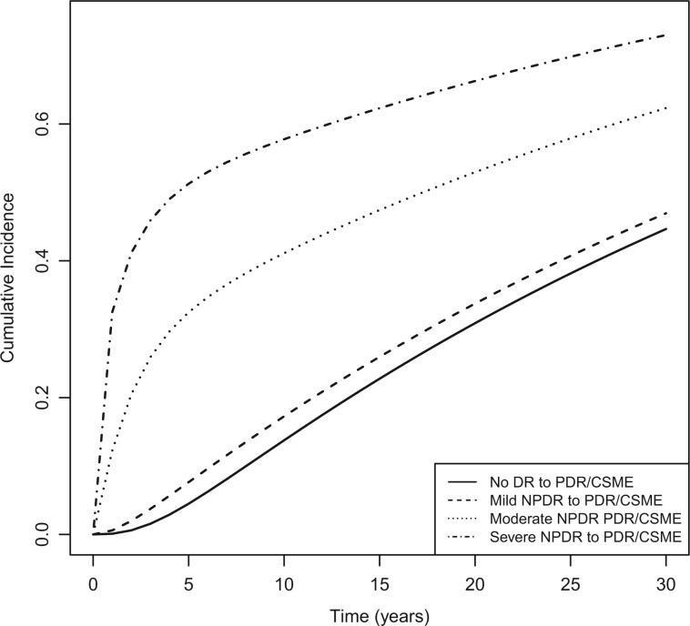
  in (2.1). The cumulative incidence functions of reaching the absorbing state (PDR/CSME) are obtained using the eigenvalues and eigenvectors of
  and are depicted in Figure 2. As expected, these are lower when starting from no or mild retinopathy compared to starting from Moderate NPDR and Severe NPDR.
Fig. 2.
Incidence functions for PDR/CSME, stratified by the initial state.
Further details on the fitted Markov model and checking model assumptions are reported in Nathan and others (2017).
5.1 Fixed schedule
For illustration, we compare annual visits versus biennial (every other year) visits. Using (3.1), the expected undetected time for annual visits is 0.153 years, while for biennial visits is 0.303 years. Annual screening leads to approximately 18.37 expected visits over 20 years, while for biennial visits yields approximately 9.76 visits. Using (3.4), annual visits are less costly than visits every two years if the ratio  is 33.86 or higher.
5.2 Personalized schedule
The first approach is illustrated using  , and
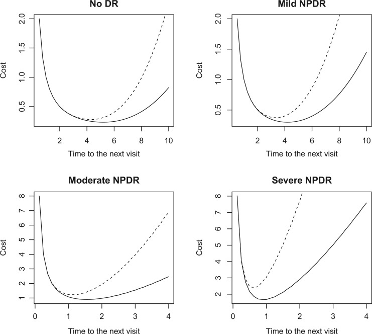
, and  . The total costs are computed using Equation (4.1), and are depicted in Figure 3 for various values of the time to the next visit
  . The optimal screening is given by the values that minimize the total cost in Figure 2, namely (5, 4, 1.5, 1) for
  and (4.5, 3.5, 1.25, 0.63) for
  . More frequent visits are required as the severity of the current retinopathy state increases. Furthermore, the larger the ratio
  , the shorter the optimal time to the next visit.
Fig. 3.
Total cost as a function of the time to the next visit for  (solid line) and
  (dashed line), stratified by the current retinopathy state. Note that the axes differ for each intermediate state to best display the change in costs as the screening interval changes.
To illustrate the second approach, first notice (Figure 2) there is less than 5% chance of reaching PDR or CSME within 4 years when starting from no DR, and less than 5% chance within 3 years when starting from Mild NPDR. One option is then to schedule the next visit in 4 years for a subject with no retinopathy, in 3 years for a subject with mild retinopathy, and (as per current guidelines) every year for those with Moderate NPDR or Severe NPDR, which leads to the screening schedule  (4, 3, 1, 1). This schedule will be compared with annual screening.
The conditional probabilities  ’s of reaching the PDR/CSME state under the (4, 3, 1, 1) screening are given by (0.056, 0.073, 0.238, 0.633), while for annual visits these are (0.002, 0.013, 0.270, 0.715).
The undetected time among those reaching the absorbing state under a (4, 3, 1, 1) screening schedule is 0.684 years, while for annual visits it is 0.606 years. Since most transitions to the absorbing state occur when subjects currently have Moderate NPDR and Severe NPDR, more frequent visits from those states will lead to lower  . For example, a (4, 3, 0.5, 0.25) schedule leads to an expected undetected time of 0.415 years.
Using (4.3), there is 67.43% chance the undetected time will exceed  years for a (4, 3, 1, 1) schedule, 65.60% chance for annual screening, but only 18.15% chance for a (4, 3, 0.5, 0.25) schedule.
The average number of visits with a 20-year horizon for a  schedule is 6.73 visits, for
  is 18.38, while a
  schedule leads to an average of 7.65 visits.
Notice that the  schedule dominates the
  schedule both in terms of effectiveness with an expected 0.19 years lower average undetected time, and costs with an expected 10.7 fewer number of visits over up to 20 years of follow-up.
Given an acceptable probability of progressing to PDR/CSME, the time to the next visit is determined based on the current retinopathy level such that the probability of reaching PDR/CSME is below (approximately) that threshold value. For illustration, using 5% as the acceptable risk, Table 1 presents the time to the next visit based on the current retinopathy state.
Table 1.
Time to next visit (years) as a function of the current state such that the risk of reaching PDR/CSME is approximately 5%
| Current State | Time to Next Visit | Probability |
|---|---|---|
| No retinopathy | 5.250 | 0.049 |
| Mild NPDR | 3.583 | 0.048 |
| Moderate NPDR | 0.333 | 0.045 |
| Severe NPDR | 0.083 | 0.057 |
5.3 Covariates
The effect of covariates can be incorporated using 2.3), and this is illustrated using HbA1c, entered as a time-dependent covariate (Table 2). High levels of HbA1c were associated with higher risk of worsening from no retinopathy to Mild NPDR, from Mild NPDR to Moderate NPDR, from Moderate NPDR to Severe NPDR and from Severe NPDR to PDR/CSME, and with lower risk of improvement from Mild NPDR to no retinopathy and from Moderate NPDR to Mild NPDR (Nathan and others, 2017).
Table 2.
Time to next visit (years) as a function of the current state such that the risk of reaching PDR/CSME is approximately 5% for various values of HbA1c, along with the average undetected time  (years) and the expected number of visits
| HbA1c (%) | No retinopathy | Mild NPDR | Moderate NPDR | Severe NPDR |

|

|
|---|---|---|---|---|---|---|
| 6 | 14.41 | 11.91 | 0.58 | 0.41 | 3.05 | 2.11 |
| 8 | 6.08 | 4.33 | 0.41 | 0.08 | 1.12 | 5.77 |
| 10 | 3.16 | 2.08 | 0.25 | 0.08 | 0.42 | 14.46 |
With regards to the screening schedule, more frequent visits are required for larger values of HbA1c. With a 5% acceptable risk of reaching the PDR/CSME state, the next visit for a patient with no retinopathy at the current visit is scheduled in 14.4 years for an HbA1c of 6% and in 3.2 years for an HbA1c of 10%, while for a patient with Severe NPDR they are scheduled in 0.42 years for an HbA1c of 6% and in 0.08 years for an HbA1c of 10%.
Other covariates were also considered (gender, age, duration of diabetes, hypertension), but did not have a significant role in determining a screening schedule (Nathan and others, 2017).
6. Discussion
A Markov model in continuous time is employed to describe transitions among health states over time, with particular interest in the incidence of an absorbing state which requires intervention. The goal is to investigate personalized cost-effective screening schedules. The cost associated with a given schedule has two components: the number of visits and the length of time between the onset of the treatable condition and the visit when it is actually diagnosed (the undetected time). A more frequent screening schedule leads to more visits (more costly) and lower undetected time (less costly).
We first investigated fixed screening schedules (e.g., annual vs. biennial visits), and then considered tailoring the time to the next visit based on the current health state. The methods were illustrated using the progression of retinopathy in the DCCT/EDIC cohort, and the proposed approaches compared these personalized screening schedules with the current guidelines which recommend annual visits. It was shown that having more frequent visits for subjects at high risk and fewer visits for subjects at low risk leads to cost-effective screening schedules. In our example, a (4, 3, 0.5, 0.25) schedule for a subject starting in the no retinopathy state resulted in a 58% reduction in the number of visits compared to annual visits over a 20-year follow-up, while at the same time reducing the undetected time by 31%. The screening schedule can also take into account the effect of various risk factors, which is illustrated using HbA1c.
Besides the clinical benefit of early detection of progression, adopting the personalized screening schedule for retinopathy may lead to important savings. Assume the US population of T1D is approximately 1 million, 10% of which have already reached the PDR/CSME state. Of the remaining 90%, approximately 25% have no retinopathy, 25% have Mild NPDR, 30% Moderate NPDR, and 20% Severe NPDR. Assuming  200 per fundus photography, annual visits will require approximately
  2.46B over 20 years, while the (4,3,0.5,0.25) schedule requires
  1.39B, for a saving of
  1.07B, or 43%.
In the DCCT/EDIC retinopathy example, the standard therapy for PDR is photocoagulation and for CSME is either photocoagulation or anti-VEGF, the costs of which are well established. There is no literature, however, on the costs associated with delayed detection and treatment (the untreated time,  ), or the savings associated with early detection and treatment. Thus, these costs may be more or less fixed, at least over short periods of a few months that PDR/CSME might go undetected. However, for retinopathy and other conditions, there could be substantial costs associated with an increase in the period that progression goes undetected. Retinopathy progression doesn’t stop when PDR/CSME occurs. Rather, if untreated subjects can progress to more severe levels, such as the development of what are termed “high risk characteristics” that place a subject at markedly higher risk of blindness. Thus, delayed detection could increase the costs associated with the initial treatment (e.g., photocoagulation) owing to a more severe condition upon detection, and could increase the costs associated with the treatment of a more rapid deterioration in the patient’s condition. Thus, the above models could be further extended to incorporate temporal and other covariates into the cost function
  .
.
As suggested by the Associated Editor and the two reviewers, our work can be extended in several ways. While a constant cost per unit of undetected time (i.e.,  ) was considered in our work, it may be possible to consider a time-varying cost function for the undetected time instead. Also notice that the screening schedule derived in Section 4.1 is obtained by minimizing the expected cost at the next visit; while this approach provides a local optimum, there is no guarantee it will be a global optimum. However, while of clear theoretical interest, both these generalizations rely on assigning a monetary value to the undetected time, which in most applications may be problematic. Yet another extension could address situations when there may be multiple states (not necessarily all absorbing) which require interventions.
It should be noted that the calculation of undetected time only depends on the cumulative distribution of the time to the absorbing state. Therefore, the results presented here apply to non-homogeneous Markov models as well, where the transition probabilities are obtained as solutions of the nonlinear differential equations corresponding to the Kolmogorov equations (Titman, 2011).
The setup considered for determining the optimal screening schedule by minimizing the expected total cost (Section 4.1) is somewhat similar to a discrete-time Markov decision process (MDP) with deterministic history dependent decision rules (Puterman, 2005). However, in the retinopathy application, the time horizon ( ) refers to the time elapsed since the first visit, and therefore the number of visits up to and including
  is random. In contrast, an MDP with finite time-horizon typically refers to a fixed (finite) number of transitions, visits in our case. Another difference is that the ultimate goal is to obtain screening schedules that are meaningful for each patient, rather than cost-effective from a payer perspective, which is addressed by limiting the risk of undetected time (Section 4.2).
Other authors have considered the problem of optimal scheduling examinations (Zelen, 1993; Parmigiani, 1997). Three health states were considered (no disease, pre-clinical state and the clinical state), and the goal was to determine screening schedules that lead to early detection of disease. Unlike the retinopathy application considered herein where the status can either improve or worsen over time, the disease (e.g., cancer) was assumed progressive (i.e., no transitions from the pre-clinical state to the healthy state), and these methods are not directly applicable to our problem.
Clinical management of a chronic disease such as diabetes is a complex task requiring periodic evaluation of various potential complications, such as retinopathy, nephropathy and neuropathy. It is important to help provide informed guidelines on the screening for these complications which are tailored to the risk profile of the individual patient. The present work shows that personalized screenings have the potential to be cost-effective relative to fixed screenings.
Supplementary material
Supplementary material with the R code implementing the proposed methods is available online at http://biostatistics.oxfordjournals.org.
Supplementary Material
Acknowledgments
The authors are grateful to the Associate Editor and the two reviewers for their thoughtful questions and suggestions. Conflict of Interest: None declared.
7. Appendix
The number of visits with a time horizon  is obtained by discretizing the problem based on the smallest interval of time which can be considered between two consecutive visits. In the retinopathy progression example, these are monthly intervals, and the time scale is changed accordingly (e.g.,
  is in months). Since we are interested in the number of visits with an
  -year follow up, we define an imbedded Markov chain which captures information on both the clinical state and the follow up time. Given the clinical states
  , where
  is the absorbing state, the imbedded Markov model will have states
  , where a subject is in state
  if the clinical state is
  (
( ) at time
  (
( ), while
  is the absorbing state of the imbedded state defined as either reaching the absorbing clinical state
  or the follow up time exceeding the time horizon
  .
.
Transitions over time in the imbedded model occur with probabilities obtained based on the actions  , since the actions only depend on the clinical state. Transitions to a non-absorbing state (
 ) are given by
| (7.1) |
while the transition probability to the absorbing state is
| (7.2) |
The expected number of visits before absorption in the initial Markov model  with a time horizon
  is the time to absorption in the imbedded chain with state set
  and transition probabilities matrix
  with components defined in (7.1) and (7.2). It follows that the expected number of visits within the first
  years is given by
  (Iosifescu, 1980), where
  is a vector of ones and
  is the fundamental matrix of the imbedded chain, with
  .
.
Funding
Diabetes Control and Complications Trial/Epidemiology of Diabetes Interventions and Complications (DCCT/EDIC) study (U01-DK-094176 and U01-DK-094157), and Glycemia Reduction Approaches in Diabetes: A Comparative Effectiveness (GRADE) Study (U01-DK-098246), all from the National Institute of Diabetes, Digestive and Kidney Diseases (NIDDK), NIH. Samuel W. Greenhouse Biostatistics Research Enhancement Award (to I.B.).
References
- Aiello L. P., Sun W., Das A., Gangaputra S., Kiss S., Klein R., Cleary P. A., Lachin J. M., Nathan D. M. and DCCT/EDIC Research Group. (2015). Intensive diabetes therapy and ocular surgery in type 1 diabetes. New England Journal of Medicine 372, 1722–1733. [DOI] [PMC free article] [PubMed] [Google Scholar]
- Cox D. R. and Miller H. D. (1965). The Theory of Stochastic Processes. London: Methuen. [Google Scholar]
- Dasbach E. J., Fryback D. G., Newcomb P. A., Klein R. and Klein B. E. K. (1991). Cost-effectiveness of strategies for detecting diabetic retinopathy. Medical Care 29, 20–39. [DOI] [PubMed] [Google Scholar]
- The DCCT/EDIC Research Group (DCCT). (2015). Effect of intensive diabetes therapy on the progression of diabetic retinopathy in patients with type 1 diabetes: 18 years of follow-up in the DCCT/EDIC. Diabetes 64: 631–642. [DOI] [PMC free article] [PubMed] [Google Scholar]
- Early Treatment Diabetic Retinopathy Study Research Group (ETDRS). (1991). Early photocoagulation for diabetic macula retinopathy: ETDRS report number 9. Ophthalmology 98, 766–785. [PubMed] [Google Scholar]
- Fu J. C. and Lou W. Y. W. (2003). Distribution Theory of Runs and Patterns and its Applications: A Finite Markov Cain Imbedding Approach. New Jersey: World Scientific. [Google Scholar]
- Iosifescu M. (1980). Finite Markov Processes and Their Applications. New-York: Wiley. [Google Scholar]
- Jackson C. H. (2011). Multi-state models for panel data: the $msm$ package for R. Journal of Statistical Software 38, 1–29. [Google Scholar]
- Kalbfleisch J. D. and Lawless J. F. (1985). The analysis of panel data under a Markov assumption. Journal of the American Statistical Association 80, 863–871. [Google Scholar]
- Morris A. D., Doney A. S., Leese G. P., Pearson E. R. and Palmer C. N. (2013). Glycemic exposure and blood pressure influencing progression and remission of diabetic retinopathy: a longitudinal cohort study in GoDARTS. Diabetes Care 36, 3979–3984. [DOI] [PMC free article] [PubMed] [Google Scholar]
- Marshall G. and Jones R. H. (1995). Multi-state Models and Diabetic Retinopathy. Statistics in Medicine 14, 1975–1983. [DOI] [PubMed] [Google Scholar]
- Nathan D. M., Bebu I., Hainsworth D., Klein R., Tamborlane W., Lorenzi G., Gubitosi-Klug R., Lachin J. M. and DCCT/EDIC Research Group. (2017). Evidence-based screening frequency for retinopathy in type 1 diabetes. New England Journal of Medicine, in press. :10.1056/NEJMoa1612836. [Google Scholar]
- Parmigiani G. (1997). Timing medical examinations via intensity functions. Biometrika 84, 803–816. [Google Scholar]
- Puterman M. L. (2005). Markov Decision Processes: Discrete Stochastic Dynamic Programming. New York: Wiley. [Google Scholar]
- Titman A. C. (2011). Flexible nonhomogeneous markov models for panel observed data. Biometrics 67, 780–787. [DOI] [PubMed] [Google Scholar]
- Zelen M. (1993). Optimal scheduling of examinations for the early detection of disease. Biometrika 80, 279–293. [Google Scholar]
Associated Data
This section collects any data citations, data availability statements, or supplementary materials included in this article.


