Abstract
DNA charge transport chemistry involves the migration of charge over long molecular distances through the aromatic base pair stack within the DNA helix. This migration depends upon the intimate coupling of bases stacked one with another, and hence any perturbation in that stacking, through base modifications or protein binding, can be sensed electrically. In this review, we describe the many ways DNA charge transport chemistry has been utilized to sense changes in DNA, including the presence of lesions, mismatches, DNA-binding proteins, protein activity, and even reactions under weak magnetic fields. Charge transport chemistry is remarkable in its ability to sense the integrity of DNA.
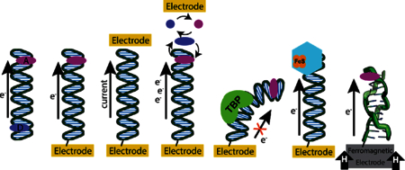
Here, we describe a variety of studies carried out in our laboratory probing the DNA duplex and DNA-binding partners using DNA-mediated charge transport (DNA CT). Over the past three decades, we have explored this chemistry and its application in sensing DNA.1–5 Additionally, we have focused on how nature makes use of this chemistry for DNA sensing and for long-range signaling across the nucleus of the cell.6,7
Through a full range of strategies and platforms, we and others have characterized this chemistry in detail. Two critical characteristics of this chemistry have been established. DNA CT can occur over long molecular distances. In fact, while ground state DNA CT has been documented to occur over 34 nm,8 the distance limit for DNA CT has yet to be established. Many recent experiments suggest that DNA CT occurs over kilobase distances, but DNA CT can occur over long distances only if the DNA duplex is well stacked. Small perturbations in DNA stacking perturb DNA CT. Thus, base mismatches, lesions, and even DNA-binding proteins that perturb the DNA base stack can be sensitively detected electrochemically.9,10 Using these parameters, DNA CT chemistry provides a powerful means to sense DNA and the small and large molecules that interact with the DNA duplex.
1. PLATFORMS FOR MEASURING DNA CT
There are many different platforms that have been used to measure DNA-mediated charge transport (DNA CT), illustrated in Figure 1. Early experiments testing DNA CT were performed in solution and involved a photoexcited charge donor that transfers charge to an acceptor through a DNA bridge.11 A wide variety of donors and acceptors were used in these experiments, ranging from transition metal complexes to purely organic moieties, base analogs, and proteins.12–14 More recent experiments have been conducted using electrodes, typically gold or graphite, modified with a self-assembled monolayer of DNA.15–18 Here, DNA duplexes are linked to the surface using a covalent modification on the phosphate backbone (alkane-thiols for gold or pyrene for graphite) that allows the duplexes to stand upright,17 facilitating interaction with DNA-binding molecules in solution. Redox molecules, either noncovalently or covalently attached to the DNA, can then be reduced or oxidized by applying a potential across the electrode surface.3
Figure 1.
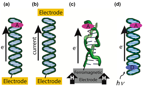
Platforms for the study of DNA-mediated charge transport (DNA CT). (a) DNA is covalently tethered to an electrode surface with an intercalated redox probe. A cyclic increasing or decreasing potential is applied that results in charge being transported through the DNA either to or from the electrode, which can be measured as a change in the current during a potential sweep. (b) DNA is covalently tethered between two electrodes. This type of setup is used to measure the current between the two electrodes in conductive AFM and STM break junction methods. (c) A ferromagnetic electrode influences the yield of charge transport through DNA in different conformations, such as the Z-form shown above. (d) Donor and Acceptor molecules (ovals) are intercalated into a DNA duplex. Transition metal complexes, Ru metallointercalators, Rh metalloinsertors, intercalating organic dyes, and fluorescent base analogs are commonly used as donor and/or acceptor molecules. Photoexcitation initiates charge transport through the DNA bridge and is measured using spectroscopy or other means generally probing the donor or acceptor.
The advantage to these electrochemical studies is that they allow measurements of ground state CT, rather than CT through excited state photochemistry. Moreover, while the chemistry is occurring on the electrode surface, in all respects it appears that the chemistry is like that in solution; proteins bind to their specific cognate sites and carry out their various enzymatic reactions with their specific nucleic acid substrates. One disadvantage is that the rates of CT through the DNA duplex cannot be determined electrochemically, because for all studies thus far conducted, even using DNA 100-mers, the rates of CT have been limited by transport through the alkane linker.19 It is this linker that keeps the duplex “upright.”17 Measurements of base–base DNA CT in solution, using photoexcitation of 2-aminopurine, nonetheless, show DNA CT to be on the picosecond time scale and gated by the motion of the DNA bases.20–24 Indeed, this chemistry provides a sensor also for the dynamics of DNA.
Other experimental setups have allowed for measurements of DNA conductivity. Conductive atomic force microscopy has been used to create metal–DNA–metal junctions that can be used as a circuit to measure the current–voltage characteristics of DNA.25 The scanning tunneling microscopy break junction technique measures the conductivity as the tip is pushed toward and retracted away from the surface, apparently hybridizing and dehybridizing the duplex.26 The current is measured as a function of the distance of the tip from the surface with the assumption that the stretch of separation where the current is constant represents the conductivity of a DNA duplex bridge. Single DNA molecule circuits have also been made that tether DNA between a nanotube gap and measure the change in current that passes through the circuit; this experimental setup provides a measurement of conductivity relative to that of the carbon nanotube.27
2. DNA CT CHARACTERISTICS FOR DNA SENSING
For all of these measurements, the “connection” to the DNA duplex is critical. For experiments where DNA CT is monitored using small-molecule probes,3 the careful selection of the redox-active probe is essential. DNA-mediated charge transport occurs efficiently only with redox-active molecules that couple effectively to the π-stack. Thus, intercalators that stack well with the DNA duplex have been our most effective probes. Intercalative redox probes such as methylene blue that are able to insert themselves into the π-stack undergo efficient DNA-mediated charge transport.28 Other molecules, like the positively charged ruthenium hexammine, associate electrostatically to the phosphate backbone and are unable to access DNA CT.29 In some cases, as with methylene blue, different DNA binding modes are available. At low micromolar concentrations, methylene blue primarily intercalates into DNA where it can undergo efficient DNA CT, but at higher concentrations it can bind electrostatically where it cannot utilize DNA CT. Screening these electrostatic interactions with increased salt concentrations promotes primarily intercalative binding.
The importance of this coupling or connection to the DNA π-stack was highlighted in comparing DNA CT in experiments monitoring photooxidation of guanine using two fluorescent base analogs, 2-aminopurine, which stacks well with the duplex, versus etheno-adenine, which does not.13 The differences in CT rates and distance dependences were remarkable. Even for electrochemistry experiments, probing the same DNA construct with molecules that do and do not couple to the π-stack shows a significant difference in yield.3 It is also possible to use redox probes that selectively target mismatches or abasic sites and stack within the open site, so that a DNA-mediated signal is found only if that mismatch or abasic site is present. Critically, then, for all these experiments, DNA CT is only rapid and over long-range if the connection is truly to the base pair stack.4
It is also important when working with DNA to be aware that small changes in preparation can have dramatic influences on the heterogeneity of samples and, therefore, the reproducibility of experiments, especially when probing DNA that must be in the duplex form and fully stacked. We have, for example, always utilized duplex DNA for initial modifications of the electrode surface; single stranded DNA binds avidly to the gold surface and cannot be easily displaced. Other concerns relate to the formation of DNA self-assembled monolayers and the electrode attachments; these constructs may vary depending on the chemistry used to attach the DNA, which include thiols for gold electrodes,30 alkynes for azide-terminated electrodes,31 and pyrenes for graphite electrodes,32 and can include many less typical linkers depending on the desired surface attachment.33 In all cases, it is important that the linker allow the DNA to be positioned roughly perpendicular to the electrode surface during experiments. The packing density of DNA is also rationally controlled depending on the desired experiment, because, for example, proteins will not be able to access and bind to DNA in a monolayer that is too densely packed.18 Most importantly, new electrochemical surfaces require characterization, particularly with respect to quantitation of surface loading with the nucleic acid, and surface accessibility. Just as packing densities may be too high for protein binding experiments, very low loading densities usually indicate surface contamination or DNA damage. For sensitive experiments to detect DNA, the sensor needs to be chemically well-defined.3
It is also essential to conduct experiments that verify that charge transport is DNA-mediated, that the charge migrates through the DNA helix, for any new DNA sensor design. Ideally, these experiments will disrupt DNA CT in a way that is recoverable or in a way that has minimal other differences from the sensing experiments. One of the strongest confirmations that charge transport occurs through DNA is by the inclusion of a single base mismatch or abasic site that will disrupt the π-stacking. The main benefit of this method is that it changes very little about the DNA structure that may influence other parts of the experiment but should have a dramatic effect on charge transport that is mediated by the π-stack of the duplex. Incorporation of a particularly ruinous mismatch, such as CC or CA, will result in a significant decrease in the yield of DNA CT.34 Guanine-containing mismatches tend to be poor choices for this confirmation because they do not attenuate CT as dramatically. An abasic site will have a more significant effect, but it is also a larger structural change to the helix. If the experiments are run at relatively high temperatures, an abasic site is often a better choice than a mismatch, because increasing the temperature decreases the attenuation caused by a mismatch and potentiates the attenuation caused by an abasic site.35 Larger scale structural changes such as dehybridization or melting of the duplex may be used to provide necessary confirmation of DNA CT in some context, especially when they are used in the sensing experiment that is being established.36 Careful use of multiple redox probes, some that are able to undergo DNA CT, such as an intercalator, and others that are unable to undergo DNA CT, for example ruthenium hexammine, can also be used to confirm a DNA-mediated signal.
We developed a multiplexed chip that allows for measurements of four different DNA monolayers on a single surface with 4-fold redundancy.16 This multiplexing facilitates carrying out the important controls in parallel. As an illustration of how this device may be used, DNA-mediated CT to a covalently tethered Nile Blue redox probe was simultaneously measured through four monolayers of DNA: 100 bp and 17 bp DNA duplexes with no mismatch, and the same duplexes containing a single base mismatch.8 The rate of electron transport was calculated to be between 25 and 40 s−1 for both duplex lengths, significantly less than the 1010 s−1 rate of DNA CT found with picosecond spectroscopy,22,37 because the rate is limited by tunneling through the alkanethiol that tethers the DNA to the electrode.19 Even so, substantial signal attenuation was observed for both duplex lengths upon introduction of an intervening single base-pair mismatch in the DNA duplex. This set of experiments demonstrates that the DNA-mediated CT is responsible for the redox signals from Nile Blue on this multiplexed chip, even for a 100-mer, and also, remarkably, that such an electrochemical biosensor utilizing DNA-modified electrodes can be used to identify a single base error in a 100-mer.
It is more complicated to verify charge transport that is mediated by the DNA π-stack in dried samples and DNA in other conditions that do not have known structures. Dehydration or exposure to nonaqueous solvents can eliminate efficient DNA-mediated charge transport. DNA is stabilized by a variety of hydrophobic and hydrophilic interactions; changing these interactions can significantly change the resulting structure.38 The precise structural changes caused by dehydration or exposure to most nonaqueous solvents are not very well characterized, but it is clear that the equilibrium conformation of DNA is changed.
Some experiments conducted under nonaqueous conditions do show efficient DNA CT or conductivity. Scanning tunneling microscope studies describing DNA conductivity are generally performed under a vacuum to intentionally remove water that can make such experiments difficult,39 though some scanning tunneling microscope studies have been conducted in humid environments with different results.40 Other measurements, including many conductive atomic force microscopy experiments, have shown varying degrees of conductivity after rehydrating DNA that was deposited in a vacuum or washed with apolar solvents.25–27 Chemistry mediated by DNA CT in hydrated ionic liquids has also been reported with careful consideration of the influence this environment has on the duplex structure.41 In these extreme conditions, the structure of DNA is unclear, so it is difficult to make claims regarding DNA-mediated charge transport that are clearly deconvolved from other aspects of the experiment. For example, ionic conduction through water may be what is measured instead of DNA-mediated CT.42 Thus, to properly understand the structure that is being tested, it is essential to keep DNA hydrated with appropriate salt content during all steps of preparation and experimentation, to characterize the DNA after procedures that may change the structure, and to verify that the charge transport observed is mediated by DNA.
3. STACKING IS ESSENTIAL FOR DNA CT
The ability for DNA to mediate CT depends completely upon base stacking. It is thus not surprising that some variations in DNA CT arise with the different DNA duplexes, the A, B, and Z forms, all of which stack, albeit somewhat differently.43 Using photoinduced CT where rates can be measured, both the A and B forms of DNA show picosecond rates for the DNA-mediated charge transport process.44 Interestingly, in comparing the A and B forms, using base–base CT, the A form shows rapid interstrand transport, because of interstrand base overlap in the A form, whereas the B form, with no interstrand base overlap, shows only rapid intrastrand CT. In electrochemical experiments, the A, B, and Z forms all display long-range CT.43 Here too, the intensity depends upon stacking and, particularly, the coupling of the redox probe with the differing duplex conformations through stacking. The A-form duplex shows the most intense DNA CT.43 The B form follows next. The Z form, which has the poorest π stacking of the duplex structures, exhibits significantly less efficient yield of DNA CT to the intercalated redox probes; the peak current for B-form DNA is over 3 times larger than for Z-form DNA, and the total yield of DNA CT in a single potential sweep differs by more than an order of magnitude; here, however, the different coupling of the redox probe with each conformation needs to be taken into account. Indeed, the yield for Z-form DNA is comparable to A- and B-form DNA using photooxidation assays and a different redox probe.45 Most importantly, single stranded DNA, if present in an unstacked conformation which does not have an ordered π-stacked structure, does not facilitate efficient charge transport. This phenomenon has been confirmed with electrochemical, photooxidation, and direct conductivity studies.22,27,43 It is important, however, to consider the sequence for these experiments with single stranded DNA, since the extent of stacking varies enormously depending upon the sequence.
Perhaps the most unique characteristics of DNA CT is that even a small local disruption of the π-stacking diminishes the efficiency of DNA CT. We have probed all of the mismatches in DNA and, remarkably, all of the mismatches can be detected, even the GT mismatch, which has a thermal stability equal to that of an AT base pair.27,46 Interestingly, the presence of mismatches lowers the rate and yield of DNA CT in a way that correlates with base pair lifetime,46 and this disruption occurs even though mismatched base pairs do not cause significant structural changes.47 Most remarkable is that the attenuating effects of mismatches are evident independent of the sequence context.48 These assays are not simply measurements of thermal stabilization. Hence, DNA CT provides an exquisitely sensitive and valuable assay for DNA mismatches, where different sequences may be tested under the same experimental conditions.
Abasic sites and destabilizing lesions, such as 8-oxoguanine, also significantly diminish DNA CT.34,49 8-oxoG-A and 8-oxoG-C both destabilize the duplex structure, despite the different locations of their modification, which is sufficient to attenuate DNA CT.34 It is because these base lesions are so easily detectable that the idea that nature might also use this chemistry for detection inside the cell became reasonable to consider. Indeed, a whole family of DNA repair enzymes has been found to contain [4Fe4S] clusters, redox cofactors featured commonly in proteins, and many experiments we have carried out support the idea that these repair proteins utilize DNA CT chemistry in their search for lesions within the cell.5,50 Significant kinks to DNA caused by protein binding, such as the TATA-binding protein,51 or chemical interactions with molecules such as cisplatin will also disrupt DNA CT. Again, it is the stacking of bases that must be preserved for long-range CT, so that anything that perturbs that base stacking turns off charge transport mediated by DNA.
It is worth noting that not all modifications to DNA structure diminish DNA CT. A dephosphorylation of the backbone does not have a measurable effect on yield or efficiency,52 nor does a full break in the DNA backbone, as long as base pair stacking is preserved.52,53 Some changes in structure, such as methylation to generate 5-methylcytosine, do not significantly influence DNA CT.34 Also, proteins that do not interfere with the DNA stacking upon DNA binding, as found with helix–turn–helix proteins1 or even histones,54 do not interfere with DNA CT. We think of chromatin as packing up the DNA library to keep it undamaged, yet long-range guanine oxidation can still occur in the nucleosome. For a chromatin-bound DNA duplex, while the DNA duplex is wrapped gradually around the histone core, the DNA base pairs are still well-stacked.
4. SENSING MISMATCHES/MUTATIONS AND DNA BASE LESIONS
The exquisite sensitivity of DNA-mediated charge transport to the structure of DNA allows for it to be used to sense electrically phenomena that disrupt or alter the DNA duplex structure. Depending on the design of the experiment and what is being tested, this relationship can be utilized in a variety of ways to sense protein activity, the presence of specific nucleotide sequences, or any changes in DNA structure.
Many different lesions and modifications can be detected electrochemically via their disruption of DNA CT.34,48,55 Damage products such as thymine dimers, O4-methyl-thymine, O6-methyl-guanine, 8-oxo-guanine, and 5-hydroxy-cytosine all disrupt DNA CT. For the thymine dimer, it is clear that dimerization interrupts base stacking, but for the various base lesions, it can be difficult to predict the degree to which they will disrupt DNA CT, because it is not directly related to their influence on the thermodynamic stability of the helix. More generally, structural modifications that disrupt the hydrogen bonding in Watson–Crick base pairing will significantly disrupt DNA CT. Bulky modifications to bases or changes to general conformation can have a large influence. Smaller changes, such as the addition or subtraction of methyl groups that do not disrupt hydrogen bonding, do not appear to have a large effect on DNA CT.34
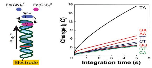
A chip-based technology utilizing DNA-functionalized electrodes was developed by the Barton lab.48 This chip-based technology uses DNA CT chemistry to probe the integrity of double-stranded DNA sequences and detect single-base mismatches for early diagnosis of genetic diseases.48 This device uses an electrocatalytic cycle with [Fe(CN)6]3− and methylene blue to amplify the difference in the yield of DNA CT for DNA with and without a lesion (Figure 2). DNA CT to methylene blue occurs rapidly, and with reduced yield when a lesion is present. [Fe(CN)6]3− regenerates the methylene blue, which allows it to be rereduced via DNA CT. This redox cycling behavior amplifies any difference in CT yield through DNA, making lesions even more apparent. This method has been applied to distinguish commonly found DNA lesions and mutations from well-matched duplexes with no damage. A two-electrode patterning and detection platform was further developed to enhance spatial resolution of patterned DNA arrays and optimization of DNA lesion detection through DNA-mediated CT with electrocatalysis.31 This methodology enables very sensitive discrimination.
Figure 2.
Detection of single base mismatches (red) in DNA duplexes by monitoring the DNA CT yield.48 The redox signal of a redox-active DNA-intercalating molecule, methylene blue (MB), is amplified via redox cycling with Fe(CN)63−/4− that oxidizes the reduced form of MB, leucomethylene blue (LB), in solution. Chronocoulometry (right) is used to quantify the attenuation with each intervening mismatch.
Several other approaches exist for the detection of nucleic acids, some of which can take advantage of DNA CT to signal substrate capture. These approaches tend to rely on hybridization of either neutral peptide nucleic acid (PNA) or negatively charged DNA or RNA to a target oligonucleotide, which have been reported to achieve sensitivities for their target nucleic acid ranging from picomoles to zeptomoles (40 zmol in 4 μL samples).56–59 Hybridization of a targeted nucleic acid with its complement on a sensor surface will restore the DNA CT-capable duplex form, which upon addition of a redox active intercalator, will result in different redox activity compared to the unhybridized DNA. The sensitivity of DNA CT to mismatches can allow sensors to detect single nucleotide polymorphisms, with high sensitivity, and use conditions that are independent of sequence, e.g., do not depend on thermal melting.
5. ELECTRICALLY MONITORING PROTEIN BINDING AND ACTIVITY
The sensitivity of DNA charge transport to structural perturbations has allowed for unique insight into the activity of many protein–DNA interactions. DNA-modified films used to assay protein–DNA interactions are different from monolayers used to assay DNA lesions. The first of these experiments involved the use of a low-density DNA monolayer containing a covalently linked daunomycin probe near the duplex terminus and away from the protein binding site.1 The low density of the DNA film is essential for allowing access to the DNA-binding proteins. Chronocoulometry on these DNA-modified surfaces in the presence and absence of different DNA-binding proteins found a significant difference in the yield of charge transport to daunomycin, which was electrocatalytically regenerated by oxygen, that was directly related to the way in which these proteins interacted with the DNA monolayers.
The methyltransferase HHa1 catalyzes the methylation of cytosine in 5′-GCGC-3′ by first flipping a cytosine out from the duplex then inserting Gln 237 into the void left in the base stack.60–62 Incubating HHa1 with the DNA monolayer without S-adenosylmethionine, which is necessary for enzymatic activity, is enough to greatly diminish the yield of charge transport (Figure 3).63–65 DNA-binding by a mutant of HHa1, Q237W, that inserts an aromatic Trp into the base-pair stack shows significantly less attenuation of charge transport; here the Trp inserted in the stack serves to restore CT. When the film is incubated with a protein that does not bind DNA, such as BSA, there is no change in current. Together, these data show that the yield of DNA CT on this modified electrode depends upon the DNA π-stack.
Figure 3.
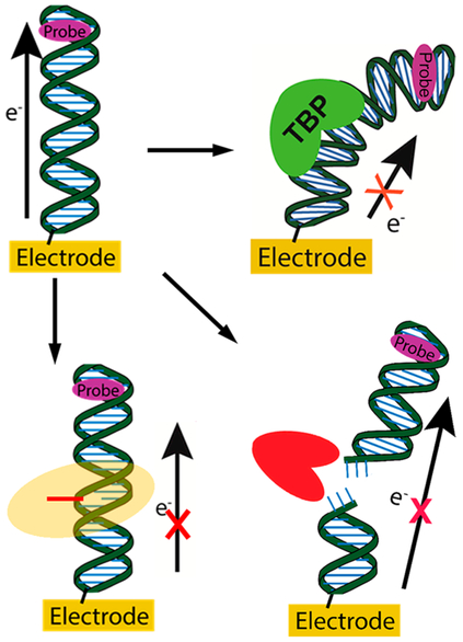
DNA CT monitoring enzymatic activity. Signal is first established for the DNA using a redox probe in the absence of protein. (Top right) Upon binding of a TATA-binding protein (TBP, green), the DNA CT signal to the intercalated redox probe (purple) decreases. (Bottom left) Upon flipping out a base (red line) by a base-flipping protein (orange halo), the yield of DNA CT decreases. (Bottom right) DNA CT to an intercalated redox probe occurring through duplex DNA decreases upon cutting the DNA duplex using a restriction enzyme (red).
This DNA CT assay is sensitive to other types of DNA–protein interactions aside from base flipping, so long as the protein perturbs the π-stacking of bases (Figure 3). The TATA-box binding protein (TBP) does not flip bases, but rather kinks DNA ~90° upon binding to its target site.66,67 This interaction disrupts base stacking, but not base pairing, and is enough to significantly diminish the yield of DNA CT.1,51
As would be expected, more dramatic changes in DNA structure such as cutting with restriction enzymes can also be monitored via this method. The binding of a restriction enzyme, such as the endonuclease PvuII, which does not significantly perturb the DNA base stack,68 does not have a significant influence on the charge transport yield.1 Upon restriction, however, the daunomycin redox probe is released from the surface, which significantly decreases the yield of DNA CT (Figure 3). Similar results have been observed with many other restriction enzymes and redox probes.1,8,69,70
Experiments with Escherichia coli photolyase show that DNA-modified films are also able to monitor the activity of proteins that restore the π-stacked structure of DNA.55 Cyclobutane pyrimidine dimers (CPD) are lesions which form as a result of a photoinduced [2 + 2] cycloaddition between two adjacent pyrimidines on the same DNA strand. Upon photolyase binding, the CPD is flipped out of the DNA helix into the protein’s active site, where a reductive catalytic cycle is initiated upon blue light irradiation of a flavin cofactor that repairs the CPD into individual pyrimidines. After repair, the monomer pyrimidines are returned to the DNA, thus restoring its π-stacked structure.71–73 Conveniently, DNA CT is able to access the redox-active flavin, which allows for DNA duplex integrity to be observed via CT to the flavin, without need for an additional redox probe. Upon photoactivation of photolyase bound to a DNA-modified electrode surface, an increase in DNA CT is observed, indicating that this platform is electrochemically monitoring the repair of the CPD.55
DNA-binding protein activity can also be used to sense reactions to modify DNA where the modification itself does not affect DNA CT, as with base methylation. A recent DNA-based biosensor was constructed to monitor the methylation of DNA by DNMT1, the human DNA (cytosine-5)-methyltransferase, by using DNA CT and a methylation-specific restriction enzyme (Figure 4).69 DNMT1 preferentially methylates hemimethylated DNA when it has access to the cofactor S-adenosyl-l-methionine (SAM).74–77 BssHII is a restriction enzyme that will cleave hemimethylated but not fully methylated DNA. Thus, if the DNA is methylated by DNMT1, the duplex will be protected from restriction by BssHII and retain high yield DNA CT. If there is no DNMT1 activity, BssHII will cleave the DNA and decrease DNA CT yield. Here too, then, a sensitive probe for DNMTI activity can be obtained.
Figure 4.
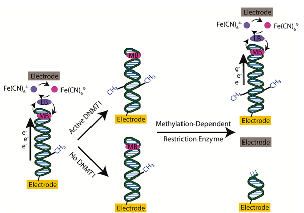
Two-electrode setup for detecting DNMT1 in physiological solutions.69 Current is measured by a reporting electrode (top, gray) near the DNA-modified electrode, via charge transferred by Fe(CN)63− that can then electrocatalytically regenerate methylene blue that was reduced via DNA CT. Active DNMT1 is able to methylate a hemimethylated DNA substrate. Incubation with a methylation-dependent restriction enzyme leads to cleavage of DNA that was not exposed to active DNMT1, thereby decreasing the current measured by the reporting electrode. DNA that is methylated by DNMT1 will retain its structure and retain a high measured current.
6. TWO-ELECTRODE PLATFORM FOR SENSITIVE DETECTION
A goal with sensitive detection is to be able to monitor protein activity in cellular samples without protein purification. Improvements in sensor design involving a two-electrode setup allow for DNA CT to sense DNMT1 activity in crude lysate with minimal purification.69,78,79 This bioanalytical platform utilizes two working-electrode arrays separated by a thin layer of solution to detect biomolecules, nucleic acids, and DNA-binding proteins (Figure 4). The primary electrode is modified with a DNA monolayer, which has a constant applied potential to reduce intercalated methylene blue. The methylene blue then diffuses in solution and is oxidized by ferricyanide, thereby regenerating the methylene blue to be reduced via DNA CT in an electrocatalytic cycle. A second, reporting electrode is held at a potential to oxidize the generated ferrocyanide, and the current at this electrode can be measured to report the yield of DNA CT. This setup enables the reporting electrode to operate with minimal background current, and the electrocatalysis increases the number of DNA CT events that are possible, which together increase the sensitivity of this DNA CT sensing platform.
A recent DNA-based biosensor for enzymatic activity of DNMT1 that has been linked to tumorigenesis shows that this chemistry can be used to observe protein–DNA interactions from biological samples with minimal purification.78,79 Crude lysate from a colorectal tumor and adjacent healthy tissue were incubated with a hemimethylated duplex DNA substrate. Increased DNMT1 activity in the tumor samples further methylated the hemimethylated substrate DNA and prevented the fully methylated form of the DNA from being cut by subsequent exposure to restriction enzymes. The samples exposed to DNMT1 retain efficient DNA CT, and those not exposed to DNMT1 are cut by restriction enzymes and have attenuated DNA CT, thus allowing DNA CT to be used as a sensor for aberrant DNMT1 activity associated with colorectal tumors. These experiments were conducted with pure DNMT1, DNMT1 added to cell lysate, colorectal tumor tissue, and healthy colorectal tissue. The presence of cell lysate did not diminish the sensitivity of this assay to nanomolar concentrations of DNMT1, indicating its potential for assaying biological samples with minimal purification. Indeed, this assay was able to distinguish the increased DNMT1 activity in colorectal tumor tissue from adjacent healthy colorectal tissue without need for purification.
It is noteworthy that what was key in these studies was the application of copper-activated click chemistry to create an open monolayer. Activation occurred with control using the second electrode. As a result, the DNA duplexes could be positioned with control, so that the DNAs were not clumped together, permitting access of the many proteins in the cell lysate to the DNA. With this methodology and the two-electrode platform, cell samples could be easily probed.
7. SENSING BY REDOX [4FE4S] CLUSTERS IN PROTEINS
Many DNA-processing enzymes have been shown to contain [4Fe4S] clusters that are redox-active and able to be reduced and oxidized via DNA CT.5,50,80 These proteins have a wide variety of functions including those involved in base excision repair, nucleotide excision repair, as well as helicases, DNA primase, and DNA polymerases.81–87 Binding to DNA shifts the redox potential of the [4Fe4S] clusters by about 200 mV and activates the clusters toward oxidation, which allows the [4Fe4S]2+/3+ redox couple to be close to +80 mV vs NHE, a potential accessible under physiological conditions.80,88
Proteins with reduced and oxidized [4Fe4S] clusters have significant differences in affinity for DNA.89 Calculations based on the shift in redox potential caused by DNA binding suggest that the affinity of proteins with an oxidized cluster is at least 2 orders of magnitude stronger than proteins with a reduced cluster. Recently, experiments were conducted that systematically varied the oxidation state of the [4Fe4S] cluster and measured how the redox state of the metallocofactor influenced DNA binding affinity.89 Electrophoretic mobility shift assays, isothermal titration calorimetry, and microscale thermophoresis were used to probe the nonspecific DNA binding of Endonuclease III, a base excision repair glycosylase that repairs oxidized pyrimidines in Escherichia coli. The protein with the oxidized cluster showed significantly stronger affinity for DNA. Microscale thermophoresis, which was able to be performed under anaerobic conditions and best prevent extraneous oxidation, shows an affinity for the oxidized state that is at least 550-fold greater than the protein with the reduced cluster. Biophysical modeling suggests that this difference in affinity can be explained primarily by changes in the electrostatic interactions between the cluster and the DNA phosphate backbone without significant changes in the protein structure.
The difference in affinity of the different redox states of [4Fe4S] clusters combined with DNA CT can be utilized in a strategy to rapidly detect and localize near DNA damage.80,90–92 A basic model of genome scanning involving only facilitated diffusion and instantaneous interrogation of the DNA integrity indicates that it is insufficient to probe the entire E. coli genome within its doubling time. Intriguingly, DNA CT provides a means to hasten this search. DNA CT between proteins bound to DNA occurs rapidly on the picosecond time scale but only in cases where DNA π-stacking is unperturbed (Figure 5). In situations where an intervening mismatch or other lesion disrupts the π-stack, DNA CT is unable to occur efficiently between proteins. Since the reduction of oxidized proteins will decrease their affinity for DNA and allow them to release and scan elsewhere, charge transport can serve as an effective first step to aid protein binding where they are needed. Altogether DNA CT decreases the amount of time for a repair protein to find its substrate lesion and redistributes the repair protein in the vicinity of lesions. Proteins that have shown the ability to participate in this redox-mediated damage search include DinG, MutY, EndoIII, and XPD.80,90∓92 Proteins that contain [4Fe4S] clusters are able to communicate with one another despite their origin or repair pathway, indicating the generality of this mechanism for proteins to aid one another in their damage search.
Figure 5.
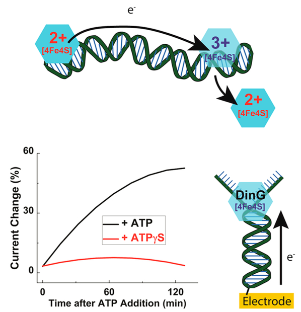
DNA damage sensing by repair proteins containing [4Fe4S] clusters. (Top) DNA CT can occur between proteins when there is no intervening lesion. The charge effectively scans the DNA for lesions and, if the DNA integrity is conserved, the reduced protein will dissociate from the DNA allowing the repair protein to search for damage elsewhere. If there is an intervening lesion between proteins, the charge is unable to be transported, which allows the oxidized protein to stay in the vicinity of the damage and locate it more quickly. (Below) DNA-modified electrodes can be used to monitor helicase activity of DinG through the redox signal of its [4Fe4S] cluster in DinG, which becomes better coupled with ATP but not with ATPγS, with which there is no helicase activity.
Mutations can be used to modify both the redox characteristics of proteins containing [4Fe4S] clusters and their enzymatic activity. By using DNA-modified platforms, electrochemical “sensing” of these proteins provides a means to probe those characteristics. XPD, for example, is a 5′-3′ helicase that is a key member in the nucleotide excision repair process.86 By utilizing a DNA-modified Au surface, wild-type XPD exhibits a redox wave centered at −120 mV vs NHE.93 The XPD L325V mutant displays an electrochemical signal that is less than half that of WT XPD, suggesting that the mutant is deficient at performing DNA CT and can therefore be identified using DNA-modified Au electrodes. Similar inhibited CT behavior is also observed for the Y82A mutant of E. coli endonuclease III (EndoIII), another [4Fe4S] cluster-containing DNA repair protein.92,94 Other mutants of EndoIII, including E200K, Y205H, K208E, and other DNA glycosylases, including WT MutY and UDG,18 display similar redox potentials, despite electrostatic perturbations in the vicinity of the cluster, suggesting that binding to the DNA polyanion is the dominant influence tuning the redox potential of the [4Fe4S].
This DNA electrochemistry can also be used to monitor the biochemical activity of [4Fe4S] cluster-containing helicases, such as XPD and DinG. Upon the addition of ATP, the redox signal corresponding to the [4Fe4S] cluster in DinG increases substantially in magnitude (Figure 5).91 Essentially, this electrochemical signal serves to “sense” enzymatic activity. Presumably, this signal is associated with increased coupling of the cluster to the DNA on reaction. Similar ATP-dependent electrochemical signaling was found in XPD.93
Most recently, we were able to monitor a DNA-binding redox-switch in DNA primase.95 In eukaryotes, both DNA primase and DNA polymerase α contain [4Fe4S] clusters.96 When the protein domain, p58C, containing the cluster in primase was added to a DNA-modified electrode, no signal was evident despite the fact that the domain was known to bind this substrate as part of its activity. However, when the loosely associated domain was oxidized electrochemically, a signal quickly emerged. Upon reduction, however, the signal was again lost. In fact, these results pointed to primase utilizing a redox switch in its cluster for substrate binding using DNA CT.95 Here, primer initiation was proposed to be associated with oxidation of the p58C cluster with electron transfer through DNA CT from polymerase α to primase, with rereduction, dissociation, and handoff once the primer DNA/RNA was complete.
8. SENSING MAGNETIC FIELDS
Recent work has enabled the use of DNA CT in interesting ways to report changes in its magnetic environment.97 First work was conducted to establish chirality-induced spin selectivity.25 On the basis of that work, we utilized DNA CT in the presence of magnetic fields to probe the ability of the DNA duplex to filter spin. Aqueous DNA monolayers were formed on a ferromagnetic electrode substrate capped with a thin layer of gold. Magnetizing the electrode generates a spin-polarized current when the applied potential is the negative of the reduction potential of DNA-bound probe molecules. The sign of the polarization can be switched by changing the direction of the applied magnetic field without influencing its magnitude. When the redox probe is intercalated and undergoes DNA CT, a difference in probe reduction yield is observed for the two magnetic field directions, indicating that one spin moves through the duplex with higher yield than the other (Figure 6). For a 60 bp B-form duplex with covalently tethered Nile Blue, there was a 29% increase in methylene blue reduction when the magnetic field was pointing up, which indicates that the duplex causes at least a 55% spin polarization of electrons that are transported through. There is no magnetic field effect found when DNA is not present, when using single stranded DNA, or when using redox probes that are near a duplex but not undergoing DNA CT. Utilizing 5-methylcytosine (mC) to create DNA oligomers, d(mCG)n, allows for a duplex to undergo a reversible B-to-Z transition under conditions that allow for methylene blue to intercalate into both the B and Z DNA. Remarkably, this switch in DNA helicity changes the magnetic field direction that results in higher DNA CT yield. We find an upward magnetic field causing at least 36% spin polarization for the right-handed B form, but the same duplex with the same magnetic field has −19% spin polarization when switched to the left-handed Z form. Thus, spin transport efficiency can be used to distinguish between B- and Z-form duplexes.
Figure 6.
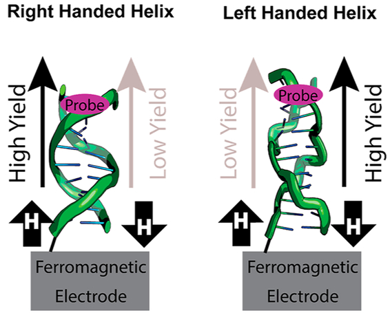
Helix-dependent spin filtering through DNA duplexes attached to ferromagnetic electrodes.97 A magnetic field influences the yield of CT to the redox probe. The magnetic field direction with higher yield CT is switched by changing from the right-handed B DNA to the left-handed Z DNA.
We also recently found that DNA CT can be used to sense the strength and direction of magnetic fields when bound by magnetosensitive proteins.70 We had earlier developed electrochemical methods to monitor the repair of cyclobutane pyrimidine dimer (CPD) lesions that disrupt DNA CT.55 As proteins such as E. coli photolyase and a modified Arabidopsis thaliana cryptochrome I bind DNA on an electrode surface and repair the pyrimidine dimer, high yield DNA CT is restored that allows efficient oxidation or reduction of the redox-active flavin cofactor within the DNA-bound protein (Figure 7).70 The repair of these CPD lesions occurs via a reductive catalytic cycle upon irradiation of the flavin cofactor with blue light. Intriguingly, this repair reaction is sensitive both to the magnetic field strength and to magnetic field angle to which the photolyase and cryptochrome are exposed, where the magnetic field generally dampens the restoration of high yield DNA CT.
Figure 7.
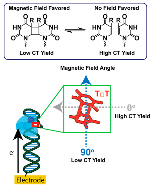
Magnetosensitive repair of cyclobutane pyrimidine dimers by photolyase and cryptochrome.70 (Top) The CPD is favored in the presence of a magnetic field, which disrupts the yield of DNA CT to the protein flavin. (Bottom) The direction of a weak magnetic field that is applied to photolyase or cryptochrome (light blue) influences the yield of repair of CPD (red square) and therefore also affects the yield of measured CT to the protein flavin (blue hexagon).
The sensitivity of this repair reaction is exquisite, allowing for the detection of magnetic fields as weak as 0.2 gauss, on the order of variations seen across the Earth. Increasing the field strength eventually saturates this effect, with 30 gauss fields showing no difference compared to 6000 gauss fields. The angle, but not the direction at which these fields is applied relative to the electrode surface, determines the DNA CT dampening effect. The largest dampening happens at fields that are perpendicular to the electrode surface, while the weakest dampening occurs at fields that are parallel to the electrode surface. By removing the applied magnetic field, the repair activity is restored and so is the yield of DNA CT to the flavin cofactor. It is important to note that this magnetosensitivity relies on the uniform orientation of the proteins on the electrode surface, as there is no observed magnetosensitivity of CPD repair in solution.70
Experiments with different DNA sequences and protein mutations were able to uncover the mechanism by which the magnetic fields influence CPD repair. First, mutations in the active site of E. coli photolyase were used to determine which part of the CPD repair pathway is magnetosensitive. Mutations E274A and M345A near the CPD eliminated magnetosensitivity, but N378C near the flavin retained magnetosensitivity, which suggested that the magnetosensitivity arises from the dimer and not from the flavin. Next, uracil-containing dimers were used to test this hypothesis, and it was found that U□U showed diminished magnetosensitivity, while T□U and U□T both had no magnetosensitivity. These characteristics, together, suggest that the magnetosensitivity arises from a radical pair involving the CPD. This chemistry harkens back to experiments conducted by Turro et al. that showed how radical pair reactions can be controlled by weak magnetic fields.98 These data show how DNA CT can be used to sense magnetic fields, and it is intriguing to consider whether nature takes advantage of this chemistry for in vivo magnetic sensing.
9. SUMMARY AND PROSPECTS
DNA-mediated charge transport is a fascinating phenomenon that relies on the π-stacked structure present in some DNA conformations. Disrupting the π-stack inhibits efficient charge transport, and recovering the π-stack can re-enable efficient charge transport. If experiments are conducted thoughtfully, this exquisite sensitivity of DNA CT to structural changes can be used confidently to sense a large variety of biological phenomena. To date, there are numerous sensor designs that take advantage of this chemistry in order to detect oligonucleotides, single nucleotide polymorphisms and lesions, protein binding, enzymatic activity, and now DNA CT can even sense weak magnetic fields. Even nature appears to use DNA CT in order to detect DNA damage and other structural changes, with implications that it enables proteins to signal one another for efficient repair and coordination.
The future use of DNA CT for sensing phenomena is not limited to the types of experiments described. Rather, the uses of DNA CT will continue to expand as more proteins that are capable of modulating function using DNA CT are uncovered, such as recent discoveries with primase and polymerase,95,99 and as more is understood about the underlying characteristics of DNA CT. Thus, as ever more intriguing uses and characteristics of DNA CT are elucidated, the potential for DNA sensing will continue to grow.
ACKNOWLEDGMENTS
We are grateful to all our co-workers and collaborators for their efforts in developing new sensing technologies. We also thank the NIH, GM61077, for financial support. E.C.M.T. acknowledges a Croucher Foundation Fellowship.
Footnotes
Present Address
Department of Chemistry and Chemical Biology, Harvard University, Cambridge, MA 02138
Notes
The authors declare no competing financial interest.
REFERENCES
- (1).Boon EM, Salas JE, and Barton JK (2002) An Electrical Probe of Protein—DNA Interactions on DNA-Modified Surfaces. Nat. Biotechnol 20, 282–286. [DOI] [PubMed] [Google Scholar]
- (2).Drummond TG, Hill MG, and Barton JK (2003) Electrochemical DNA Sensors. Nat. Biotechnol 21, 1192–1199. [DOI] [PubMed] [Google Scholar]
- (3).Barton JK, Bartels PL, Deng Y, and O’Brien E (2017) Electrical Probes of DNA-Binding Proteins, in Methods in Enzymology (Eichman BF, Ed.), vol 591, pp 355–414, Academic Press, New York. [DOI] [PMC free article] [PubMed] [Google Scholar]
- (4).Genereux JC, and Barton JK (2010) Mechanisms for DNA Charge Transport. Chem. Rev 110, 1642–1662. [DOI] [PMC free article] [PubMed] [Google Scholar]
- (5).Sontz PA, Muren NB, and Barton JK (2012) DNA Charge Transport for Sensing and Signaling. Acc. Chem. Res 45, 1792–1800. [DOI] [PMC free article] [PubMed] [Google Scholar]
- (6).Arnold AR, Grodick MA, and Barton JK (2016) DNA Charge Transport: From Chemical Principles to the Cell. Cell Chem. Biol 23, 183–197. [DOI] [PMC free article] [PubMed] [Google Scholar]
- (7).Grodick MA, Muren NB, and Barton JK (2015) DNA Charge Transport within the Cell. Biochemistry 54, 962–973. [DOI] [PMC free article] [PubMed] [Google Scholar]
- (8).Slinker JD, Muren NB, Renfrew SE, and Barton JK (2011) DNA Charge Transport over 34 nm. Nat. Chem 3, 228–233. [DOI] [PMC free article] [PubMed] [Google Scholar]
- (9).Kelley SO, Jackson NM, Hill MG, and Barton JK (1999) Long-Range Electron Transfer Through DNA Films. Angew. Chem., Int. Ed 38, 941–945. [DOI] [PubMed] [Google Scholar]
- (10).Furst AL, Hill MG, and Barton JK (2014) Electrocatalysis in DNA Sensors. Polyhedron 84, 150–159. [DOI] [PMC free article] [PubMed] [Google Scholar]
- (11).Murphy CJ, Arkin MR, Jenkins Y, Ghatlia ND, Bossmann S, Turro NJ, and Barton JK (1993) Long Range Photoinduced Electron Transfer through a DNA Helix. Science 262, 1025–1029. [DOI] [PubMed] [Google Scholar]
- (12).Delaney S, Pascaly M, Bhattacharya PK, Han K, and Barton JK (2002) Oxidative Damage by Ruthenium Complexes Containing the Dipyridophenazine Ligand or Its Derivatives: A Focus on Intercalation. Inorg. Chem 41, 1966–1974. [DOI] [PubMed] [Google Scholar]
- (13).Kelley SO, and Barton JK (1999) Electron Transfer between Bases in Double Helical DNA. Science 283, 375–381. [DOI] [PubMed] [Google Scholar]
- (14).Boon EM, Williams SD, David SS, Barton JK, and Pope MA (2002) DNA-Mediated Charge Transport as a Probe of MutY-DNA Interaction. Biochemistry 41, 8464–8470. [DOI] [PubMed] [Google Scholar]
- (15).Gorodetsky AA, and Barton JK (2006) Electrochemistry Using Self-Assembled DNA Monolayers on Highly Oriented Pyrolytic Graphite. Langmuir 22, 7917–7922. [DOI] [PubMed] [Google Scholar]
- (16).Slinker JD, Muren NB, Gorodetsky AA, and Barton JK (2010) Multiplexed DNA-Modified Electrodes. J. Am. Chem. Soc 132, 2769–2774. [DOI] [PMC free article] [PubMed] [Google Scholar]
- (17).Kelley SO, Barton JK, Jackson NM, McPherson LD, Potter AB, Spain EM, Allen MJ, and Hill MG (1998) Orienting DNA Helices on Gold Using Applied Electric Fields. Langmuir 14, 6781–6784. [Google Scholar]
- (18).Pheeney CG, Arnold AR, Grodick MA, and Barton JK (2013) Multiplexed Electrochemistry of DNA-Bound Metalloproteins. J. Am. Chem. Soc 135, 11869–11878. [DOI] [PMC free article] [PubMed] [Google Scholar]
- (19).Drummond TG, Hill MG, and Barton JK (2004) Electron Transfer Rates in DNA Films as a Function of Tether Length. J. Am. Chem. Soc 126, 15010–15011. [DOI] [PubMed] [Google Scholar]
- (20).Wan CZ, Fiebig T, Kelley SO, Treadway CR, Barton JK, and Zewail A (1999) Femtosecond Dynamics of DNA-Mediated Electron Transfer. Proc. Natl. Acad. Sci. U. S. A 96, 6014–6019. [DOI] [PMC free article] [PubMed] [Google Scholar]
- (21).Valis L, Wang Q Raytchev M, Buchvarov I, Wagenknecht H-A, and Fiebig T (2006) Base Pair Motions Control the Rates and Distance Dependencies of Reductive and Oxidative DNA Charge Transfer. Proc. Natl. Acad. Sci. U. S. A 103, 10192–10195. [DOI] [PMC free article] [PubMed] [Google Scholar]
- (22).O’Neill MA, Becker H-C, Wan C, Barton JK, and Zewail AH (2003) Ultrafast Dynamics in DNA-Mediated Electron Transfer: Base Gating and the Role of Temperature. Angew. Chem. Int. Ed 42, 5896–5900. [DOI] [PubMed] [Google Scholar]
- (23).O’Neill MA, Dohno C, and Barton JK (2004) Direct Chemical Evidence for Charge Transfer between Photoexcited 2-Aminopurine and Guanine in Duplex DNA. J. Am. Chem. Soc 126, 1316–1317. [DOI] [PubMed] [Google Scholar]
- (24).O’Neil MA, and Barton JK (2004) DNA Charge Transport: Conformationally Gated Hopping through Stacked Domains. J. Am. Chem. Soc 126, 11471–11483. [DOI] [PubMed] [Google Scholar]
- (25).Xie Z, Markus TZ, Cohen SR, Vager Z, Gutierrez R, and Naaman R (2011) Spin Specific Electron Conduction through DNA Oligomers. Nano Lett 11, 4652–4655. [DOI] [PubMed] [Google Scholar]
- (26).Kang N, Erbe A, and Scheer E (2008) Electrical Characterization of DNA in Mechanically Controlled Break-Junctions. New J. Phys 10, 023030. [Google Scholar]
- (27).Guo X, Gorodetsky AA, Hone J, Barton JK, and Nuckolls C (2008) Conductivity of a Single DNA Duplex Bridging a Carbon Nanotube Gap. Nat. Nanotechnol 3, 163–167. [DOI] [PMC free article] [PubMed] [Google Scholar]
- (28).Boon EM, Jackson NM, Wightman MD, Kelley SO, Hill MG, and Barton JK (2003) Intercalative Stacking: A Critical Feature of DNA Charge-Transport Electrochemistry. J. Phys. Chem. B 107, 11805–11812. [Google Scholar]
- (29).Yu H-Z, Luo C-Y, Sankar CG, and Sen D (2003) Voltammetric Procedure for Examining DNA-Modified Surfaces: Quantitation, Cationic Binding Activity, and Electron-Transfer Kinetics. Anal. Chem 75, 3902–3907. [DOI] [PubMed] [Google Scholar]
- (30).Kelley SO, Barton JK, Jackson NM, and Hill MG (1997) Electrochemistry of Methylene Blue Bound to a DNA-Modified Electrode. Bioconjugate Chem 8, 31–37. [DOI] [PubMed] [Google Scholar]
- (31).Furst AL, Landefeld S, Hill MG, and Barton JK (2013) Electrochemical Patterning and Detection of DNA Arrays on a Two-Electrode Platform. J. Am. Chem. Soc 135, 19099–19102. [DOI] [PMC free article] [PubMed] [Google Scholar]
- (32).Taft BJ, Lazareck AD, Withey GD, Yin A, Xu JM, and Kelley SO (2004) Site-Specific Assembly of DNA and Appended Cargo on Arrayed Carbon Nanotubes. J. Am. Chem. Soc 126, 12750–12751. [DOI] [PubMed] [Google Scholar]
- (33).Wang H, Muren NB, Ordinario D, Gorodetsky AA, Barton JK, and Nuckolls C (2012) Transducing Methyltransferase Activity into Electrical Signals in a Carbon Nanotube–DNA Device. Chem. Sci 3, 62–65. [DOI] [PMC free article] [PubMed] [Google Scholar]
- (34).Boal AK, and Barton JK (2005) Electrochemical Detection of Lesions in DNA. Bioconjugate Chem 16, 312–321. [DOI] [PubMed] [Google Scholar]
- (35).Wohlgamuth CH, McWilliams MA, and Slinker JD (2013) Temperature Dependence of Electrochemical DNA Charge Transport: Influence of a Mismatch. Anal. Chem 85, 1462–1467. [DOI] [PubMed] [Google Scholar]
- (36).McWilliams MA, Bhui R, Taylor DW, and Slinker JD (2015) The Electronic Influence of Abasic Sites in DNA. J. Am. Chem. Soc 137, 11150–11155. [DOI] [PubMed] [Google Scholar]
- (37).Arkin MR, Stemp EDA, Holmlin RE, Barton JK, Hörmann A, Olson EJC, and Barbara PF (1996) Rates of DNA-Mediated Electron Transfer between Metallointercalators. Science 273, 475–480. [DOI] [PubMed] [Google Scholar]
- (38).Saenger W (1984) Principles of Nucleic Acid Structure, Springer, New York. [Google Scholar]
- (39).Xu MS, Tsukamoto S, Ishida S, Kitamura M, Arakawa Y, Endres RG, and Shimoda M (2005) Conductance of Single Thiolated Poly(GC)-Poly(GC) DNA Molecules. Appl. Phys. Lett 87, 083902. [Google Scholar]
- (40).Kratochvílová I, Král K, Bunček M, Víšková A, Nešpůrek S, Kochalska A, Todorciuc T, Weiter M, and Schneider B (2008) Conductivity of Natural and Modified DNA Measured by Scanning Tunneling Microscopy. The Effect of Sequence, Charge and Stacking. Biophys. Chem 138, 3–10. [DOI] [PubMed] [Google Scholar]
- (41).Xuan S, Meng Z, Wu X, Wong J-R, Devi G, Yeow EKL, and Shao F (2016) Efficient DNA-Mediated Electron Transport in Ionic Liquids. ACS Sustainable Chem. Eng 4, 6703–6711. [Google Scholar]
- (42).Fink H-W, and Schönenberger C (1999) Electrical Conduction through DNA Molecules. Nature 398, 407–410. [DOI] [PubMed] [Google Scholar]
- (43).Boon EM, and Barton JK (2003) DNA Electrochemistry as a Probe of Base Pair Stacking in A-, B-, and Z-Form DNA. Bioconjugate Chem 14, 1140–1147. [DOI] [PubMed] [Google Scholar]
- (44).O’Neil MA, and Barton JK (2002) 2-Aminopurine: A Probe of Structural Dynamics and Charge Transfer in DNA and DNA: RNA Hybrids. J. Am. Chem. Soc 124, 13053–13066. [DOI] [PubMed] [Google Scholar]
- (45).Abdou IM, Sartor V, Cao H, and Schuster GB (2001) Long-Distance Radical Cation Migration in Z-Form DNA. J. Am. Chem. Soc 123, 6696–6697. [DOI] [PubMed] [Google Scholar]
- (46).Bhattacharya PK, Cha J, and Barton JK (2002) 1H NMR Determination of Base-Pair Lifetimes in Oligonucleotides Containing Single Base Mismatches. Nucleic Acids Res 30, 4740–4750. [DOI] [PMC free article] [PubMed] [Google Scholar]
- (47).Hunter WN, Brown T, and Kennard O (1987) Structural Features and Hydration of a Dodecamer Duplex Containing Two C.A Mispairs. Nucleic Acids Res 15, 6589–6605. [DOI] [PMC free article] [PubMed] [Google Scholar]
- (48).Boon EM, Ceres DM, Drummond TG, Hill MG, and Barton JK (2000) Mutation Detection by Electrocatalysis at DNA-Modified Electrodes. Nat. Biotechnol 18, 1096–1100. [DOI] [PubMed] [Google Scholar]
- (49).Buzzeo MC, and Barton JK (2008) Redmond Red as a Redox Probe for the DNA-Mediated Detection of Abasic Sites. Bioconjugate Chem 19, 2110–2112. [DOI] [PMC free article] [PubMed] [Google Scholar]
- (50).O’Brien E, Silva RMB, and Barton JK (2016) Redox Signaling through DNA. Isr. J. Chem 56, 705–723. [DOI] [PMC free article] [PubMed] [Google Scholar]
- (51).Gorodetsky AA, Ebrahim A, and Barton JK (2008) Electrical Detection of TATA Binding Protein at DNA-Modified Microelectrodes. J. Am. Chem. Soc 130, 2924–2925. [DOI] [PMC free article] [PubMed] [Google Scholar]
- (52).Liu T, and Barton JK (2005) DNA Electrochemistry through the Base Pairs Not the Sugar-Phosphate Backbone. J. Am. Chem. Soc 127, 10160–10161. [DOI] [PubMed] [Google Scholar]
- (53).Osakada Y, Kawai K, Fujitsuka M, and Majima T (2008) Charge Transfer in DNA Assemblies: Effects of Sticky Ends. Chem. Commun 0, 2656–2658. [DOI] [PubMed] [Google Scholar]
- (54).Núñez ME, Noyes KT, and Barton JK (2002) Oxidative Charge Transport through DNA in Nucleosome Core Particles. Chem. Biol 9, 403–415. [DOI] [PubMed] [Google Scholar]
- (55).DeRosa MC, Sancar A, and Barton JK (2005) Electrically Monitoring DNA Repair by Photolyase. Proc. Natl. Acad. Sci. U. S. A 102, 10788–10792. [DOI] [PMC free article] [PubMed] [Google Scholar]
- (56).Lubin AA, Lai RY, Baker BR, Heeger AJ, and Plaxco KW (2006) Sequence-Specific, Electronic Detection of Oligonucleotides in Blood, Soil, and Foodstuffs with the Reagentless, Reusable E-DNA Sensor. Anal. Chem 78, 5671–5677. [DOI] [PubMed] [Google Scholar]
- (57).Fan C, Plaxco KW, and Heeger A (2003) J. Electrochemical Interrogation of Conformational Changes as a Reagentless Method for the Sequence-Specific Detection of DNA. Proc. Natl. Acad. Sci. U. S. A 100, 9134–9137. [DOI] [PMC free article] [PubMed] [Google Scholar]
- (58).Shi H, Yang F, Li W, Zhao W, Nie K, Dong B, and Liu Z (2015) A Review: Fabrications, Detections and Applications of Peptide Nucleic Acids (PNAs) Microarray. Biosens. Bioelectron 66, 481–489. [DOI] [PubMed] [Google Scholar]
- (59).Labib M, Sargent EH, and Kelley SO (2016) Electrochemical Methods for the Analysis of Clinically Relevant Biomolecules. Chem. Rev 116, 9001–9090. [DOI] [PubMed] [Google Scholar]
- (60).Garcia RA, Bustamante CJ, and Reich NO (1996) Sequence-Specific Recognition of Cytosine C5 and Adenine N6 DNA Methyltransferases Requires Different Deformations of DNA. Proc. Natl. Acad. Sci. U. S. A 93, 7618–7622. [DOI] [PMC free article] [PubMed] [Google Scholar]
- (61).Mi S, Alonso D, and Roberts R J. (1995) Functional Analysis of Gln-237 Mutants of HhaI Methyltransferase. Nucleic Acids Res 23, 620–627. [DOI] [PMC free article] [PubMed] [Google Scholar]
- (62).Cheng X, Kumar S, Posfai J, Pflugrath JW, and Roberts RJ (1993) Crystal Structure of the Hhal DNA Methyltransferase Complexed with S-Adenosyl-L-Methionine. Cell 74, 299–307. [DOI] [PubMed] [Google Scholar]
- (63).Rajski SR, and Barton JK (2001) How Different DNA-Binding Proteins Affect Long-Range Oxidative Damage to DNA. Biochemistry 40, 5556–5564. [DOI] [PubMed] [Google Scholar]
- (64).Rajski SR, Kumar S, Roberts RJ, and Barton JK (1999) Protein-Modulated DNA Electron Transfer. J. Am. Chem. Soc 121, 5615–5616. [Google Scholar]
- (65).Wagenknecht H-A, Rajski SR, Pascaly M, Stemp EDA, and Barton JK (2001) Direct Observation of Radical Intermediates in Protein-Dependent DNA Charge Transport. J. Am. Chem. Soc 123, 4400–4407. [DOI] [PubMed] [Google Scholar]
- (66).Kim Y, Geiger JH, Hahn S, and Sigler PB (1993) Crystal Structure of a Yeast TBP/TATA-Box Complex. Nature 365, 512–520. [DOI] [PubMed] [Google Scholar]
- (67).Kim JL, Nikolov DB, and Burley SK (1993) Co-Crystal Structure of TBP Recognizing the Minor Groove of a TATA Element. Nature 365, 520–527. [DOI] [PubMed] [Google Scholar]
- (68).Cheng X, Balendiran K, Schildkraut I, and Anderson JE (1994) Structure of PvuII Endonuclease with Cognate DNA. EMBO J 13, 3927–3935. [DOI] [PMC free article] [PubMed] [Google Scholar]
- (69).Furst AL, Muren NB, Hill MG, and Barton JK (2014) Label-Free Electrochemical Detection of Human Methyltransferase from Tumors. Proc. Natl. Acad. Sci. U. S. A 111, 14985–14989. [DOI] [PMC free article] [PubMed] [Google Scholar]
- (70).Zwang TJ, Tse ECM, Zhong D, and Barton JK (2018) A Compass at Weak Magnetic Fields Using Thymine Dimer Repair. ACS Cent. Sci 4, 405–412. [DOI] [PMC free article] [PubMed] [Google Scholar]
- (71).Heelis PF, Okamura T, and Sancar A (1990) Excited-State Properties of Escherichia coli DNA Photolyase in the Picosecond to Millisecond Time Scale. Biochemistry 29, 5694–5698. [DOI] [PubMed] [Google Scholar]
- (72).Mees A, Klar T, Gnau P, Hennecke U, Eker APM, Carell T, and Essen L-O (2004) Crystal Structure of a Photolyase Bound to a CPD-Like DNA Lesion after in situ Repair. Science 306, 1789–1793. [DOI] [PubMed] [Google Scholar]
- (73).Park H, Kim S, Sancar A, and Deisenhofer J (1995) Crystal Structure of DNA Photolyase from Escherichia coli. Science 268, 1866–1872. [DOI] [PubMed] [Google Scholar]
- (74).Robert M-F, Morin S, Beaulieu N, Gauthier F, Chute IC, Barsalou A, and MacLeod AR (2002) DNMT1 Is Required to Maintain Cpg Methylation and Aberrant Gene Silencing in Human Cancer Cells. Nat. Genet 33, 61–65. [DOI] [PubMed] [Google Scholar]
- (75).Rhee I, Bachman KE, Park BH, Jair K-W, Yen R-WC, Schuebel KE, Cui H, Feinberg AP, Lengauer C, Kinzler KW, Baylin SB, and Vogelstein B (2002) DNMT1 and DNMT3b Cooperate to Silence Genes in Human Cancer Cells. Nature 416, 552–556. [DOI] [PubMed] [Google Scholar]
- (76).Pradhan S, Bacolla A, Wells RD, and Roberts RJ (1999) Recombinant Human DNA (Cytosine-5) Methyltransferase: I. Expression, Purification, and Comparison of De Novo and Maintenance Methylation. J. Biol. Chem 274, 33002–33010. [DOI] [PubMed] [Google Scholar]
- (77).Okano M, Bell DW, Haber DA, and Li E (1999) DNA Methyltransferases DNMT3a and DNMT3b Are Essential for De Novo Methylation and Mammalian Development. Cell 99, 247–257. [DOI] [PubMed] [Google Scholar]
- (78).Furst AL, Hill MG, and Barton JK (2015) A Multiplexed, Two-Electrode Platform for Biosensing Based on DNA-Mediated Charge Transport. Langmuir 31, 6554–6562. [DOI] [PMC free article] [PubMed] [Google Scholar]
- (79).Furst AL, and Barton JK (2015) DNA Electrochemistry Shows DNMT1Methyltransferase Hyperactivity in Colorectal Tumors. Chem. Biol 22, 938–945. [DOI] [PMC free article] [PubMed] [Google Scholar]
- (80).Boal AK, Yavin E, Lukianova OA, O’Shea VL, David SS, and Barton JK (2005) DNA-Bound Redox Activity of DNA Repair Glycosylases Containing [4Fe-4S] Clusters. Biochemistry 44, 8397–8407. [DOI] [PubMed] [Google Scholar]
- (81).Lukianova OA, and David SS (2005) A Role for Iron-Sulfur Clusters in DNA Repair. Curr. Opin. Chem. Biol 9, 145–151. [DOI] [PubMed] [Google Scholar]
- (82).Kuo C, McRee D, Fisher C, O’Handley S, Cunningham R, and Tainer J (1992) Atomic Structure of the DNA Repair [4Fe-4S] Enzyme Endonuclease III. Science 258, 434–440. [DOI] [PubMed] [Google Scholar]
- (83).Golinelli M-P, Chmiel NH, and David SS (1999) Site-Directed Mutagenesis of the Cysteine Ligands to the [4Fe–4S] Cluster of Escherichia coli MutY. Biochemistry 38, 6997–7007. [DOI] [PubMed] [Google Scholar]
- (84).Fu W, O’Handley S, Cunningham RP, and Johnson MK (1992) The Role of the Iron-Sulfur Cluster in Escherichia coli Endonuclease III. A Resonance Raman Study. J. Biol. Chem 267, 16135–16137. [PubMed] [Google Scholar]
- (85).Fuss JO, Tsai C-L, Ishida JP, and Tainer JA (2015) Emerging Critical Roles of Fe–S Clusters in DNA Replication and Repair. Biochim. Biophys. Acta, Mol. Cell Res 1853, 1253–1271. [DOI] [PMC free article] [PubMed] [Google Scholar]
- (86).Fan L, Fuss JO, Cheng QJ, Arvai AS, Hammel M, Roberts VA, Cooper PK, and Tainer JA (2008) XPD Helicase Structures and Activities: Insights into the Cancer and Aging Phenotypes from XPD Mutations. Cell 133, 789–800. [DOI] [PMC free article] [PubMed] [Google Scholar]
- (87).Weiner BE, Huang H, Dattilo BM, Nilges MJ, Fanning E, and Chazin WJ (2007) An iron-sulfur cluster in the c-terminal domain of the p58 subunit of human DNA primase. J. Biol. Chem 282, 33444–33451. [DOI] [PubMed] [Google Scholar]
- (88).Gorodetsky AA, Boal AK, and Barton JK (2006) Direct Electrochemistry of Endonuclease III in the Presence and Absence of DNA. J.Am. Chem. Soc 128, 12082–12083. [DOI] [PubMed] [Google Scholar]
- (89).Tse ECM, Zwang TJ, and Barton JK (2017) The Oxidation State of [4Fe4S] Clusters Modulates the DNA-Binding Affinity of DNA Repair Proteins. J. Am. Chem. Soc 139, 12784–92. [DOI] [PMC free article] [PubMed] [Google Scholar]
- (90).Sontz PA, Mui TP, Fuss JO, Tainer JA, and Barton JK (2012) DNA Charge Transport as a First Step in Coordinating the Detection of Lesions by Repair Proteins. Proc. Natl. Acad. Sci. U. S. A 109, 1856–1861. [DOI] [PMC free article] [PubMed] [Google Scholar]
- (91).Grodick MA, Segal HM, Zwang TJ, and Barton JK (2014) DNA-Mediated Signaling by Proteins with 4Fe-4S Clusters Is Necessary for Genomic Integrity. J. Am. Chem. Soc 136, 6470–6478. [DOI] [PMC free article] [PubMed] [Google Scholar]
- (92).Boal AK, Genereux JC, Sontz PA, Gralnick JA, Newman DK, and Barton JK (2009) Redox Signaling between DNA Repair Proteins for Efficient Lesion Detection. Proc. Natl. Acad. Sci. U. S. A 106, 15237–15242. [DOI] [PMC free article] [PubMed] [Google Scholar]
- (93).Mui TP, Fuss JO, Ishida JP, Tainer JA, and Barton JK (2011) ATP-Stimulated, DNA-Mediated Redox Signaling by XPD, a DNA Repair and Transcription Helicase. J. Am. Chem. Soc 133, 16378–16381. [DOI] [PMC free article] [PubMed] [Google Scholar]
- (94).Romano CA, Sontz PA, and Barton JK (2011) Mutants of the Base Excision Repair Glycosylase, Endonuclease III: DNA Charge Transport as a First Step in Lesion Detection. Biochemistry 50, 6133–6145. [DOI] [PMC free article] [PubMed] [Google Scholar]
- (95).O’Brien E, Holt ME, Thompson MK, Salay LE, Ehlinger AC, Chazin WJ, and Barton JK (2017) The [4Fe4S] Cluster of Human DNA Primase Functions as a Redox Switch Using DNA Charge Transport. Science 355, 813–822. [DOI] [PMC free article] [PubMed] [Google Scholar]
- (96).Netz DJA, Stith CM, Stümpfig M, Köpf G, Vogel D, Genau HM, Stodola JL, Lill R, Burgers PMJ, and Pierik AJ (2012) Eukaryotic DNA Polymerases Require an Iron-Sulfur Cluster for the Formation of Active Complexes. Nat. Chem. Biol 8, 125–132. [DOI] [PMC free article] [PubMed] [Google Scholar]
- (97).Zwang TJ, Hürlimann S, Hill MG, and Barton JK (2016) Helix-Dependent Spin Filtering through the DNA Duplex. J. Am. Chem. Soc 138, 15551–15554. [DOI] [PMC free article] [PubMed] [Google Scholar]
- (98).Gould IR, Turro NJ, and Zimmt MB (1984) Magnetic Field and Magnetic Isotope Effects on the Products of Organic Reactions. Adv. Phys. Org. Chem 20, 1–53. [Google Scholar]
- (99).Bartels PL, Stodola JL, Burgers PMJ, and Barton JK (2017) A Redox Role for the [4Fe4S] Cluster of Yeast DNA Polymerase δ J. Am. Chem. Soc 139, 18339–18348. [DOI] [PMC free article] [PubMed] [Google Scholar]


