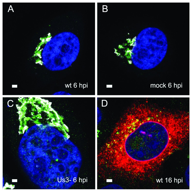
Figure 1. Immunolabelling of the Golgi complex.
Confocal microscopy of the trans-face (green) and the cis-face (white) of Vero cells, immunolabeled with anti TGN46 antibodies (green) and anti GM130 antibodies (white) at 6 h after inoculation with HSV-1 ( A), R7041(ΔUs3) ( C) after mock infection ( B), as well a at 16 hpi with wt HSV-1 ( D) together with immunolabeling of the viral glycoprotein gB (red). The Golgi complex was always in a juxtanuclear position and enormously enlarged after R7041(ΔUs3). Note the enlargement of the nucleus after infection with R7041(ΔUs3). Bars 1 µm.

