Dear Editor
Hemoglobin Lepore (Hb Lepore) is an uncommon hemoglobinopathy with δβ hybrid chains produced by 7.4 kb deletion in the β-globin gene cluster. The fusion gene results in poor of synthesis of δβ hybrid chains resulting in heterozygous β-thalassemia phenotype with mild hypochromic microcytic anemia. Homozygosity for Hb Lepore or compound heterozygosity for β-thalassemia and Hb Lepore results in the phenotype of thalassemia major or thalassemia intermedia [1–3]. Five variants of Hb Lepore have been identified, each characterized by different gene deletion breakpoints. Hb Lepore-Boston-Washington (Hb LBW), (HGVS: NG_000007.3: g.63632_71046del) is the most commonly reported variant, worldwide and in many ethnic groups from Mediterranean countries. Hb Lepore-Baltimore, (HGVS: NG_000007.3: g.63564_70978,) is reported from Southern Europe and Latin America [4–6]. Hb Lepore Hollandia (HGVS: NG_000007.3: g.63290_70702del) is mainly found among individuals of Southern and Southeast Asian origin [7, 8]. Here we report the presence of Hb Lepore (δ22/β50) for the first time in a family belonging to the Baiga tribe from Dindori District, Madhya Pradesh, India.
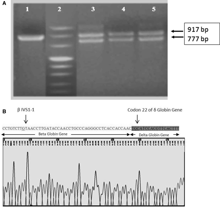
A 1.6 year old male child with complaints of fever and anemia was referred to us for confirmation and diagnosis. Table 1 summarizes the results of hematological and molecular analysis of the propositus and his parents. On cellulose acetate electrophoresis at alkaline pH 8.9, the propositus and his mother showed a slow moving band not corresponding to the controls of HbS/HbD position but marginally trailing the HbS band. Cation exchange high performance liquid chromatography (HPLC) analysis on the VARIANT II hemoglobin testing system using the β-Thalassemia Short Program (Bio-Rad Laboratories, USA) showed HbA2 of 3.2% with a RT of 3.42 min along with HbF and HbA0 of 7.6 and 82.3% respectively suggesting the presence of Hb Lepore (Fig. 1). On family screening his father was found to be normal whereas his mother was found to be slightly anemic. Her HPLC chromatogram showed a split HbA2 peak of 11.1% with a retention time of 3.43 min. Her HbF and HbA0 level was 0.9 and 79.9% respectively (Table 1). Earlier studies suggest that the retention time of Hb Lepore + HbA2 is 3.42–3.43 min. Molecular analysis by PCR showed the presence of the Hb Lepore mutation in heterozygous state in both (Fig. 2a). DNA sequencing confirmed the presence of Hb Lepore Hollandia (Fig. 2b). Alpha genotyping by GAP-PCR showed normal genotype and Xmn1 polymorphism by PCR–RFLP showed. +/− genotype in both mother and the propositus.
Table 1.
Haematological and molecular findings of the propositus and his parents
| Parameters | Propositus | Mother | Father |
|---|---|---|---|
| Hb (g/dl) | 13.4 | 11.8 | 11.2 |
| RBC (× 106/μl) | 5.89 | 5.61 | 3.94 |
| MCV (fl) | 72.7 | 72 | 86.0 |
| MCH (pg) | 22.8 | 21 | 28.4 |
| MCHC (g/dl) | 31.5 | 29.2 | 33.0 |
| RDW (%) | 22.1 | 21 | 16.9 |
| Cellulose acetate electrophoresis at alkaline pH (8.9) | Abnormal slow moving band at S/D position | Abnormal slow moving band at S/D position | Normal |
| HbA0 | 82.3 | 79.9 | 90.7 |
| HbA2 (%) | 3.2 (RT: 3.42 min) | 11.1 (RT: 3.43 min) | 2.9 |
| HbF (%) | 7.6 | 0.9 | 0.9 |
| DNA Mutation | Heterozygous for Hb Lepore Hollandia (δ22/β50) | Heterozygous for Hb Lepore Hollandia (δ22/β50) | Normal |
| α-genotype | αα/αα | αα/αα | αα/αα |
| Xmn I polymorphism | +/− | +/− | ND |
Hb haemoglobin, RBC red blood cell, MCV mean corpuscular volume, MCH mean corpuscular haemoglobin, MCHC mean corpuscular haemoglobin concentration, RDW red cell distribution width, ND not determined
Fig. 1.
HPLC chromatograms of the propositus and his mother
Fig. 2.
a Hb Lepore mutation by PCR analysis. Lane 1—Normal (917 bp). Lane 2—Marker VIII (Roche). Lane 3—Control for Hb lepore. Lanes 4 and 5—Mother and Propositus. b DNA sequencing of the mutant PCR fragment showed a δβ hybrid gene producing the Hb Lepore Hollandia (δ22/β50) genotype with reverse primer
Hemoglobinopathies particularly hemoglobin S and E (HbS, HbE) and β-thalassemia are widely present among the tribal populations [9]. On the other hand, Hb Lepore is a relatively uncommon in India and was first reported by Chouhan et al. in 1971 [10]. In the heterozygous condition, Hb Lepore generally constitutes 6–15% of the total Hb, the HbA2 levels are normal, and most subjects have increased HbF levels. Hb Lepore can be suspected using electrophoresis and HPLC analysis. DNA analysis is needed for confirmation. In the heterozygous condition, Hb Lepore produces the phenotype of heterozygous β thalassemia with slightly raised HbF [11]. Co-inheritance of Hb Lepore with other β-globin variants plays an important role in the clinical presentation.
In the present study, the propositus and his mother were found to be heterozygous for Hb Lepore Hollandia with mild to moderate anemia. The propositus had a low percentage of HbA2 as well as Hb Lepore and elevated HbF as compared to other reported cases. α-thalassemia could be one of the reason for the low level of Hb Lepore, however, in the present case the propositus had a normal α-genotype. The high HbF levels could be due to associated variations in other genes like HBS1L-MYB, KLF, BCL11A and SOX6 [12], however these were not looked into in this family.
This is the first observation of Hb Lepore in any tribal population from India. This tribal family belongs to Baiga primitive tribal group from Dindori District in Madhya Pradesh.
Compliance with Ethical Standards
Conflict of interest
The authors declare no conflict of interest.
Ethical Standards
Study was approved by Institutional Ethics Committee. All procedures performed in the study involving human participants were in accordance with the ethical standards of the institutional research committee and with the 1964 Helsinki declaration and its later amendments or comparable ethical standards.
Informed Consent
Informed consent was obtained from all individual participants included in the study.
Contributor Information
Harsha Lad, Email: harsha.bioch@gmail.com.
Manju Yadav, Email: manjusg87@gmail.com.
Pallavi Mehta, Email: sarthi710@gmail.com.
Purushottam Patel, Email: patelrmrc@rediffmail.com.
Pratibha Sawant, Email: pratibhamsawant@rediffmail.com.
Roshan B. Colah, Email: colahrb@gmail.com
Malay B. Mukherjee, Email: malaybmukherjee@gmail.com
Rajasubramaniam Shanmugam, Email: raja.rmrct@gmail.com.
References
- 1.Bozkurt G, Baysal E, Gu L-H, Huisman THJ. Thalassemia intermedia in two patients with Hb Lepore-β0-thalassemia (Frameshift codon 8,-AA) Hemoglobin. 1994;18(3):247–250. doi: 10.3109/03630269409043627. [DOI] [PubMed] [Google Scholar]
- 2.Chakova L, Spasova M, Genev E. Thalassemia intermedia in an infant. Folia Med. 1998;40(1):84–87. [PubMed] [Google Scholar]
- 3.Sreedharanunni S, Chhabra S, Kaur JH, Bansal D, Sharma P, Das R. β-thalassemia intermedia caused by compound heterozygosity for Hb Lepore-Hollandia and β-thalassemia is rare in the Indian Population. Hemoglobin. 2015;39(5):362–365. doi: 10.3109/03630269.2015.1064004. [DOI] [PubMed] [Google Scholar]
- 4.Labie D, Schroeder WA, Huisman THJ. The amino acid sequence of the δ-β chains of Hemoglobin Lepore-Augusta = Lepore Washington. Biochim Biophys Acta. 1966;127(2):428–437. doi: 10.1016/0304-4165(66)90397-7. [DOI] [PubMed] [Google Scholar]
- 5.Ostertag W, Smith EW. Hemoglobin-Lepore-Baltimore, a third type of a δβ crossover (δ50, β86) Eur J Biochem. 1969;10(2):371–376. doi: 10.1111/j.1432-1033.1969.tb00700.x. [DOI] [PubMed] [Google Scholar]
- 6.Ribeiro ML, Cunha E, Gonçalves P, et al. Hb Lepore-Baltimore (δ68Leu-β84Thr) and Hb Lepore-Washington-Boston (δ87Gln-βIVS-II-8) in central Portugal and Spanish Alta Extremadura. Hum Genet. 1997;99(5):669–673. doi: 10.1007/s004390050426. [DOI] [PubMed] [Google Scholar]
- 7.Barnabas J, Muller CJ. Hemoglobin-Lepore Hollandia. Nature. 1962;194:931–932. doi: 10.1038/194931a0. [DOI] [Google Scholar]
- 8.Viprakasit V, Pung-Amritt P, Suwanthon L, et al. Complex interactions of db hybrid hemoglobin (Hb Lepore-Hollandia) Hb E (β26(G→A)) and α+ thalassemia in a Thai family. Eur J Hematol. 2002;68(2):107–111. doi: 10.1034/j.1600-0609.2002.01637.x. [DOI] [PubMed] [Google Scholar]
- 9.Ghosh K, Colah RB, Mukherjee MB. Haemoglobinopathies in tribal populations of India. Indian J Med Res. 2015;141(5):505–508. doi: 10.4103/0971-5916.159488. [DOI] [PMC free article] [PubMed] [Google Scholar]
- 10.Chouhan D, Sharma R, Valki B, Parekh J. Hemoglobin Lepore in an Indian Family. J Indian Med Assoc. 1971;56:287–290. [PubMed] [Google Scholar]
- 11.Ropero P, Gonzalez FA, Sanchez J, Anguita E, Asenjo S, Del Acro A, Murga MJ, Ramos R, Fernandez C, Villegas A. Identification of Hb Lepore phenotype by HPLC. Haematologica. 1999;84:1081–1084. [PubMed] [Google Scholar]
- 12.Wonkam A, Ngo Bitoungui VJ, Vorster AA, Ramesar R, Cooper RS, Tayo B, et al. Association of variants at BCL11A and HBS1L-MYB with hemoglobin F and hospitalization rates among sickle cell patients in Cameroon. PLoS ONE. 2014;9(3):e92506. doi: 10.1371/journal.pone.0092506. [DOI] [PMC free article] [PubMed] [Google Scholar]


