

Abstract
Fractions obtained from HeLa cell extracts were used to study RNA polymerase III-catalyzed transcription from the human 7SK and mouse U6 RNA promoters in vitro. Although both genes depend on two almost identical core promoter elements (TATA box and PSE), different fractions were required. The 7SK promoter revealed full activity with the phosphocellulose B fraction alone. In contrast, efficient transcription from the U6 promoter depended on the additional presence of the C or D fraction. The analysis of the b1 and b2 subfractions (obtained by DEAE-Sephadex chromatography) revealed that for both promoters the b1 and the phosphocellulose D fraction were mutually interchangeable. However, while both fractions were fully equivalent for the 7SK promoter, the U6 promoter revealed an additional requirement for the C fraction in the presence of the b1 fraction. Since the b1 and the D fractions enclose two different complexes of the TATAbinding protein (TBP), B-TFIID and D-TFIID, our results indicate that functionally these two complexes are responsible for the observed differences in transcription of the 7SK and U6 genes.
The two mammalian genes coding for 7SK RNA and U6 snRNA are unique among the pol III genes (genes transcribed by RNA polymerase III) characterized so far because they are controlled by exclusively gene-external promoters (reviewed in Geiduschek and Tocchini-Valentini, 1988; Murphy et al., 1989; Palmer and Folk, 1990). Both promoters are composed of a core promoter, which is required for efficient transcription in vitro (Murphy et al., 1987; Krüger and Benecke, 1987; Kunkel and Pederson, 1988; Das et al., 1988), and an upstream element. Two functional elements have been identified within these core promoters: a TATA box at position −25, which confers RNA polymerase specificity (Murphy et al., 1987; Mattaj et al., 1988; Lobo and Hernandez, 1989) and a proximal sequence element (PSE) around position −50 (Kunkel and Pederson, 1988; Lobo and Hernandez, 1989), which reveals significant sequence homology to the PSE of the U-snRNA genes transcribed by RNA polymerase II (reviewed in Dahlberg and Lund, 1988). Together, both elements dictate the transcription start site (Goomer and Kunkel, 1992). In addition to the core promoter, efficient expression of both genes in vivo requires the presence of a distal sequence element (DSE). At first, this DSE was considered to be identical in both genes, since in both cases octamer motifs are localized at about the same distance from the core promoter. The functionality of the U6 octamer was supported by deletion analyses (Carbon et al., 1987). Furthermore, in vitro complementation analyses with purified octamer factors seemed to indicate that the same holds true for the slightly variant 7SK octamer (Murphy et al., 1989). However, a detailed mutation/deletion analysis of the octamer-like sequence element of the 7SK DSE revealed that in this case an immediately adjacent CACCC box represents the functional DSE motif (Kleinert et al., 1990), while the octamer-like sequence has no detectable function in vivo (Kleinert et al, 1991). These results provided the first evidence that the two seemingly identical 7SK and U6 promoters are regulated differently.
By structure and sequence element composition, the 7SK and U6 promoters strongly resemble classical pol II promoters. Other pol III promoters, such as those of the human 7SL (Bredow et al., 1990) or Epstein-Barr virus-encoded small RNA (EBER; Howe and Shu, 1989) genes, were also found to contain transcription factor binding sites well known from the pol II system. Moreover, recent analyses of pol III transcription have revealed that the promoters of these two eukaryotic transcription systems are generally very similar. Consequently, it is important to ask whether both transcription systems also use identical transcription factors. In this context, it is interesting to note that, as a general transcription factor, the TATA-binding protein (TBP) is engaged in all three eukaryotic transcription systems (reviewed in Sharp 1992). Moreover, several authors recently described an intimate association of TBP with the basic pol III transcription factor TFIIIB (Taggert et al., 1992; Lobo et al., 1992; White and Jackson, 1992; Kassavetis et al., 1992; Simmen et al., 1992), as reviewed by Rigby (1993).
In our attempts to identify and characterize the protein factors required for 7SK RNA transcription in vitro, cell extracts were fractionated by conventional procedures. In contrast to all other genes studied so far, maximal in vitro transcription of the human 7SK gene required only a single phosphocellulose fraction, i.e., the B fraction In contrast, the U6 snRNA promoter, which looks so much alike, revealed only basal level activity with the B fraction alone. Together, the experiments reported here demonstrate that the activity of the seemingly identical 7SK and U6 core promoters depends on the formation of different initiation complexes, depending on which of the two TBP complexes is involved.
Materials and methods
Templates
The human 7SK RNA gene (Krüger and Benecke, 1987) and the mouse U6 snRNA gene (kindly provided by Dr. Ram Reddy, Houston; Das et al., 1988) have been described previously. In order to preclude endogenous RNA, hybrid genes were used to detect specific transcripts by S1 nuclease protection analysis. The 7SK construct has been described in detail by Kleinert et al. (1988). Briefly, the 7SK promoter (−245 to −3) was fused to the 189 bp EcoR I–EcoR V fragment of the pAT 153 vector supplemented with the 7SK termination region (Hind III–EcoR I fragment containing the last 62 nt of the 7SK RNA sequence). By filling the Cla I site of the pAT 153 fragment with Klenow enzyme, a 3′-labeled DNA was obtained, which gave rise to a 225 nt DNA sequence protected against S1 nuclease by specific transcripts. The U6 construct was generated by fusing the 5′ end of the mouse U6 gene (−253 to +60) via a short polylinker sequence to a 91 bp fragment of the VA1 gene supplemented with the 7SK termination region. In the SI analysis, a fragment 3′-labeled at the Xba I site of the polylinker sequence gave rise to a specific band of 158 nt. A DSE deletion mutant of the 7SK promoter (−111 mutant) was obtained by Bal 31 exonuclease digestion (Kleinert et al., 1988). The corresponding 5′ deletion of the U6 promoter was constructed by deletion of the EcoR I (−150)–Cla I (−253) fragment of clone pUEA-244 (Das et al., 1988). A human 7SL RNA gene construct was used for normalization of 7SK and U6 transcripts obtained upon transfection into intact cells. This construct was obtained by fusing the 7SK termination region (see above) to the Tth111 I site (+292) of the 7SL coding region (the Hind III and the Tth111 I sites were filled with the Klenow enzyme).
In vitro transcription
S100 extracts (15 mg/ml of protein) were prepared from HeLa cells according to Weil et al. (1979). Subsequent phosphocellulose chromatography (Whatman P11) resulted in the A, B, C, and D fractions described by Segall et al. (1980). In vitro transcription assays with 1 μg of plasmid DNA template and 20 μl of extract (or 5 μl subfraction) in a total volume of 50 μl were performed under the optimal conditions described for 7SK RNA (Kleinert et al., 1988) or U6 snRNA (Das et al., 1988), respectively. Briefly, 7SK is transcribed in the presence of 70 mM KC1, whereas U6 is optimal at 100 mM KC1. Electrophoretic analysis in 8 M urea-10% polyacrylamide gels of transcripts obtained in the presence of 0.5 μg/ml of α-amanitin has been described in detail by Kleinert et al. (1990). Extracts from HeLa cell nuclei were prepared as described by Dignam et al. (1983).
Transfections and S1 nuclease analysis of transcripts
7SK and U6 wild-type or DSE deletion mutant gene constructs were transfected into hepatoma (HepG2) monolayer cells by the DNA-calcium phosphate co-precipitation technique (Graham and van der Eb, 1973) using 25 μg of plasmid DNA/9-cm plate. S1 nuclease protection analysis after hybridization with labeled DNA fragments and the analysis of reference transcripts for normalization have been described in detail by Kleinert et al. (1991).
Western blot analysis
50 μg of protein from extracts or subfractions were separated in SDS-Laemmli gels (10% acrylamide) and transferred electrophoretically (2.5 V/cm; 100 mA; 16 hours) to a nitrocellulose membrane (BA85, Schleicher and Schuell). Membranes were blocked with 5% nonfatty milk powder in 50 mM Tris-HCl, pH 7.9 for 1 hour at room temperature. The blot was incubated with a 1:2000 dilution of the 3G3 antibody (kindly provided by Dr. J.-M. Egly, Strasbourg) in phosphate-buffered saline including 2.5% nonfatty milk powder and 0.05% Tween 20. Detection of TBP was with the anti-mouse IgG (Promega) and the enhanced chemiluminescence system (Amersham), followed by a 30 second exposure to Fuji X-ray film.
Results
In vitro transcription of the 7SK and U6 RNA genes does not require the distal sequence element
RNA polymerase III (pol III)-catalyzed transcription of the human 7SK RNA and the mouse U6 snRNA gene is controlled by exclusively gene-external promoters (see Introduction). As is evident from the sequence comparison in Figure 1, these promoters consist of a core promoter with a TATA element at position −28 and a proximal sequence element (PSE) around position −60. In addition, an upstream modulating element (i.e., the DSE) is located about 160 bp upstream from the core promoter at position −220. Although this upstream region of both genes contains an octamer motif only the U6 DSE function is mediated by the octamer element, whereas functionality of the 7SK DSE depends exclusively on the integrity of the CACCC box (Kleinert et al., 1990).
Figure 1.
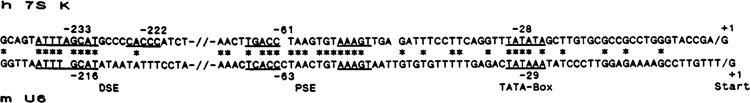
Schematic representation of the known functional promoter elements of the human 7SK (h 7SK) and the mouse U6 snRNA (m U6) genes. Consensus sequences are underlined, nucleotides conserved between the two promoters are indicated by asterisks, and +1 indicates the transcription start site.
The activity of the 7SK and U6 promoters in vivo is strictly dependent on the presence of the DSE. This is evident from S1 nuclease protection analyses of transcripts obtained with DSE deletion mutants in comparison to the wild-type promoter constructs upon transfection into intact cells. As is seen from the comparison of lanes 1 and 2 of Figure 2A, the DSE deletion mutant of the 7SK promoter no longer supported the synthesis of the specific transcript (observed as a protected fragment of 225 nt) obtained with the wild-type promoter (lane 1). The same was true for the DSE deletion mutant (lane 4) of the mouse U6 promoter (protected fragment of 158 nt, lane 3). The prominent band of 305 nt observed in lanes 1 to 4 of Figure 2A provided a normalization standard for transfection efficiency and represented labeled DNA protected against SI nuclease digestion by transcripts from a co-transfected 7SL RNA gene construct.
Figure 2.

Expression of 7SK and U6 constructs with and without the distal sequence element (DSE). A. S1 nuclease protection analysis of transcripts obtained in vivo upon transfection of hepatoma cells with the 7SK (lanes 1, 2) or the U6 (lanes 3, 4) constructs. Lanes 1 and 3 show transcripts from the full promoter, whereas lanes 2 and 4 represent the activity of DSE deletion mutants. The prominent band of 305 nt represents a normalization standard of a co-transfected 7SL RNA gene construct. B. In vitro transcription with HeLa S100 extract of the 7SK (lanes 1, 2) or U6 RNA wild-type genes (lanes 3, 4), in the presence (lanes 1, 3) or absence (lanes 2, 4) of the DSE. In contrast to part A, RNA labeled in vitro was directly analyzed by gel electrophoresis, m: marker of labeled DNA fragments with the respective lengths indicated at the left and right.
A different picture was obtained if both promoters were analyzed for transcription in vitro. In this case, deletion of the DSE region did not show any effect on promoter activity. This is evident from the direct analysis of RNA transcribed from both wild-type genes in vitro (Fig. 2B), with lanes 1 and 2 representing the 7SK RNA (331 nt) and lanes 3 and 4 the U6 RNA (106 nt), respectively. The observed heterogeneity of in vitro synthesized U6 snRNA is reproducibly obtained and has been observed by others before (Das et al., 1988; Simmen et al., 1991); that is why in the experiments described below the constructs of Figure 2A were used as templates and the in vitro synthesized RNA analyzed by SI nuclease protection. Thus, upon removal of the DSE, in vitro transcription of both deletion mutants was as accurate and efficient as that observed with the 7SK and U6 wild-type genes under our conditions.
Different combinations of S100 subfractions are necessary for in vitro transcription from the 7SK and U6 promoters
In order to purify and characterize transcription factors essential for expression of both genes, HeLa cell S100 extracts were fractionated and individual fractions tested for their ability to support efficient transcription in vitro. So far, most in vitro transcription studies with the pol III genes have been performed with extract subfractions obtained by phosphocellulose chromatography. In fact, the entire nomenclature of eukaryotic transcription factors originally emerged from their association with the four step-eluted fractions A, B, C, and D obtained from phosphocellulose at 0.1 M, 0.35 M, 0.6 M, and 1.0 M KC1, respectively (Segall et al., 1980).
Therefore, we studied in vitro transcription from the 7SK and U6 RNA promoters by using these four phosphocellulose fractions. In these experiments, transcripts obtained from the gene constructs described in Materials and Methods were analyzed in SI nuclease protection assays, as in Figure 2A. As is evident from Figure 3A, extract fractions were able to support 7SK transcription to the same extent as the complete S100 extract (lane 1; protected band of 225 nt). From the results shown in lanes 5 (A + B), 7 (B + C), and 8 (A + B + C) of Figure 3A, it is obvious that the B fraction is always required for efficient transcription from the 7SK promoter. To our surprise, however, we observed that the same efficiency was obtained with the B fraction alone (lane 3). In contrast, neither the A fraction (lane 2) nor the C fraction (lane 4) alone was able to support active transcription of the 7SK gene construct. Since in any combination, addition of these two fractions failed to stimulate further the activity already obtained with the B fraction alone, we conclude that all transcription factors required for maximal expression of the 7SK gene in vitro are associated with the phosphocellulose B fraction. It should be noted that this is the first example of a single phosphocellulose fraction being sufficient to transcribe a eukaryotic gene in vitro, both accurately and with full efficiency.
Figure 3.

In vitro transcription of constructs with extract fractions. A. The 7SK construct was transcribed in the presence of total S100 extract (lane 1) or with individual phosphocellulose fractions and combinations thereof, as indicated on top. In contrast to Figure 2B, in this and all further in vitro RNA synthesis experiments, analysis of transcripts was by the S1 nuclease protection method using the same labeled DNA fragments as in the in vivo experiment of Figure 2A. B. The same fractions as in A were used for transcription from the U6 promoter, and transcript analysis was by the same method. The size markers were as in Figure 2.
Since one could not exclude the possibility that our conditions of extract preparation and/or phosphocellulose chromatography might be slightly different from those described by others, we used the same extract fractions (and combinations thereof) to transcribe the mouse U6 snRNA gene in parallel. The results (shown in part B of Figure 3) revealed that in addition to the complete S100 system (lane 1), only two combinations, the B + C fractions or the A + B + C fractions, were able to support active transcription in vitro from the U6 promoter, as indicated by a protected band of 158 nt. The other combinations, A + B and A + C, were found to be inactive (Fig. 3B, lanes 5 and 6). Likewise, the three individual fractions A, B, and C (lanes 2, 3, 4) revealed no activity with the U6 promoter. This result is in full agreement with data obtained before with the human U6 snRNA gene (Lobo et al., 1991). Therefore, in contrast to the 7SK promoter, the activity of the U6 snRNA promoter requires two phosphocellulose fractions for in vitro transcription and consequently depends on a different factor combination.
The data shown in Figure 2 demonstrates that S100-mediated in vitro transcription of these two pol III genes depends on the core promoter only. Therefore, one would assume that the observed difference in factor requirement is related to the proximal elements only. However, one cannot exclude the possibility that the use of extract subfractions created specific conditions under which the distal sequence elements, which were demonstrated to be different in the two genes (Kleinert et al., 1990), now acquire a certain functionality in modulating the in vitro transcription activity. For this reason, upstream deletion mutants were also transcribed with the phosphocellulose fractions. This analysis (Fig. 4) was performed, however, with only the B fraction or the B + C combination. In agreement with the results shown in Figure 3, transcription from the 5′ deleted (−111) 7SK promoter stayed the same with B fraction alone (Fig. 4, lane 1), compared to the B + C combination (lane 2). In contrast, transcription from the corresponding U6 deletion (−150) again was not obtained with the B fraction alone (Fig. 4, lane 3) but required the additional presence of the C fraction (lane 4). This result confirms our conclusion that the observed difference in transcription initiation of both genes is indeed related to their respective core promoters.
Figure 4.

Transcription analysis from the 7SK and U6 core promoters either separately (lanes 1–4) or simultaneously in the same reaction (lanes 5–8). Transcription was either with the phosphocellulose B fraction alone (lanes 1, 3, 5, 7) or with the B + C combination (lanes 2, 4, 6, 8). Templates used were 1.0 μg of the 7SK construct (lanes 1, 2) or the U6 construct (lanes 3, 4). In lanes 5 to 8, 0.5 μg of both constructs were transcribed simultaneously, under conditions optimal either for 7SK (lanes 5, 6) or for U6 (lanes 7, 8) RNA transcription in vitro. Analysis of transcripts with S1 nuclease was as above.
In vitro transcription of the 7SK and U6 RNA genes requires slightly different ionic conditions, as outlined in Materials and Methods. That is why most analyses were performed separately with each of the two genes. To determine whether the simultaneous presence of both promoters might alter the observed factor requirements—for example, by internal competition—the 7SK and U6 constructs were transcribed simultaneously in the same reaction (Fig. 4, lanes 5–8). For this, 0.5 μg of each gene construct was analyzed with the B fraction alone (lanes 5 and 7) or the B + C combination (lanes 6 and 8) either under optimal 7SK conditions (lanes 5 and 6) or those optimal for U6 transcription (lanes 7 and 8). The analysis (by S1 nuclease) of individual transcripts obtained under these conditions confirmed that the selectivity for different factor combinations was not influenced by the presence of the other core promoter.
The exchangeability for U6 transcription of the C and D fractions is not related to the presence of the TATA-binding protein (TBP)
As described above, two well-established sequence elements have been identified within the 7SK (Murphy et al., 1987) and U6 (Kunkel, 1991) core promoters: the TATA box at −25 and the PSE around −50. In order to get a first indication of which of these two elements might be responsible for the observed difference in transcription factor requirements between these two pol III genes, we analyzed the in vitro transcription from the U6 promoter with different factor combinations as before. However, this experiment aimed at a putative involvement of the well-characterized TFIID complex, associated with the phosphocellulose D fraction (eluted at 1.0 M KC1). As shown in Figure 5, the B, C, and D fractions alone (lanes 2, 3, and 4, respectively) were found inactive again. Yet, in agreement with the requirements observed for the human U6 promoter (Lobo et al., 1991), comparison of lanes 5 and 6 of Figure 5 revealed that the activity of the mouse U6 promoter observed with the B + C combination (lane 5) was also obtained with the B + D combination (lane 6). In contrast, the C + D fractions were not active (lane 7) with this promoter. Since the B + C + D combination did not increase further the in vitro transcription activity (Fig. 5, lane 8) over that observed with either the B + C or the B + D fractions, this data seemed to indicate that the C or D fractions are functionally equivalent in complementation of the B fraction for efficient transcription from the mouse U6 promoter. Therefore, one might conclude that both fractions contain the same functional factor component—for example, the TBP complex (TATA-binding protein). This conclusion seemed to be supported by the finding that for complementation of the B fraction, the C or D fractions could each be replaced by recombinant TBP (Lobo et al., 1991). However, the analysis performed by Timmers and Sharp (1991) with a TBP antibody revealed that no detectable amounts of TBP were associated with the C fraction. This result was confirmed by a Western blot analysis of our individual fractions (Fig. 6), using a monoclonal TBP antibody (kindly provided by Dr. J.-M. Egly, Strasbourg). The results presented in Figure 6, lanes 1 to 6, demonstrate that in addition to the unfractionated extracts (nuclear extract, lane 1; S100 extract, lane 6) only the B and D fractions (lanes 3 and 5, respectively) contained detectable amounts of TBP, indicating that the C and D fraction are not at all equivalent with respect to the TATA-binding transcription factor.
Figure 5.

In vitro transcription from the U6 promoter with phosphocellulose fractions, including the D fraction. Transcription with full extract (lane 1) or with the individual fractions and combinations indicated on top was as in Figure 3B.
Figure 6.

Western blot analysis with a monoclonal TBP antibody of extract fractions. Proteins (50 μg each) of nuclear extract (lane 1), S100 extract (lane 6), or the fractions indicated on top were separated by gel electrophoresis and electroblotted onto a nitrocellulose membrane. The blot was incubated with anti-TBP, and specific binding was demonstrated by chemiluminescence, using a second antibody. Exposure was for 30 seconds. A longer exposure (2 minutes, not shown) revealed that no TBP was detectable in either the A or the C fraction.
The TBP complexes associated with the b1 or the D fraction are not functionally equivalent
In this context it is interesting to note that two structurally different complexes of TBP have recently been characterized (Timmers and Sharp, 1991; Timmers et al., 1992), one being associated with the D fraction (D-TFIID) and the other found within the B fraction (B-TFIID). Therefore, we wanted to analyze whether or not these two complexes are functionally equivalent in conjunction with the two 7SK and U6 core promoters. For this, the phosphocellulose B fraction was further fractionated by DEAE-Sephadex-A25 chromatography. Subsequently, the flow-through fraction (b1, 50 mM ammonium sulfate, containing the B-TFIID form; Timmers and Sharp, 1991) and the eluted fraction b2 (obtained at 200 mM ammonium sulfate, containing TFIIIB; Waldschmidt et al., 1988) were assayed for their capacity to support 7SK transcription in vitro in the presence of exogenous RNA polymerase III. As shown in Figure 7A, the combination of b1 plus b2 fraction (lane 2) revealed a level of activity comparable to that of S100 extract (lane 1). In contrast, neither the b1 fraction (Fig. 7A, lane 3) nor the b2 fraction (lane 4) alone supported active transcription when supplemented with exogenous RNA polymerase III. However, the inactive b2 fraction acquired full activity when supplemented with the phosphocellulose D fraction, containing the D-TFIID complex (Fig. 7A, lane 5). This indicates that transcription initiation from the 7SK core promoter in vitro can be equally mediated by both the B-TFIID and the D-TFIID complex.
Figure 7.

In vitro transcription with extract subfractions obtained by DEAE-Sephadex chromatography. The phosphocellulose B fraction was subjected to DEAE-Sephadex chromatography resulting in subfractions b1 (flow-through at 50 mM ammonium sulfate) and b2 (eluted at 0.2 M ammonium sulfate). A. Comparison of 7SK transcription in S100 extract (lane 1) with the sub-fractions and combinations indicated on top. B. Same as A, but with the U6 construct as template and the fractions again indicated on top. Analysis of transcripts was as in Figure 3.
On the other hand, the inactivity of the U6 gene with the phosphocellulose B fraction alone, which contains the B-TFIID complex (Timmers and Sharp, 1991), the general pol III transcription factor TFIIIB (Segall et al., 1980; Shastry et al., 1982; Kassavetis et al., 1990), the PSE-binding protein (PBP; Simmen et al., 1992), and RNA polymerase III, indicated that—under identical conditions—the U6 promoter is not able to cooperate with the B-TFIID complex. This is demonstrated by the experiment shown in Figure 7B. In contrast to the 7SK promoter, only basal level activity was obtained with the b1 plus b2 combination (Fig. 7B, lane 5). Comparable to Figure 2, full activity of the U6 promoter again was observed only upon addition of the C fraction to b1 + b2 (lane 2). The slight increase over basal level transcription of the b2 + C combination (Fig. 7B, lane 3) is in agreement with the very small amount of B-TFIID detected in lane 8 of Figure 6. However, the C fraction was not required any more if the b1 fraction was replaced by the D fraction, resulting in a b2 + D combination (Fig. 7B, lane 7). Under these conditions, that is to say, only in the presence of the D fraction, the factor requirement was the same for the 7SK and U6 promoter. In summary, as opposed to the 7SK promoter, in the case of the U6 promoter, no simple exchangeability was observed between the b1 fraction and the D fraction. We conclude that in case of the U6 promoter two different modes for formation of a functional initiation complex can be followed, depending on the TBP complex used. If the B-TFIID complex of the bi fraction is involved, the U6 promoter depends on the additional presence of a C fraction polypeptide that remains to be identified. In contrast, this compound is not required if the D-TFIID complex is involved.
Together, these in vitro transcription analyses, performed with the two pol III promoters 7SK and U6, provide strong support for a functional difference between the two recently isolated TBP complexes, B-TFIID and D-TFIID, which were characterized by Timmers et al. (1992) and which are associated with the b1 and the D fractions, respectively, as also shown in lanes 7 and 5 of Figure 6.
Discussion
In recent years, analyses of several genes transcribed by RNA polymerase III (pol III genes) have significantly contributed to our under-standing of this process and of eukaryotic transcription in general. In classical pol III genes (such as 5S and tRNA), transcription initiation is controlled by promoter elements located downstream (3′) from the transcription start site. In contrast, analysis of U6, 7SK, 7SL, and EBER RNA genes (reviewed in Murphy et al., 1989) has provided firm evidence that pol III promoters, like those of the pol II and pol I transcription systems, may also be localized either partly (7SL, EBER RNA genes) or exclusively (7SK, U6 genes) upstream from the start site Furthermore, a significant number of promoter elements identified within the upstream region of these pol III genes was found to be similar, or even identical, to those of genes transcribed by RNA polymerase II. These results support a much closer evolutionary relationship among the three eukaryotic transcription machineries than originally anticipated. This notion was substantiated by the recent finding that at least one factor, the TBP, is apparently involved in transcription initiation by all three RNA polymerases (reviewed in Sharp, 1992). Together, these findings seemed to favor a rather simple model for the mechanism of transcription initiation in eukaryotes. However, the complex pattern of differential gene expression requires an additional level of complexity. Most likely, this level is provided by the existence of extended families of transcription factors, recognizing identical, or at least very similar, sequence elements. In addition, the variation in complex formation of individual transcription factors, such as the TBP with TAFs (TBP-associated factors) as observed in the B-TFIID and D-TFIID complexes, further increases the level of diversity in gene-specific regulation.
The results described here demonstrate that two seemingly identical core promoters allow the formation of functionally different transcription factor complexes. This is in strong support of protein–protein interactions contributing to flexibility in initiation complex formation. Such flexibility should be particularly important for a general transcription factor, such as TBP, which is involved in different RNA polymerase systems and which therefore has to provide the capacity to interact with a large number of different auxiliary factors (reviewed in Greenblatt, 1992).
Initially, the interpretation of the results presented in Figure 3 appeared to be rather simple: two different promoters, although looking almost identical with respect to sequence and structural arrangement of their two functional elements (PSE and TATA box), nevertheless depend on at least one different transcription factor. In case of U6, this gene-specific factor appeared to be associated with the phosphocellulose C fraction. Since for U6 transcription, the C fraction could be replaced by the D fraction (Fig. 5), one might argue that the U6-specific factor was also present within the phosphocellulose D fraction. This assumption in turn would support the conclusion that the two fractions are functionally equivalent.
However, this simple picture was obscured when the phosphocellulose B fraction was further fractionated on DEAE-Sephadex A 25, resulting in the subfractions b1 and b2. As expected, the b2 fraction, which is known to contain the transcription factor TFIIIB (Waldschmidt et al., 1988), was essential in all combinations tested. This is in agreement with the notion that TFIIIB represents the general transcription factor of the pol III system (Kassavetis et al., 1990). Interestingly, several recent publications have stated that a close association of TFIIIB with TBP complexes is required for pol III transcription (Taggert et al., 1992; Lobo et al., 1992; White and jackson, 1992; Kassavetis et al., 1992; Simmen et al., 1992). These data suggest that B-TFIID may be part of TFIIIB but is not identical with it (discussed in detail by Rigby, 1993).
In contrast to the results presented in Figure 5, however, supplementation of the b2 fraction with either the C or D fractions demonstrated that these two phosphocellulose fractions were not fully equivalent (compare lanes 3 and 7 of Fig. 7B) when analyzed with the U6 promoter. On the other hand, the D fraction in turn was able to replace the b1 fraction for efficient transcription from the 7SK promoter (Fig. 7A, lanes 2 and 5). Only in this case the bi fraction was functionally equivalent to the phosphocellulose D fraction in supplementing the b2 fraction for efficient transcription. In contrast, if for transcription from the U6 promoter the b2 fraction was supplemented with either the b1 or the D fraction, no simple exchangeability between the two fractions was observed anymore (compare Fig. 7B, lanes 5 and 7). For full activity, supplementation of the b2 subfraction with b1 now required the additional presence of the C fraction (Fig. 7B, lanes 2 and 5), which was dispensable when the b2 fraction was supplemented with the D fraction alone (lane 7).
Our interpretation of this complex pattern is as follows: the b1 and D fractions have been shown to contain the TATA-binding protein (Fig. 6, lanes 5 and 7; Timmers and Sharp, 1991), forming two complexes which differ in structure and function (Timmers et al., 1992). By using TBP antibodies, it was also shown that the C fraction does not contain detectable amounts of TBP (Timmers and Sharp, 1991; Fig. 6 of this article). The two different TBP complexes can be separated by phosphocellulose chromatography, as described by Timmers et al. (1992). One complex is found in the B fraction (therefore B-TFIID) and the other one in the D fraction (D-TFIID). Furthermore, upon DEAE-Sephadex fractionation, the B-TFIID complex is associated with the b1 subfraction. Now, our experiments demonstrate that the two pol III promoters studied here can obviously follow two alternative paths to form a functional initiation complex in vitro. Although the complexes have not yet been further purified, our data indicate that both promoters select either the B-TFIID (b1 fraction) or the D-TFIID (D fraction) complex for initiation. Only for the 7SK promoter are both TBP complexes fully equivalent. In contrast, the U6 promoter discriminates between these two TBP complexes and requires an additional factor for efficient use of B-TFIID. This additional factor is provided by the C fraction. We would like to emphasize, however, that these alternatives observed in vitro do not exclude the possibility that both genes select the same TBP complex under in vivo conditions.
Our conclusion that the active compound responsible for the observed differences in U6 transcription is identical with the B-TFIID complex of the b1 fraction is strongly supported by the result in lane 3 of Figure 7B. We expected the b2 + C combination to be inactive. However, although much less active than the b1 + b2 + C combination (lane 2), the b2 + C combination showed a significant increase over the basal level activity of lanes 4 and 5. Since the Western blot analysis of Fig. 6 (lane 8) revealed a slight contamination of the b2 fraction with TBP, this correlation provides strong evidence that the limiting factor of the b2 + C combination was the TBP complex.
At present we cannot exclude the possibility that for the alternate use of the two TBP complexes different PSE factors may also be required, instead of the postulated U6-specific additional C fraction protein. The PSE factor was purified from the C fraction (Simmen et al., 1992), but significant amounts are also associated with the B fraction (K. H. Seifart, personal communication). This is supported by our finding that the additional presence of the C fraction (which is not required at all for 7SK) did not further stimulate transcription from the 7SK promoter by the B fraction, indicating that the PSE factor is not present within the B fraction in limiting amounts.
In summary, the factor combinations described point to a functional difference in pol III transcription of the two active TBP complexes. It remains to be seen whether or not this effect can be attributed directly to different TAFs. Alternatively, our results might reflect a selective interaction with different bridging proteins and/or different PSE factors of the two TBP complexes when establishing the TATA-PSE cooperation for the initiation reaction.
Acknowledgments
We wish to thank Dr. I. Grummt (Heidelberg) for help with the Western blot analysis and Dr. J.-M. Egly (Strasbourg) for providing the TBP antibody.
This work was supported by a grant from the Deutsche Forschungsgemeinschaft to B.-J. Benecke.
The costs of publishing this article were defrayed in part by the payment of page charges. This article must therefore be hereby marked “advertisement” in accordance with 18 USC Section 1734 solely to indicate this fact.
Sebastian Bredow is currently in the Department of Physiology and Biophysics, College of Medicine, University of Tennessee, Memphis, TN 38163.
References
- Bredow S., Kleinert H., and Benecke B.-J. (1990), Gene 86, 217–225. [DOI] [PubMed] [Google Scholar]
- Carbon P., Murgo S., Ebel J.-P., Krol A., Tebb G., and Mattaj I. W. (1987), Cell 51, 71–79. [DOI] [PubMed] [Google Scholar]
- Dahlberg J. E. and Lund E. (1988), in Structure and Function of Major and Minor Small Nuclear Ribonucleoprotein Particles (Birnstiel M. L., ed.), Springer Verlag, Berlin, pp. 38–70. [Google Scholar]
- Das G., Henning D., Wright D., and Reddy R. (1988), EMBO J 7, 503–512. [DOI] [PMC free article] [PubMed] [Google Scholar]
- Dignam J. D., Lebovitz R. M., and Roeder R. G. (1983), Nucleic Acids Res 11, 1474–1489. [DOI] [PMC free article] [PubMed] [Google Scholar]
- Geiduschek E. P. and Tocchini-Valentini G. P. (1988), Annu Rev Biochem 57, 873–914. [DOI] [PubMed] [Google Scholar]
- Goomer R. S. and Kunkel G. (1992), Nucleic Acids Res 18, 4903–4912. [DOI] [PMC free article] [PubMed] [Google Scholar]
- Graham F. L. and van der Eb A. J. (1973), Virology 52, 456–457. [DOI] [PubMed] [Google Scholar]
- Greenblatt J. (1992), Nature 360, 16–17. [DOI] [PubMed] [Google Scholar]
- Howe J. G. and Shu M.-D. (1989), Cell 57, 825–834. [DOI] [PubMed] [Google Scholar]
- Kassavetis G. A., Braun B. R., Nguyen L. H., and Geiduschek E. P. (1990), Cell 60, 235–245. [DOI] [PubMed] [Google Scholar]
- Kassavetis G. A., Joazeiro C. A. P., Pisano M., Geiduschek E. P., Colbert T., Hahn S., and Blanco J. A. (1992), Cell 71, 1055–1064. [DOI] [PubMed] [Google Scholar]
- Kleinert H. and Benecke B.-J. (1988), Nucleic Acids Res 16, 1319–1331. [DOI] [PMC free article] [PubMed] [Google Scholar]
- Kleinert H., Bredow S., and Benecke B.-J. (1990), EMBO J 9, 711–718. [DOI] [PMC free article] [PubMed] [Google Scholar]
- Kleinert H., Assert R., and Benecke B.-J. (1991), J Biol Chem 266, 23872–23877. [PubMed] [Google Scholar]
- Krüger W. and Benecke B.-J. (1987), J Mol Biol 195, 31–41. [DOI] [PubMed] [Google Scholar]
- Kunkel G. and Pederson T. (1988), Genes Dev 2, 196–204. [DOI] [PubMed] [Google Scholar]
- Kunkel G. (1991), Biochim Biophys Acta 1088, 1–9. [DOI] [PubMed] [Google Scholar]
- Lobo S. M. and Hernandez N. (1989), Cell 58, 55–67. [DOI] [PubMed] [Google Scholar]
- Lobo S. M., Lister J., Sullivan M., and Hernandez N. (1991), Genes Dev 5, 1477–1489. [DOI] [PubMed] [Google Scholar]
- Lobo S. M., Tanaka M., Sullivan M. L., and Hernandez N. (1992), Cell 71, 1029–1040. [DOI] [PubMed] [Google Scholar]
- Mattaj I. W., Dathan N. A., Parry H. D., Carbon P., and Krol A. (1988), Cell 55, 435–442. [DOI] [PubMed] [Google Scholar]
- Murphy S., Di Liegro C., and Melli M. (1987), Cell 51, 81–87. [DOI] [PubMed] [Google Scholar]
- Murphy S., Moorefield B., and Pieler T. (1989), Trends Genet 5, 122–126. [DOI] [PubMed] [Google Scholar]
- Murphy S., Pierani A., Scheidereit C., Melli M., and Roeder R. G. (1989), Cell 59, 1071–1080. [DOI] [PubMed] [Google Scholar]
- Palmer J. M. and Folk W. A. (1990), Trends Biochem Sci 15, 300–304. [DOI] [PubMed] [Google Scholar]
- Rigby P. W. (1993), Cell 72, 7–10. [DOI] [PubMed] [Google Scholar]
- Segall J., Matsui T., and Roeder R. G. (1980), J Biol Chem 255, 11986–11991. [PubMed] [Google Scholar]
- Shastry B. S., Ng S. Y., and Roeder R. G. (1982), J Biol Chem 257, 12979–12986. [PubMed] [Google Scholar]
- Simmen K. A., Bernués J., Parry H. D., Stunnenberg H. G., Berkenstam A., Cavallini B., Egly J.-M., and Mattaj I. W. (1991), EMBO J 10, 1853–1862. [DOI] [PMC free article] [PubMed] [Google Scholar]
- Simmen K. A., Bernués J., Lewis J. D., and Mattaj I. D. (1992), Nucleic Acids Res 20, 5889–5898. [DOI] [PMC free article] [PubMed] [Google Scholar]
- Taggert A. K. P., Fisher T. S., and Pugh B. F. (1992), Cell 71, 1015–1028. [DOI] [PubMed] [Google Scholar]
- Timmers H. Th. M. and Sharp P. A. (1991), Genes Dev 5, 1946–1956. [DOI] [PubMed] [Google Scholar]
- Timmers H. Th. M., Meyers R. E., and Sharp P. A. (1992), Proc Natl Acad Sci USA 89, 8140–8144. [DOI] [PMC free article] [PubMed] [Google Scholar]
- Waldschmidt R., Jahn D., and Seifart K. H. (1988), J Biol Chem 263, 13350–13356. [PubMed] [Google Scholar]
- Weil P. A., Segall J. H., Harris B., Ng S. Y., and Roeder R. G. (1979), J Biol Chem 254, 6163–6173. [PubMed] [Google Scholar]
- White R. J. and Jackson S. P. (1992), Cell 71, 1041–1053. [DOI] [PubMed] [Google Scholar]