Abstract
Candida albicans is one of the most common causes of hospital-acquired urinary tract infections (UTIs). However, azoles are poorly active against biofilms, echinocandins do not achieve clinically useful urinary concentrations, and amphotericin B exhibits severe toxicities. Thus, novel strategies are needed to prevent Candida UTIs, which are often associated with urinary catheter biofilms. We previously demonstrated that cranberry-derived proanthocyanidins (PACs) prevent C. albicans biofilm formation in an in vitro urinary model. To elucidate functional pathways unique to urinary biofilm development and PAC inhibition, we investigated the transcriptome of C. albicans in artificial urine (AU), with and without PACs. C. albicans biofilm and planktonic cells were cultivated with or without PACs. Genome-wide expression analysis was performed by RNA sequencing. Differentially expressed genes were determined using DESeq2 software; pathway analysis was performed using Cytoscape. Approximately 2,341 of 6,444 total genes were significantly expressed in biofilm relative to planktonic cells. Functional pathway analysis revealed that genes involved in filamentation, adhesion, drug response and transport were up-regulated in urinary biofilms. Genes involved in carbon and nitrogen metabolism and nutrient response were down-regulated. In PAC-treated urinary biofilms compared to untreated control biofilms, 557 of 6,444 genes had significant changes in gene expression. Genes downregulated in PAC-treated biofilms were implicated in iron starvation and adhesion pathways. Although urinary biofilms share key features with biofilms formed in other environments, many genes are uniquely expressed in urinary biofilms. Cranberry-derived PACs interfere with the expression of iron acquisition and adhesion genes within urinary biofilms.
Introduction
Candida albicans and related species are the third leading cause of hospital-associated urinary tract infections (UTIs), and are a marker of increased mortality and healthcare costs [1–4]. Candida UTIs disproportionately impact ICU patients, burn patients, neonates, and those with immunosuppression, malignancy, diabetes, and urologic abnormalities [5,6]. Most cases are associated with the use of urinary catheters [7]. Of note, the Centers for Medicare and Medicaid Services are no longer reimbursing healthcare systems for hospital-acquired UTIs. Although three major classes of antifungal therapy are available, each has important limitations. Echinocandins do not achieve effective urinary concentrations, lipid amphotericin causes substantial renal and other toxicities, and fluconazole can be limited by drug resistance, particularly within biofilms. Furthermore, urinary concentrations of posaconazole, voriconazole, and itraconazole are very low [8]. Our understanding of C. albicans urinary pathogenesis is surprisingly limited [8,9], and there is a lack of strategies to prevent development of Candida UTIs [10]. Thus, discovery of effective methods to prevent Candida UTIs, based on a greater understanding of Candida urinary molecular pathogenesis, would represent an important advance.
C. albicans readily forms biofilms on both biotic and abiotic surfaces, a major attribute that contributes to pathogenesis [11,12]. Although less well understood than biofilms formed in intravascular catheters, urinary biofilms are a key component of urinary pathogenesis [13–15]. As demonstrated in a rat urinary catheter model, C. albicans urinary biofilm formation occurs over 24–48 h, followed by pyuria, acute cystitis, and bladder tissue invasion with hyphae [16]. The initial stage of biofilm formation relies on adhesion, which is dependent on both non-specific bio-physical interactions and specific expression of adhesins and other cell-surface proteins [17]. Interestingly, a C. albicans adhesion mutant lacking the adhesins Als1p and Als3p was less virulent in this rat urinary catheter model [16]. Therefore, inhibition of adherence by C. albicans is one attractive strategy for disrupting the initial stages of urinary biofilm formation.
While short and long-term urinary catheters are still overused in clinical practice, in many instances they are unavoidable (e.g. trauma, urinary obstruction, spinal cord injury, etc.). Because urinary catheter flow needs to be maintained, antimicrobial lock therapies are not feasible. Silver-impregnated and other coated urinary catheters have been used for two decades, and while they have reduced the short-term incidence of asymptomatic bacteriuria, they have proven to be of marginal (or no) value for prevention of catheter-associated UTIs [18]. In contrast, there is an extensive body of literature demonstrating the preventative value of cranberry juice against Escherichia coli UTIs [19–22]. Anti-adherence and iron chelation mechanisms of cranberry juice extract against E. coli are well-described [23–29]. Cranberry-derived proanthocyanidins (PACs) exhibit both non-biospecific activity against adherence [25], and biospecific activity, including reduced expression of E. coli adhesion genes [27]. PACs also reduce E. coli adherence to synthetic materials such as PVC and polytetrafluoroethylene [25]. Cranberry PACs decrease C. albicans adherence to oral epithelial cells and reduce biofilm formation and inflammatory responses in vitro [30]. Furthermore, PACs have been shown to cause iron starvation in uropathogenic E. coli by iron chelation [28,29]. These studies highlight the potential of cranberry PACs for prevention of UTIs via inhibition of adherence and iron acquisition.
In contrast, there is extremely limited data on the role of cranberry juice and PACs for the prevention of Candida UTIs. Further, there is little specific data on the molecular pathogenesis of urinary biofilm formation, candiduria, and infection of the urinary tract. We have previously demonstrated that cranberry PACs prevent the development of C. albicans urinary biofilms formed on polystyrene or silicone in an in vitro model of biofilm formation in artificial urine (AU) [31]. Mechanistically, we have shown that PACs inhibit C. albicans adhesion, and that iron supplementation partially reverses inhibition of biofilm formation [31]. In these experiments, we utilized genome-wide transcriptional analysis to further define the mechanism of action of cranberry PACs, and gain more understanding of features unique to C. albicans biofilm formation in an artificial urinary environment.
Materials and Methods
Strains, media, and reagents
Cranberry PACs were isolated from cranberry fruit (Vaccinium macrocarpon Ait.) using solid-phase chromatography as previously described [23,32,33]. The cranberry PACs were derived from a mixture of the cranberry varieties ‘Stevens’ and ‘Early Black’ grown in New Jersey at the Marucci Center for Blueberry Cranberry Research, Rutgers University, New Jersey, USA. All experiments were completed using C. albicans strain SC5314 (gift from W. Fonzi, Georgetown University, Washington DC, USA) [34]. Initial cell cultures were grown in YPD (1% yeast extract, 2% peptone, and 2% glucose), supplemented with 80 μg/mL uridine to promote better growth. Artificial urine (AU, a defined medium composed of 0.65 g/L CaCl2, 0.65 g/L MgCl2, 4.6 g/L NaCl, 2.3 g/L Na2SO4, 0.65 g/L sodium citrate, 0.02g/L sodium oxalate, 2.8 g/L KH2PO4, 1.6 g/L KCl; 1.0 g/L NH4Cl, 25.0 g/L urea, 1.1 g/L creatinine, 0.34 g/L YNB without amino acids, 0.04 g/L CSM-URA, 80 g/L D-glucose, and 80 μg/mL uridine) was prepared using a published recipe [13].
Treatment conditions and RNA extraction
Cells were grown under four separate conditions: planktonic (or free-living) cells, planktonic cells treated with 256 μg/mL cranberry PACs, biofilm cells, and biofilm cells treated with 256 μg/mL cranberry PACs. The concentration of 256 μg/mL was picked as it is the lowest dose with a significant effect on both biofilm formation and biofilm treatment [31]. For each condition, RNA was extracted from three separate biological replicates. Biofilm formation was completed using a previously published static microplate model [35] with several modifications. Firstly, biofilms were formed in six-well polystyrene plates rather than 96-well plates to increase RNA yield. Secondly, biofilms were grown for 48h rather than 24h to allow artificial urine biofilms to fully mature, as previously described [13]. Preliminary experiments were conducted to determine the optimal cell concentration and volume in 6-well plates to maximize biofilm formation; a starting cell inoculum of 5×10⁵ cells/mL and a volume of 6 mL per well generated the most robust biofilms. Using the optimized inoculum, planktonic and biofilm cells were prepared for RNA extraction. Briefly, C. albicans SC5314 cells from overnight cultures in YPD were washed three times in 1XPBS and added to AU ± 256 μg/mL cranberry PACs to a concentration of 5×10⁵ cells/mL. For planktonic treatments, 5 mL of cells in AU ± cranberry PACs were grown in a 50mL conical tube at 30°C, 250 rpm for 48h. For biofilm treatments, a 6 mL volume of cells in AU ± cranberry PACs was added to the wells of a six-well plate; the plate was incubated in a 37°C static incubator for 48h. After the 48h incubation, AU was aspirated from the biofilm wells via pipetting. Biofilm cells were collected by adding 1mL ice-cold RNase-free water (Life Technologies) to each well and scraping adherent cells from the bottom of each well. The scrape-collection step was repeated a second time to ensure full collection of biofilm cells. Collected biofilm cells in ice-cold water were pelleted via centrifugation at 250×g, 4°C for ten minutes. RNA was extracted from both biofilm and planktonic treatments ± cranberry PACs using the Ambion RiboPure™ yeast RNA Purification Kit (ThermoFisher Scientific) according to the manufacturer’s protocol.
RNA-sequencing
RNA libraries were prepared using standard Illumina TruSeq library kits. A PolyA selection step was performed as part of the library protocol to enrich for messenger RNA. Prepared libraries were then sequenced on an Illumina HiSeq 2000 instrument to generate 50nt single end reads. Raw sequence reads were post-processed to remove Illumina adapters/primer sequences. Sequence data generated has been deposited at NCBI under bioproject number PRJNA338054.
Transcriptome analysis
Post-processed high quality reads for each sample were aligned to C. albicans SC5314 Assembly 22 using GSNAP (version released on 2014_12_29) with indel penalty set to 2, maximum mismatches set to 0.06 and all other parameters set to default [36]. Read counts were generated using the Alpheus pipeline developed at NCGR [37]. Gene expression was quantified as the total number of reads for each sample that uniquely aligned to the reference, binned by annotated gene coordinate. Annotation file in GFF format associated with the assembly in this study was used for gene coordinate information and to bin the reads. C. albicans is a diploid organism; since both haplotypes were present in the assembly file, read counts for each gene were computed by adding all the multi-mapping reads for both alleles of the gene. Differential expression and related quality control analyses was investigated using the Bioconductor package, DESeq2 [38] with the DEApp tool [39]. Raw gene read-count values were normalized for differences in sequencing depth and composition using strategies implemented in DESeq2, enabling gene expression comparisons across samples. Differential expression analysis of the per-sample, normalized read counts were assessed with the negative binomial test as implemented in DESeq2 with the Benjamini-Hochberg false discovery rate (FDR) adjustment [40] applied for multiple testing corrections. For our analysis, a false discovery rate of 0.05 was applied and any candidate with a p-adjusted value of less than or equal to 0.05 and a fold-change of 1.5 or higher was defined as significantly up- or down- regulated.
Venny (http://bioinfogp.cnb.csic.es/tools/venny/index.html), an online Venn diagram tool, was used to identify shared differentially-expressed genes between the three treatments as compared to planktonic cells in AU, namely, control biofilm, PAC-treated biofilms and PAC-treated planktonic cells [41].
The top differentially-expressed genes across all treatments as compared to planktonic cells in AU were selected using DESeq2 with the DEApp tool, with an increased cut-off defined as a p-adjusted value of less than or equal to 0.01 and a fold-change of 5 or higher. The top differentially-expressed genes were visualized using the Heatmapper web tool [42].
Pathway analysis
Biological information in the context of pathways was obtained using ClueGo, a plug-in developed for Cytoscape [43,44]. ClueGo annotates genes into biological process, molecular function and cellular components. Significantly expressed genes, split as up- or down- expressed, were used as input data for functional analysis. For figures, the Candida Genome Database GOSlim annotations, which collapse GO terms into broad, Candida-specific categories, were utilized [45]. For tables, the full set of GO terms were used.
Results
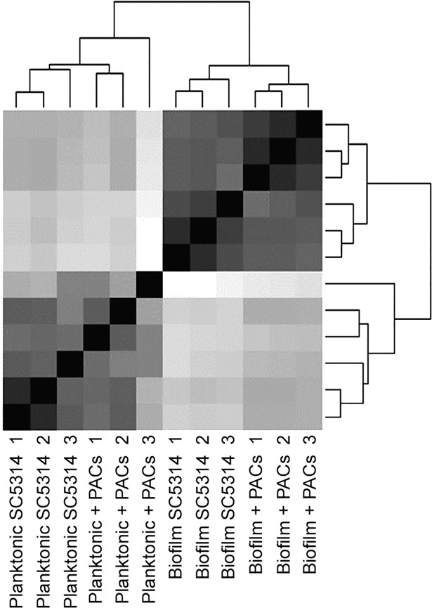
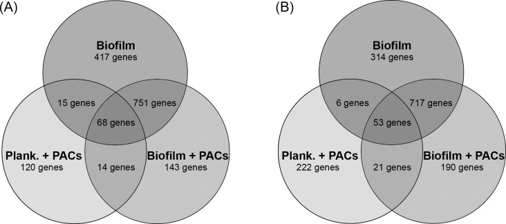
PACs were isolated from cranberry fruit (Vaccinium macrocarpon Ait.) using solid-phase chromatography as described [23]. RNA was isolated after 48h from planktonic and biofilm cells in AU both with and without cranberry PACs. We next used RNA-Seq to profile genome-wide changes in transcription in (i) C. albicans urinary biofilms compared to planktonic cells, to elucidate functional pathways unique to biofilm development in the artificial urinary environment, and (ii) urinary biofilms treated with PAC compared to untreated urinary biofilms, to elucidate pathways involved in the response to proanthocyanidins. All experiments were completed at the late biofilm stage (48h). Sequencing yielded between 25,152,600 and 30,903,560 high-quality reads per sample (Table 1). High correlation existed between all three replicates within each sample, which was represented as a dendrogram, generated using a hierarchical clustering approach integrated as part of the DESeq tool (Fig 1). A total of 6444 genes had any kind of expression in all conditions, which is a measure of any read aligning to gene features in the Candida Genome Database. Differentially-expressed genes were defined as genes with a fold-change ≥1.5 and a p-value ≤0.05 as compared to planktonic cells grown in AU. Of the genes that were significantly upregulated, 49.1% were common between biofilms with and without PAC treatment (Fig 2A). In contrast, only 0.91% of genes were upregulated in both biofilms and planktonic cells treated with PACs. 9.36% of upregulated genes were unique to PAC-treated biofilms; 7.85% of upregulated genes were unique to PAC-treated planktonic cells; and 27.3% of upregulated genes were unique to biofilms formed in AU without PACs. A similar distribution was observed amongst down-regulated genes (Fig 2B). In both planktonic and biofilm cells treated with PACs, more genes were down-regulated than upregulated.
Table 1. Sequencing yield per sample.
Four treatments were sequenced using Illumina HiSeq2000 with three replicates each.
| Sample | Number of reads per sample |
|---|---|
| Planktonic SC5314 1 | 28,614,219 |
| Planktonic SC5314 2 | 28,079,324 |
| Planktonic SC5314 3 | 30,903,560 |
| Biofilm SC5314 1 | 28,474,183 |
| Biofilm SC5314 2 | 28,146,364 |
| Biofilm SC5314 3 | 28,598,751 |
| Planktonic + PACs 1 | 26,913,821 |
| Planktonic + PACs 2 | 28,634,731 |
| Planktonic + PACs 3 | 33,308,787 |
| Biofilm + PACs 1 | 29,288,331 |
| Biofilm + PACs 2 | 25,152,600 |
| Biofilm + PACs 3 | 27,178,053 |
Fig 1. Dendrogram representing hierarchical sample clustering representing high correlation between the samples.
Sample clustering was performed as part of the DESeq tool. The dist function was applied to the transpose of transformed count matrix to obtain sample-to-sample distances. A heat map of this distance matrix highlights the differences and similarities between samples.
Fig 2. Venn diagram analysis of gene expression.
Genes differentially expressed between the three conditions, namely, untreated biofilms, PAC-treated biofilms and PAC-treated planktonic cells were determined as compared to planktonic cells in AU. A) Venn diagram illustrating the number of upregulated genes within each comparison and shared genes between different conditions. B) Venn diagram illustrating the number of downregulated genes within each comparison and shared genes between different conditions.
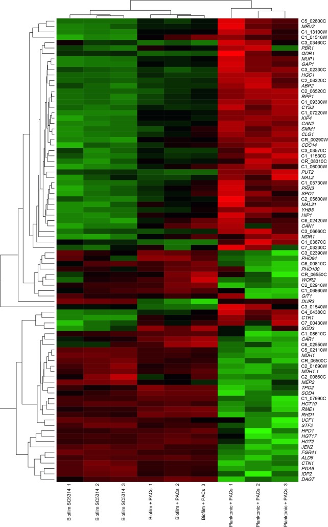
The top differentially-expressed genes in all three treatments (biofilm cells, planktonic cells + PACs, and biofilm cells + PACs) compared to planktonic cells in AU are represented via heatmap (Fig 3). Hierarchical clustering revealed that gene expression patterns were more similar between biofilm cells and PAC-treated biofilm cells than between PAC-treated planktonic cells and PAC-treated biofilm cells. This is suggestive of a strong biofilm-specific transcriptional profile; this profile was present but markedly down-regulated upon PAC treatment (Fig 3).
Fig 3. Heatmap of the top differentially-expressed genes across all treatments.
The top differentially-expressed genes across all treatments as compared to planktonic cells in AU were selected using DESeq2 with the DEApp tool, with an increased cut-off defined as a p-adjusted value of less than or equal to 0.01 and a fold-change of 5 or higher. The top differentially-expressed genes were visualized using the Heatmapper web tool [42]. Red coloring indicates down-regulation as compared to planktonic cells in AU; black indicates no change in expression compared to planktonic cells in AU; green indicates up-regulation as compared to planktonic cells in AU. Dendrogram indicates hierarchical clustering between samples (top) or genes (right).
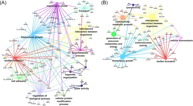
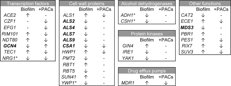
A total of 2341 out of 6434 genes had significant changes in expression in biofilm cells relative to planktonic cells (S1 and S2 Tables). Functional pathway analysis revealed that genes involved in filamentous growth, adhesion, drug response, stress response, and oxidation-reduction processes were up-regulated in biofilms (Fig 4A, S1 Table). Many of these differentially-expressed genes have previously been shown to play roles in C. albicans biofilm formation (Fig 5). Several transcription factors involved in hyphal formation were upregulated in our dataset. These included TEC1, ACE2, RIM101 and OFI1 [46–49]. Other upregulated genes with more direct roles in hyphal biology included HWP1 and ECE1 [50,51]. Genes such as PBR1 (unknown function) and ALS1 (adhesion) have been implicated in biofilm formation and are thought to be involved in the adherence stage of biofilm formation [52–54]. Amino acid transport was the most significantly enriched GO term in this dataset (S1 Table); upregulated amino acid transport genes included ALP1, CAN1, and the general amino acid permeases GAP1, GAP4, GAP5 and GAP6. Notably, methionine biosynthesis and phosphate transport pathways are up-expressed in early biofilm formation [55], and methionine biosynthetic pathways remain prominently up-expressed in late stage biofilms [56]. Methionine biosynthetic pathway genes were also up-expressed in late stage urinary biofilms (genes included MET1, MET3, MET10, and MET15). In addition, the high-affinity phosphate transporter PHO84 [57] and the putative phosphate transporter PHO87 [58] were up-expressed in biofilm compared to planktonic cells. Lipid metabolism genes were also upregulated in urinary biofilms, including genes involved in ergosterol, cholesterol, sphingolipid, and oleic acid synthesis (S1 Table). Among genes that were down-expressed in biofilms relative to planktonic cells, many were involved in endocytosis, vesicle-mediated transport, nitrogen and carbon metabolism, oxidoreductase activity, and nutrient response (Fig 4B, S2 Table). The downregulation of metabolic processes is not surprising as biofilm cells were harvested at 48hr, and metabolic genes are down-regulated in mature biofilms [59,60]. A complete list of enriched GO terms that are down-expressed in biofilms compared to age-matched planktonic cells is provided in S2 Table.
Fig 4. Pathway analysis of differentially-expressed genes as determined by RNA Seq in urinary biofilms.
Analysis was completed using the ClueGO plugin for Cytoscape and the Candida Genome Database GOSlim annotation. (A) Pathway analysis of differentially-expressed genes in urinary biofilms compared to planktonic cells in AU. Genes involved in adhesion, filamentation biofilm formation, transport, lipid metabolism and response to stress were up-regulated in urinary biofilms. (B) Genes involved in carbohydrate metabolism, cellular homeostasis, oxidoreductase activity, and lyase activity were down-regulated in urinary biofilms.
Fig 5. Differentially-expressed biofilm-related genes.
Genes previously shown to have roles in biofilm formation were broken into different functional categories as in Finkel and Mitchell [53]. Genes up- or down-regulated in biofilms as compared to planktonic cells or in PAC-treated biofilms as compared to biofilms alone are indicated by arrows. Negative regulators of biofilm formation are indicated by asterisks (*). Genes indicated in bold are those with potentially unexpected expression changes compared to biofilms formed in serum-like conditions.
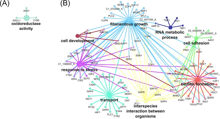
When PAC-treated urinary biofilms were compared with untreated urinary biofilms, 556 of 6434 genes had significant changes in gene expression (S4 and S5 Tables). Up-regulated genes in biofilm cells treated with PACs relative to biofilm control cells were involved in oxidoreductase activity, transmembrane transport, and alcohol biosynthesis (Fig 6A, S4 Table). Genes down-regulated were implicated in adhesion, filamentation, biofilm formation, drug transport, and iron metabolism (Fig 6B, S5 Table). Complete gene lists associated with their GO terms are available as supplemental data (S4 and S5 Tables). Specific down-regulated genes included the secreted aspartyl protease SAP9 (down-regulated five-fold), and the major adhesin ALS1 (down-regulated three-fold). In addition, FRE30 and FRE7 have sequence similarity to ferric reductases [58], and FET31 and SIT1 are known iron-starvation related genes [61,62]. Other genes involved in iron regulation included RIM101 (down-regulated 2.5-fold) which is required for expression of iron uptake genes in neutral or alkaline conditions [63]; FTR2, which encodes a high-affinity iron permease [64] whose transcript is induced in low iron [65]; FLC1, which encodes an iron-starvation-regulated heme uptake protein [66], and MRS4, an iron transporter that mediates Fe2+ transport across the inner mitochondrial membrane under low iron conditions [67]. Overall, iron uptake genes are down-regulated upon treatment with PACs. As artificial urine is a low-iron environment, this indicates that PAC treatment inhibits the expression of iron acquisition genes in response to low environmental iron levels. Thus, pathways related to adhesion and to inhibition of adaptations to a low-iron environment are important in the response to PAC inhibition of C. albicans urinary biofilms. Overall, PACs induced changes in adhesion and iron metabolism pathways within late stage urinary biofilms.
Fig 6. Pathway analysis of differentially-expressed genes as determined by RNA Seq in PAC-treated biofilms.
Analysis was completed using the ClueGO plugin for Cytoscape and the Candida Genome Database GOSlim annotation. (A) Genes that are up-expressed in PAC-treated biofilms were enriched for oxidoreductase-activity related genes. (B) Genes that are down-expressed in PAC treated biofilms and the genes mainly fall under processes such as regulation of filamentous growth, adhesion and biofilm formation.
We also analysed gene expression in PAC-treated planktonic cells as compared to untreated planktonic cells to differentiate between PAC response in planktonic cells versus biofilms (S6–S9 Tables). Like PAC-treated biofilms, genes downregulated in PAC-treated planktonic cells were enriched for genes involved in iron homeostasis (S7 Table). Several of the specific genes down-regulated in PAC-treated biofilms were also down-regulated in PAC-treated planktonic cells; these genes included FET31, FRE7, FRE30, CTR1, and the secreted aspartyl proteases SAP4 and SAP5 (S9 Table). Interestingly, the number of differentially-expressed genes shared between PAC-treated biofilms and PAC-treated planktonic cells was low (14 upregulated genes and 21 downregulated genes; see Fig 2). Thus, cranberry PACs induce distinct transcriptomic landscapes in biofilms and planktonic cells.
Discussion
The global transcriptional changes involved in the transition of C. albicans from planktonic growth to biofilms in bloodstream-like conditions have been studied in detail [55,56,68]. In contrast, the transcriptional changes regulating urinary biofilm development are largely unknown, although clear differences in urinary biofilm structure are evident compared to biofilms in bloodstream-like conditions [69]. Overall, comparatively little is known about C. albicans urinary pathogenesis in general. In these studies, we determined that urinary biofilm formation shares core pathways with biofilms formed under other environmental conditions, including several master transcription factors [68] as well as genes involved in the critical processes of adhesion, filamentation, maturation and dispersal [53]. Genes involved in the process of filamentation, a key component of C. albicans pathogenesis, including well-described transcription factors involved in hyphal formation, were up-expressed. Adhesion related genes were also up-expressed in urinary biofilms. Additional pathways common to serum-like and urinary biofilms included those involved in amino acid transport, including methionine biosynthesis which is well-described in biofilm formation. Phosphate transporter was also up-expressed. Down-expressed pathways in urinary biofilms included various metabolic pathways and house-keeping functions, which is consistent with the relatively quiescent state of cells within biofilms. However, much of the gene expression patterns in urinary biofilms were not observed under other conditions; this subset of genes included a large number of genes of unknown function.
To delve further into understanding genes and pathways unique to urinary biofilms, we used annotations from the Candida Genome Database (CGD) to compare our dataset with previous biofilm studies, including gene expression data from two previous studies of mature biofilms. Nobile et al. (2012) analysed differential gene expression in 48h biofilms formed in Spider medium as compared to planktonic cells [68]. 275 upregulated genes and 148 downregulated genes were shared between biofilms formed in Spider media, and biofilms formed in AU in this study (S3 Table). Of note, Nobile et al. described six master transcriptional regulators, BCR1, BRG1, EFG1, NDT80, ROB1 and TEC1, of C. albicans biofilm formation. Of these, only NDT80 and TEC1 were upregulated in late urinary biofilms in this study. Nett et al. (2009) compared expression of mature in vivo rat catheter biofilms to in vitro planktonic cells [70]. 171 upregulated genes and 105 downregulated genes were shared between rat catheter biofilms and our urinary biofilms (data not shown). Of these, 84 upregulated genes and 34 downregulated genes were common to all three studies, potentially indicating a core set of genes involved in biofilm formation across varying environmental conditions (S3 Table). These shared genes included ECE1, TEC1, and ACE2. We further excluded any genes previously annotated in CGD with roles in biofilm formation. Many differentially-expressed genes in our dataset have not been previously implicated in biofilm formation, indicating a gene expression pattern unique to mature biofilms formed in artificial urine (824 uniquely upregulated genes and 874 uniquely downregulated genes, S3 Table). Functional pathway analysis revealed that these genes were not significantly enriched for any gene ontology terms, with a full 30.3% of genes corresponding to genes of unknown function. Some of these genes may have roles specific to the ecological niche of the urinary tract, an understudied area in C. albicans commensalism and pathogenesis. The top upregulated gene in this urinary biofilm-specific dataset was OFI1 (log2FC = 5.42), a putative transcription factor previously shown to be involved in hyphal morphogenesis [68]. These genes of unknown function appear to be unique to urinary biofilm formation and are consequently of great interest for further study. The extensive differences observed between gene expression in urinary biofilms compared to previously studied biofilms are likely due to environmental differences, including nutrient availability and external pH. Likewise, there have been no previous studies of the transcriptomic response of C. albicans biofilms to cranberry-derived PACs. In previous work, we demonstrated that cranberry PACs inhibit urinary biofilm formation, but have minimal effects on planktonic cell growth [31]. We also assayed expression of selected key adhesin genes within urinary biofilms subjected to PACs in early urinary biofilms via qRT-PCR and discovered biofilm-induced gene expression changes (unpublished data). Based upon these preliminary observations, we then used RNA-Seq to study the global transcriptional response of late biofilms subjected to PAC inhibition, and found that PAC treatment induced down-regulation of genes involved in iron uptake, adhesion, filamentation, and biofilm formation. Notable biofilm-related genes that were down-expressed included the secreted aspartyl protease SAP9, and the adhesin ALS1. Prominently, a number of genes related to iron starvation and iron regulation were down-regulated in urinary biofilms exposed to PACs. Taken together, cranberry-derived PACs inhibit response to low iron, adhesion, and multiple key biofilm-specific processes. Previously, we showed that addition of exogenous iron to AU biofilms partially restores C. albicans biofilm-forming capacity [31]. These transcriptomic data indicate that cranberry PACs interfere with C. albicans tolerance of low-iron, but not iron-replete, environments.
It is quite interesting that cranberry-derived PACs inhibit both E. coli and C. albicans adherence and iron acquisition in vitro. PACs can be expected to chelate exogenous iron, thus impacting iron availability and consequently iron metabolism in any organism. It is difficult to say whether organism-specific mechanisms also contribute, though study of various C. albicans iron uptake mutants may help further elucidate the responsible mechanisms. Interference of non-specific adhesion could also be expected to occur regardless of the organism. However, the explanation for shared adhesion mechanisms across kingdoms is not immediately intuitive. We speculate that over the course of host-pathogen evolution, mechanisms of pathogen adherence to host substrates have evolved convergently. Thus, although well beyond the scope of this study to investigate, we suspect that cross-kingdom inhibition of adherence by PACs is due to common evolutionary demands, such that distinct adhesion molecules have converged enough in structure and function to allow PAC inhibition of adherence even in organisms that evolved pathogenicity independently.
The overall goal of these experiments is to develop a novel strategy for prevention of Candida UTIs. Whether this involves different types of PACs, more potent PAC derivatives, or PACs in combination with other preventative agents or strategies remains to be determined. From a mechanistic standpoint, further studies incorporating genetic mutants of genes in pathways of potential importance to C. albicans urinary biofilm formation could be studied in relation to the effect of PACs. Further, we intend to use dual RNA-Seq [71] to simultaneously investigate the pathogen and mammalian cell transcriptomes during simulated C. albicans urinary infection. These studies will include comparisons in conditions including PACs to further elucidate the biological pathways relevant to Candida UTIs and PAC prevention. Next, studies using animal models of urinary infection could be used to confirm the efficacy of cranberry-derived PACs in prevention of catheter-associated UTIs due to C. albicans and other Candida species. Finally, we hope to study cranberry-derived PACs for prevention of Candida UTIs in catheterized patients. Thus, by understanding the mechanisms of activity of cranberry-derived PACs, we hope to develop PACs or PAC derivatives as a novel therapeutic strategy for the prevention of catheter-associated Candida UTIs. Taken together, these studies suggest that cranberry-derived PACs may have promise as a clinically viable therapeutic option to prevent development of fungal urinary biofilms, particularly in patients with indwelling urinary catheters.
Supporting information
Sheet 1 contains gene expression information. Sheet 2 contains GO term enrichment analysis.
(XLSX)
Sheet 1 contains gene expression information. Sheet 2 contains GO term enrichment analysis.
(XLSX)
(XLSM)
Sheet 1 contains gene expression information. Sheet 2 contains GO term enrichment analysis.
(XLSX)
Sheet 1 contains gene expression information. Sheet 2 contains GO term enrichment analysis.
(XLSX)
Sheet 1 contains gene expression information. Sheet 2 contains GO term enrichment analysis.
(XLSX)
Sheet 1 contains gene expression information. Sheet 2 contains GO term enrichment analysis.
(XLSX)
Sheet 1 contains gene expression information. Sheet 2 contains upregulated genes shared between PAC-treated planktonic cells and PAC-treated biofilm cells relative to WT planktonic cells, and GO term enrichment analysis of this data.
(XLSX)
Sheet 1 contains gene expression information. Sheet 2 contains downregulated genes shared between PAC-treated planktonic cells and PAC-treated biofilm cells relative to WT planktonic cells, and GO term enrichment analysis of this data.
(XLSX)
Acknowledgments
We thank William Fonzi (Georgetown University) for providing strain SC5314.
Part of these data were previously presented at the 13th ASM Conference on Candida and Candidiasis, 2016: Sundararajan A, Rane H, Sena J, Schilkey FD, Lee SA. “Investigating the effects of cranberry-derived proanthocyanidins on Candida albicans urinary biofilms using RNA-seq.” 13th ASM Conference on Candida and Candidiasis, Apr 13–17, 2016, Seattle, WA.
Data Availability
RNA sequence data generated have been deposited at the NCBI database under bioproject number PRJNA338054. All other relevant data are within the paper and its Supporting Information files.
Funding Statement
This work was supported in part by grants from the Department of Veterans' Affairs (Merit Award 5I01BX001130-03 to SAL), and Biomedical Research Institute of New Mexico (to SAL). RNA-Seq data was obtained through a pilot award funded by New Mexico INBRE through an Institutional Development Award (IDeA) from NIH-NIGMS grant P20GM103451.
References
- 1.Gardner A, Mitchell B, Beckingham W, Fasugba O. A point prevalence cross-sectional study of healthcare-associated urinary tract infections in six Australian hospitals. BMJ Open. 2014;4: e005099–e005099. 10.1136/bmjopen-2014-005099 [DOI] [PMC free article] [PubMed] [Google Scholar]
- 2.Tambyah PA, Knasinski V, Maki DG. The direct costs of nosocomial catheter-associated urinary tract infection in the era of managed care. Infect Control Hosp Epidemiol. 2002;23: 27–31. 10.1086/501964 [DOI] [PubMed] [Google Scholar]
- 3.Bouza E, San Juan R, Muñoz P, Voss A, Kluytmans J, Co-operative Group of the European Study Group on Nosocomial Infections. A European perspective on nosocomial urinary tract infections II. Report on incidence, clinical characteristics and outcome (ESGNI-004 study). Clin Microbiol Infect. 2001;7: 532–542. [DOI] [PubMed] [Google Scholar]
- 4.Kauffman CA, Vazquez JA, Sobel JD, Gallis HA, McKinsey DS, Karchmer AW, et al. Prospective multicenter surveillance study of funguria in hospitalized patients. The National Institute for Allergy and Infectious Diseases (NIAID) Mycoses Study Group. Clin Infect Dis. 2000;30: 14–18. 10.1086/313583 [DOI] [PubMed] [Google Scholar]
- 5.Achkar JM, Fries BC. Candida infections of the genitourinary tract. Clin Microbiol Rev. 2010;23: 253–273. 10.1128/CMR.00076-09 [DOI] [PMC free article] [PubMed] [Google Scholar]
- 6.Kauffman CA. Diagnosis and management of fungal urinary tract infection. Infect Dis Clin North Am. 2014;28: 61–74. 10.1016/j.idc.2013.09.004 [DOI] [PubMed] [Google Scholar]
- 7.Sobel JD, Fisher JF, Kauffman CA, Newman CA. Candida urinary tract infections—epidemiology. Clin Infect Dis. 2011;52: S433–436. 10.1093/cid/cir109 [DOI] [PubMed] [Google Scholar]
- 8.Fisher JF. Candida urinary tract infections—epidemiology, pathogenesis, diagnosis, and treatment: executive summary. Clin Infect Dis. 2011;52: S429–S432. 10.1093/cid/cir108 [DOI] [PubMed] [Google Scholar]
- 9.Lee SA, Andriole VT. Clinical significance and management of Candida infections of the urinary system. Todays Ther Trends. 2004;22: 97–114. [Google Scholar]
- 10.Fisher JF, Sobel JD, Kauffman CA, Newman CA. Candida urinary tract infections—treatment. Clin Infect Dis. 2011;52: S457–S466. 10.1093/cid/cir112 [DOI] [PubMed] [Google Scholar]
- 11.Ramage G, Saville SP, Thomas DP, López-Ribot JL. Candida biofilms: an update. Eukaryot Cell. 2005;4: 633–638. 10.1128/EC.4.4.633-638.2005 [DOI] [PMC free article] [PubMed] [Google Scholar]
- 12.Bonhomme J, d’Enfert C. Candida albicans biofilms: building a heterogeneous, drug-tolerant environment. Curr Opin Microbiol. 2013;16: 398–403. 10.1016/j.mib.2013.03.007 [DOI] [PubMed] [Google Scholar]
- 13.Jain N, Kohli R, Cook E, Gialanella P, Chang T, Fries BC. Biofilm formation by and antifungal susceptibility of Candida isolates from urine. Appl Environ Microbiol. 2007;73: 1697–1703. 10.1128/AEM.02439-06 [DOI] [PMC free article] [PubMed] [Google Scholar]
- 14.Wang X, Fries BC. A murine model for catheter-associated candiduria. J Med Microbiol. 2011;60: 1523–1529. 10.1099/jmm.0.026294-0 [DOI] [PMC free article] [PubMed] [Google Scholar]
- 15.Maki DG, Tambyah PA. Engineering out the risk for infection with urinary catheters. Emerg Infect Dis. 2001;7: 342–347. 10.3201/eid0702.700342 [DOI] [PMC free article] [PubMed] [Google Scholar]
- 16.Nett JE, Brooks E, Cabezas-Olcoz J, Sanchez H, Zarnowski R, Marchillo K, et al. Rat indwelling urinary catheter model of Candida albicans biofilm infection. Infect Immun. 2014; 10.1128/IAI.02284-14 [DOI] [PMC free article] [PubMed] [Google Scholar]
- 17.Ramage G, Mowat E, Jones B, Williams C, Lopez-Ribot J. Our current understanding of fungal biofilms. Crit Rev Microbiol. 2009;35: 340–355. 10.3109/10408410903241436 [DOI] [PubMed] [Google Scholar]
- 18.Johnson JR, Kuskowski MA, Wilt TJ. Systematic review: antimicrobial urinary catheters to prevent catheter-associated urinary tract infection in hospitalized patients. Ann Intern Med. 2006;144: 116–126. [DOI] [PubMed] [Google Scholar]
- 19.Foxman B, Cronenwett AEW, Spino C, Berger MB, Morgan DM. Cranberry juice capsules and urinary tract infection after surgery: results of a randomized trial. Am J Obstet Gynecol. 2015;213: 194.e1–194.e8. 10.1016/j.ajog.2015.04.003 [DOI] [PMC free article] [PubMed] [Google Scholar]
- 20.Wang C-H, Fang C-C, Chen N-C, Liu SS-H, Yu P-H, Wu T-Y, et al. Cranberry-containing products for prevention of urinary tract infections in susceptible populations: a systematic review and meta-analysis of randomized controlled trials. Arch Intern Med. 2012;172: 988–996. 10.1001/archinternmed.2012.3004 [DOI] [PubMed] [Google Scholar]
- 21.Tempesta M, Barrett M. Cranberry products prevent urinary tract infections in women: clinical evidence In: Cooper R, Kronenberg F, editors. Botanical Medicine: From Bench To Beside. New Rochelle, NY: Mary Ann Liebert, Inc.; 2009. pp. 203–211. [Google Scholar]
- 22.Raz R, Chazan B, Dan M. Cranberry juice and urinary tract infection. Clin Infect Dis. 2004;38: 1413–1419. 10.1086/386328 [DOI] [PubMed] [Google Scholar]
- 23.Howell AB, Reed JD, Krueger CG, Winterbottom R, Cunningham DG, Leahy M. A-type cranberry proanthocyanidins and uropathogenic bacterial anti-adhesion activity. Phytochemistry. 2005;66: 2281–2291. 10.1016/j.phytochem.2005.05.022 [DOI] [PubMed] [Google Scholar]
- 24.Gupta K, Chou MY, Howell AB, Wobbe C, Grady R, Stapleton AE. Cranberry products inhibit adherence of p-fimbriated Escherichia coli to primary cultured bladder and vaginal epithelial cells. J Urol. 2007;177: 2357–2360. 10.1016/j.juro.2007.01.114 [DOI] [PMC free article] [PubMed] [Google Scholar]
- 25.Eydelnant IA, Tufenkji N. Cranberry derived proanthocyanidins reduce bacterial adhesion to selected biomaterials. Langmuir. 2008;24: 10273–10281. 10.1021/la801525d [DOI] [PubMed] [Google Scholar]
- 26.Howell AB, Botto H, Combescure C, Blanc-Potard A-B, Gausa L, Matsumoto T, et al. Dosage effect on uropathogenic Escherichia coli anti-adhesion activity in urine following consumption of cranberry powder standardized for proanthocyanidin content: a multicentric randomized double blind study. BMC Infect Dis. 2010;10. [DOI] [PMC free article] [PubMed] [Google Scholar]
- 27.Hidalgo G, Chan M, Tufenkji N. Inhibition of Escherichia coli CFT073 fliC expression and motility by cranberry materials. Appl Environ Microbiol. 2011;77: 6852–6857. 10.1128/AEM.05561-11 [DOI] [PMC free article] [PubMed] [Google Scholar]
- 28.Hidalgo G, Ponton A, Fatisson J, O’May C, Asadishad B, Schinner T, et al. Induction of a state of iron limitation in uropathogenic Escherichia coli CFT073 by cranberry-derived proanthocyanidins as revealed by microarray analysis. Appl Environ Microbiol. 2011;77: 1532–1535. 10.1128/AEM.02201-10 [DOI] [PMC free article] [PubMed] [Google Scholar]
- 29.Lin B, Johnson BJ, Rubin RA, Malanoski AP, Ligler FS. Iron chelation by cranberry juice and its impact on Escherichia coli growth. Biofactors. 2010;37: 121–130. 10.1002/biof.110 [DOI] [PubMed] [Google Scholar]
- 30.Feldman M, Tanabe S, Howell AB, Grenier D. Cranberry proanthocyanidins inhibit the adherence properties of Candida albicans and cytokine secretion by oral epithelial cells. BMC Complement Altern Med. 2012;12. [DOI] [PMC free article] [PubMed] [Google Scholar]
- 31.Rane HS, Bernardo SM, Howell AB, Lee SA. Cranberry-derived proanthocyanidins prevent formation of Candida albicans biofilms in artificial urine through biofilm- and adherence-specific mechanisms. J Antimicrob Chemother. 2013; 10.1093/jac/dkt398 [DOI] [PMC free article] [PubMed] [Google Scholar]
- 32.Foo LY, Lu Y, Howell AB, Vorsa N. A-Type proanthocyanidin trimers from cranberry that inhibit adherence of uropathogenic P-fimbriated Escherichia coli. J Nat Prod. 2000;63: 1225–1228. [DOI] [PubMed] [Google Scholar]
- 33.Foo LY, Lu Y, Howell AB, Vorsa N. The structure of cranberry proanthocyanidins which inhibit adherence of uropathogenic P-fimbriated Escherichia coli in vitro. Phytochemistry. 2000;54: 173–181. [DOI] [PubMed] [Google Scholar]
- 34.Fonzi WA, Irwin MY. Isogenic strain construction and gene mapping in Candida albicans. Genetics. 1993;134: 717–728. [DOI] [PMC free article] [PubMed] [Google Scholar]
- 35.Ramage G, López-Ribot JL. Techniques for antifungal susceptibility testing of Candida albicans biofilms. Methods Mol Med. 2005;118: 71–79. 10.1385/1-59259-943-5:071 [DOI] [PubMed] [Google Scholar]
- 36.Wu TD, Nacu S. Fast and SNP-tolerant detection of complex variants and splicing in short reads. Bioinformatics. 2010;26: 873–881. 10.1093/bioinformatics/btq057 [DOI] [PMC free article] [PubMed] [Google Scholar]
- 37.Miller NA, Kingsmore SF, Farmer A, Langley RJ, Mudge J, Crow JA, et al. Management of high-throughput DNA sequencing projects: Alpheus. J Comput Sci Syst Biol. 2008;1: 132 [DOI] [PMC free article] [PubMed] [Google Scholar]
- 38.Love MI, Huber W, Anders S. Moderated estimation of fold change and dispersion for RNA-seq data with DESeq2. Genome Biol. 2014;15 10.1186/s13059-014-0550-8 [DOI] [PMC free article] [PubMed] [Google Scholar]
- 39.Li Y, Andrade J. DEApp: an interactive web interface for differential expression analysis of next generation sequence data. Source Code Biol Med. 2017;12 10.1186/s13029-017-0063-4 [DOI] [PMC free article] [PubMed] [Google Scholar]
- 40.Hochberg Y, Benjamini Y. More powerful procedures for multiple significance testing. Stat Med. 1990;9: 811–818. [DOI] [PubMed] [Google Scholar]
- 41.Oliveros JC. VENNY. An interactive tool for comparing lists with Venn Diagrams. [Internet]. 2007. Available: http://bioinfogp.cnb.csic.es/tools/venny/index.html
- 42.Babicki S, Arndt D, Marcu A, Liang Y, Grant JR, Maciejewski A, et al. Heatmapper: web-enabled heat mapping for all. Nucleic Acids Res. 2016;44: W147–153. 10.1093/nar/gkw419 [DOI] [PMC free article] [PubMed] [Google Scholar]
- 43.Bindea G, Mlecnik B, Hackl H, Charoentong P, Tosolini M, Kirilovsky A, et al. ClueGO: a Cytoscape plug-in to decipher functionally grouped gene ontology and pathway annotation networks. Bioinforma Oxf Engl. 2009;25: 1091–1093. 10.1093/bioinformatics/btp101 [DOI] [PMC free article] [PubMed] [Google Scholar]
- 44.Shannon P, Markiel A, Ozier O, Baliga NS, Wang JT, Ramage D, et al. Cytoscape: a software environment for integrated models of biomolecular interaction networks. Genome Res. 2003;13: 2498–2504. 10.1101/gr.1239303 [DOI] [PMC free article] [PubMed] [Google Scholar]
- 45.Skrzypek MS, Binkley J, Binkley G, Miyasato SR, Simison M, Sherlock G. Candida Genome Database [Internet]. Available: http://www.candidagenome.org/
- 46.Schweizer A, Rupp S, Taylor BN, Röllinghoff M, Schröppel K. The TEA/ATTS transcription factor CaTec1p regulates hyphal development and virulence in Candida albicans. Mol Microbiol. 2000;38: 435–445. [DOI] [PubMed] [Google Scholar]
- 47.Kelly MT, MacCallum DM, Clancy SD, Odds FC, Brown AJP, Butler G. The Candida albicans CaACE2 gene affects morphogenesis, adherence and virulence. Mol Microbiol. 2004;53: 969–983. 10.1111/j.1365-2958.2004.04185.x [DOI] [PubMed] [Google Scholar]
- 48.Davis D. Adaptation to environmental pH in Candida albicans and its relation to pathogenesis. Curr Genet. 2003;44: 1–7. 10.1007/s00294-003-0415-2 [DOI] [PubMed] [Google Scholar]
- 49.Du H, Li X, Huang G, Kang Y, Zhu L. The zinc-finger transcription factor, Ofi1, regulates white-opaque switching and filamentation in the yeast Candida albicans. Acta Biochim Biophys Sin. 2015;47: 335–341. 10.1093/abbs/gmv011 [DOI] [PubMed] [Google Scholar]
- 50.Staab JF, Bahn Y-S, Tai C-H, Cook PF, Sundstrom P. Expression of transglutaminase substrate activity on Candida albicans germ tubes through a coiled, disulfide-bonded N-terminal domain of Hwp1 requires C-terminal glycosylphosphatidylinositol modification. J Biol Chem. 2004;279: 40737–40747. 10.1074/jbc.M406005200 [DOI] [PubMed] [Google Scholar]
- 51.Birse CE, Irwin MY, Fonzi WA, Sypherd PS. Cloning and characterization of ECE1, a gene expressed in association with cell elongation of the dimorphic pathogen Candida albicans. Infect Immun. 1993;61: 3648–3655. [DOI] [PMC free article] [PubMed] [Google Scholar]
- 52.Araújo D, Henriques M, Silva S. Portrait of Candida species biofilm regulatory network genes. Trends Microbiol. 2017;25: 62–75. 10.1016/j.tim.2016.09.004 [DOI] [PubMed] [Google Scholar]
- 53.Finkel JS, Mitchell AP. Genetic control of Candida albicans biofilm development. Nat Rev Microbiol. 2011;9: 109–118. 10.1038/nrmicro2475 [DOI] [PMC free article] [PubMed] [Google Scholar]
- 54.Gulati M, Nobile CJ. Candida albicans biofilms: development, regulation, and molecular mechanisms. Microbes Infect. 2016;18: 310–321. 10.1016/j.micinf.2016.01.002 [DOI] [PMC free article] [PubMed] [Google Scholar]
- 55.Murillo LA, Newport G, Lan C-Y, Habelitz S, Dungan J, Agabian NM. Genome-wide transcription profiling of the early phase of biofilm formation by Candida albicans. Eukaryot Cell. 2005;4: 1562–1573. 10.1128/EC.4.9.1562-1573.2005 [DOI] [PMC free article] [PubMed] [Google Scholar]
- 56.García-Sánchez S, Aubert S, Iraqui I, Janbon G, Ghigo J-M, d’Enfert C. Candida albicans biofilms: a developmental state associated with specific and stable gene expression patterns. Eukaryot Cell. 2004;3: 536–545. 10.1128/EC.3.2.536-545.2004 [DOI] [PMC free article] [PubMed] [Google Scholar]
- 57.Liu N-N, Flanagan PR, Zeng J, Jani NM, Cardenas ME, Moran GP, et al. Phosphate is the third nutrient monitored by TOR in Candida albicans and provides a target for fungal-specific indirect TOR inhibition. Proc Natl Acad Sci U S A. 2017;114: 6346–6351. 10.1073/pnas.1617799114 [DOI] [PMC free article] [PubMed] [Google Scholar]
- 58.Skrzypek MS, Binkley J, Binkley G, Miyasato SR, Simison M, Sherlock G. The Candida Genome Database (CGD): incorporation of Assembly 22, systematic identifiers and visualization of high throughput sequencing data. Nucleic Acids Res. 2017;45: D592–D596. 10.1093/nar/gkw924 [DOI] [PMC free article] [PubMed] [Google Scholar]
- 59.Yeater KM, Chandra J, Cheng G, Mukherjee PK, Zhao X, Rodriguez-Zas SL, et al. Temporal analysis of Candida albicans gene expression during biofilm development. Microbiol Read Engl. 2007;153: 2373–2385. 10.1099/mic.0.2007/006163-0 [DOI] [PubMed] [Google Scholar]
- 60.Fox EP, Bui CK, Nett JE, Hartooni N, Mui MC, Andes DR, et al. An expanded regulatory network temporally controls Candida albicans biofilm formation. Mol Microbiol. 2015;96: 1226–1239. 10.1111/mmi.13002 [DOI] [PMC free article] [PubMed] [Google Scholar]
- 61.Eck R, Hundt S, Härtl A, Roemer E, Künkel W. A multicopper oxidase gene from Candida albicans: cloning, characterization and disruption. Microbiology. 1999;145: 2415–2422. 10.1099/00221287-145-9-2415 [DOI] [PubMed] [Google Scholar]
- 62.Lan C-Y, Rodarte G, Murillo LA, Jones T, Davis RW, Dungan J, et al. Regulatory networks affected by iron availability in Candida albicans. Mol Microbiol. 2004;53: 1451–1469. 10.1111/j.1365-2958.2004.04214.x [DOI] [PubMed] [Google Scholar]
- 63.Bensen ES, Martin SJ, Li M, Berman J, Davis DA. Transcriptional profiling in Candida albicans reveals new adaptive responses to extracellular pH and functions for Rim101p. Mol Microbiol. 2004;54: 1335–1351. 10.1111/j.1365-2958.2004.04350.x [DOI] [PubMed] [Google Scholar]
- 64.Ramanan N, Wang Y. A high-affinity iron permease essential for Candida albicans virulence. Science. 2000;288: 1062–1064. [DOI] [PubMed] [Google Scholar]
- 65.Knight SAB, Lesuisse E, Stearman R, Klausner RD, Dancis A. Reductive iron uptake by Candida albicans: role of copper, iron and the TUP1 regulator. Microbiol Read Engl. 2002;148: 29–40. 10.1099/00221287-148-1-29 [DOI] [PubMed] [Google Scholar]
- 66.Protchenko O, Rodriguez-Suarez R, Androphy R, Bussey H, Philpott CC. A screen for genes of heme uptake identifies the FLC family required for import of FAD into the endoplasmic reticulum. J Biol Chem. 2006;281: 21445–21457. 10.1074/jbc.M512812200 [DOI] [PubMed] [Google Scholar]
- 67.Xu N, Cheng X, Yu Q, Zhang B, Ding X, Xing L, et al. Identification and functional characterization of mitochondrial carrier Mrs4 in Candida albicans. FEMS Yeast Res. 2012;12: 844–858. 10.1111/j.1567-1364.2012.00835.x [DOI] [PubMed] [Google Scholar]
- 68.Nobile CJ, Fox EP, Nett JE, Sorrells TR, Mitrovich QM, Hernday AD, et al. A recently evolved transcriptional network controls biofilm development in Candida albicans. Cell. 2012;148: 126–138. 10.1016/j.cell.2011.10.048 [DOI] [PMC free article] [PubMed] [Google Scholar]
- 69.Uppuluri P, Dinakaran H, Thomas DP, Chaturvedi AK, Lopez-Ribot JL. Characteristics of Candida albicans biofilms grown in a synthetic urine medium. J Clin Microbiol. 2009;47: 4078–4083. 10.1128/JCM.01377-09 [DOI] [PMC free article] [PubMed] [Google Scholar]
- 70.Nett JE, Lepak AJ, Marchillo K, Andes DR. Time course global gene expression analysis of an in vivo Candida biofilm. J Infect Dis. 2009;200: 307–313. 10.1086/599838 [DOI] [PMC free article] [PubMed] [Google Scholar]
- 71.Westermann AJ, Förstner KU, Amman F, Barquist L, Chao Y, Schulte LN, et al. Dual RNA-seq unveils noncoding RNA functions in host-pathogen interactions. Nature. 2016;529: 496–501. 10.1038/nature16547 [DOI] [PubMed] [Google Scholar]
Associated Data
This section collects any data citations, data availability statements, or supplementary materials included in this article.
Supplementary Materials
Sheet 1 contains gene expression information. Sheet 2 contains GO term enrichment analysis.
(XLSX)
Sheet 1 contains gene expression information. Sheet 2 contains GO term enrichment analysis.
(XLSX)
(XLSM)
Sheet 1 contains gene expression information. Sheet 2 contains GO term enrichment analysis.
(XLSX)
Sheet 1 contains gene expression information. Sheet 2 contains GO term enrichment analysis.
(XLSX)
Sheet 1 contains gene expression information. Sheet 2 contains GO term enrichment analysis.
(XLSX)
Sheet 1 contains gene expression information. Sheet 2 contains GO term enrichment analysis.
(XLSX)
Sheet 1 contains gene expression information. Sheet 2 contains upregulated genes shared between PAC-treated planktonic cells and PAC-treated biofilm cells relative to WT planktonic cells, and GO term enrichment analysis of this data.
(XLSX)
Sheet 1 contains gene expression information. Sheet 2 contains downregulated genes shared between PAC-treated planktonic cells and PAC-treated biofilm cells relative to WT planktonic cells, and GO term enrichment analysis of this data.
(XLSX)
Data Availability Statement
RNA sequence data generated have been deposited at the NCBI database under bioproject number PRJNA338054. All other relevant data are within the paper and its Supporting Information files.


