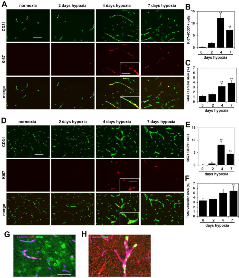
Figure 1.
Chronic mild hypoxia (CMH) stimulates vascular remodeling in spinal cord blood vessels. Dual-IF was performed on frozen sections of spinal cord white matter (A) or gray matter (D) taken from mice exposed to normoxia or 2, 4 or 7 days hypoxia using antibodies specific for CD31 (AlexaFluor-488) and Ki67 (Cy-3). Scale bar = 100 µm, except for the inset where scale bar = 50 µm. Note that little endothelial proliferation is seen after 2 days hypoxia, but after 4 days hypoxia a strong proliferative response is observed, before declining at later time-points. Proliferating endothelial cells are seen grouped together in a linear arrangement within a single vessel, in which an angiogenic sprout is observed (see inset). B and E. Quantification of endothelial proliferation at different time-points of CMH in white matter (B) and gray matter (E). Results are expressed as the mean ± SEM (n = 3 mice/group). C and F. Quantification of the influence of CMH on total vascular area in white matter (C) and gray matter (F). Results are expressed as the mean ± SEM in which vascular area is represented as a % of the FOV (n = 3 mice/group). Note that endothelial proliferation and vascular expansion occur both in white matter and gray matter, but vascular remodeling is more extensive in white matter, where the higher rate of endothelial proliferation translates into a greater expansion of total vascular area. * p < 0.05, ** p < 0.01 vs. normoxia (0 days hypoxia). G Triple-IF for laminin (Cy-5), Ki67 (Cy-3) and NeuN (AlexaFluor-488). H. Triple-IF for laminin (Cy-5), Ki67 (AlexaFluor-488) and GFAP (Cy-3).

