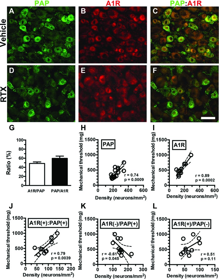
Figure 7.
Contributions of adenosine A1 receptors (A1Rs) and prostatic acid phosphatase (PAP) neurons to neuropathic pain hypersensitivity in resiniferatoxin (RTX) neuropathy. (A–F) Double-labeling immunofluorescence staining was performed on dorsal root ganglion (DRG) sections with anti-PAP (A, C, D, and F in green) and anti-A1R (B, C, E, and F in red) antisera in the vehicle group (A–C) and day 7 after RTX neuropathy (RTX group; D–F). (C and F) The images show colocalization of A1R and PAP DRG neurons in the vehicle (C) and RTX (F) groups. (G) The diagram indicates the ratios of A1R(+)/PAP(+) (open bar, n = 6) and PAP(+)/A1R(+) (filled bar, n = 6) in the vehicle group shown in (A–F). (H–L) The mechanical threshold was assessed through von Frey monofilament tests using up-and-down algorithm. The mechanical threshold was highly linearly correlated with the densities of PAP(+) (H, n = 16), A1R(+) (I, n = 11), and A1R(+):PAP(+) neurons (J, n = 11). By contrast, the density of A1R(−)/PAP(+) neurons (K, n = 11) showed a weak linear correlation with the mechanical threshold, and no linear correlation between A1R(+)/PAP(−) neurons (L, n = 11) and the mechanical threshold was evident. Bar, 50 µm.

