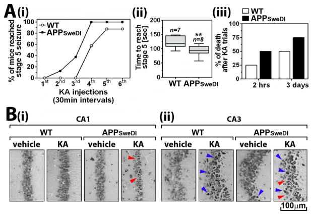
Fig. 3. Altered seizure susceptibility of APPSweDI mice (6 months old).
A. Seizure responses of mice to KA was assessed for analysis of seizure vulnerability of the mice. The mice were treated with repeated low dose of KA (5mg/kg; 30 min intervals) till each mouse reached stage 5 Racine scale. The number of KA treatment (i) and time to reach stage 5 seizure (ii) and death of mice following the KA treatment (iii) were analyzed. The columns in panel A-ii show 75% of distribution; horizontal bar in each column is median; and vertical T-bars are minimum and maximum values of the data: **. P ≤ 0.01 as compared to WT mice. B. Three days of KA treatment, hippocampal morphology and pyramidal neuronal status in CA1 (i) and CA3 (ii) regions were assessed by Nissle staining. Red and blue arrow heads indicate neuronal loss (empty areas) and degenerating neurons (dark Nissl bodies), respectively.

