Abstract
Background
Foam Rolling (FR) has steadily gained in popularity as an intervention to increase range of motion (ROM) and reduce pain. It is believed that FR can remove restrictions due to fascial adhesions, thus improving ROM. FR has been proposed as a means to increase ITB length as a means to achieve these outcomes. Previous research has focused on the effects of FR over both muscle and fascia tissue together. However, no studies have examined the effects of FR over fascial tissue not containing muscle.
Purpose
The purpose of this study was to compare the acute effect of a single bout of foam rolling (FR) over the Iliotibial Band (ITB) compared to FR over the gluteal muscle group on hip adduction passive range of motion (PROM).
Methods
Twenty-seven participants were recruited for the study. Each participant performed three sessions: FR over tissue devoid of muscle, the ITB (PFR), FR over contractile tissue, the gluteal muscles (AFR), and a session without FR (control) in a randomized order. Hip adduction PROM was measured in a pre-post manner for each session.
Results
Results of the repeated measures ANOVA showed a significant interaction across session and time (F(2, 25) = 25.202, p < 0.001,
= 0.502, 1 – β = 1.000). Post-hoc analysis showed the AFR post-test measure was significantly different from both control (p < 0.001) and PFR counterparts (p < 0.001). FR over the gluteal muscle group lead to a 14.8% improvement in hip adduction ROM, with PFR only a 2% improvement.
Conclusion
A single bout of FR over a myofascial group appears to increase PROM in healthy young adults, whereas FR over the ITB itself (primarily fascial tissue) does not. This suggests the conventional theory behind FR may need to be reevaluated.
Level of Evidence
Level 1B, laboratory study, repeated measures design
Keywords: Fascia, foam rolling, iliotibial band, Ober's Test, range of motion
INTRODUCTION
Foam rolling (FR) has gained favor as an adjunct to manual therapy to increase range of motion (ROM) 1-3 and improve outcomes with regard to pain.4-7 FR over the iliotibial band (ITB) is a popular adjunct in treatment for patellofemoral pain syndrome,8 runner's knee9 and hip bursitis.10 More recently, FR has become common practice for the general population, adopting the technique before training and exercising as a means for enhancing ROM,11 as self-massage,12 as an adjunct to warm-up,11 and to reduce pain associated with muscle soreness. 4-7
FR is proposed as a method to remove restrictions in ROM due to fascial adhesions, despite limited evidence that FR is capable of achieving this outcome.13 Evidence for FR having an effect on adhesions is limited to studies assessing changes in ROM.1-3 However, these studies have focused FR over myofascial tissue, with a significant component of the tissue being muscle, or “active” tissue. Significant skepticism remains as to whether or not FR therapy can generate sufficient pressure to remove any restrictions in ROM that may exist in passive, or non-contractile tissues,14 such as the ITB. Other fascial release techniques have limited evidence. Rolfing is a technique that combines deep manual therapy with active movement.15 Rolfers posit that Rolfing improves ROM of the fascia. However, Rolfing under anesthesia has been shown to have no change is tissue length.16
The ITB is comprised exclusively of dense connective tissue, and is devoid of muscle fibers. Its anatomical origin arises from the proximal end of the tendons of the tensor fasciae latae (TFL) and gluteus maximus muscles, converging into the proximal bands.17 It continues down the lateral femur and crosses the knee joint where most of its fibers insert on Gerdy's tubercle.18 Some of the ITB's deeper fibers insert on the linea aspera of the femur, while some distal superficial fibers blend with the lateral retinaculum of the patella.19 The TFL acts as a lateral hip stabilizer and assists the gluteal muscle group during hip extension.19
Functionally, it is believed that the orientation of fascial fibers allow for reduced activity from the gluteal muscles to maintain hip stability due to the intrinsic strength of the ITB, the orientation of the fibers and the relatively low ground reaction forces that occur during a static weight-bearing position.20 As such, the ITB serves to reduce work required by the gluteal muscles during gait, and accepts load directly.
The gluteal muscle group is traditionally divided into three distinct muscles: the gluteus maximus, gluteus medius and gluteus minimus.18 However, this division has been called into question, as Flack et al22 found poor evidence for compartmentalization based on fascial separation, innervation and individual function. Collectively, the gluteal muscle group forms a large, fan-shaped muscle whose fibers converge on the greater trochanter of the femur deeply and blend with the ITB superficially.23 The contractile elements of the muscle fibers pull on its fascial fibers, including those of the ITB, to generate extension, abduction and external rotation of the hip.22
FR over the posterior lateral hip area cannot be specific to a single type of tissue. Indeed, there is a non-contractile (fascia) tissue within this area which is similar histologically to the fascia found in the ITB.21 However, the primary difference between the fascia of the ITB and that of this gluteal muscle group is the significant presence of contractile tissue.22 As such, this area has more neurological and proproeceptive input than the ITB, as the gluteal muscles are supplied with both sensory neurons in the form on Pancinian corpuscles and Merkle's discs and mechanoreceptors. Further, the gluteal muscles are capable of actively alternating length via efferent input and GTO and muscle spindle activity. However, the ITB is unable to alter its length, as it is primarily the tendinous fascia from the tensor fascia latae and is largely devoid of motor neurons,23 therefore any changes in ITB ROM from FR are likely to be due to muscular adaptions, rather than the removal of mechanical restrictions due to fascial adhesions, as popular theory suggests.13 Previous FR studies have documented increases ROM when applying this intervention over regions of the body containing muscle.1-3 The acute effect of FR over non-muscle tissue on ROM is unknown. Therefore, the purpose of this study was to compare the acute effect of a single bout of FR over the ITB compared to FR over the gluteal muscle group on hip adduction passive range of motion (PROM).
METHODS
Subjects
Twenty-seven (14 female, 12 males) healthy adults volunteered for this study. Exclusion criteria included prior lower limb or low back injury, currently participating in a stretching program, or any use of foam rolling within the previous six weeks. One participant was eliminated from the study due to failure to comply with the testing protocol. Mean ± SD for age, height, and weight for female subjects were 21.07 ± 1.141 yrs, 166.36 ± 7.110 cm, and 68.00 ± 10.53 kg, respectively. Mean ± SD for age, height, and weight for male participants were 21.50 ± 1.243 yrs, 171.92 ± 6.640 cm, and 79.682 ± 19.573 kg, respectively.
Testing Procedure
Each participant completed three sessions (control, active foam rolling (AFR), and passive foam rolling (PFR). Sessions were randomized by numbered containers selected by the author, and concealed until all interventions were assigned. Each sessions was scheduled one week apart at the same time of day and location. Prior to participation, each participant completed an informed consent and health history questionnaire. All procedures were approved by the Coastal Carolina University Institutional Review Board, approval code #2016.41. The rights of all subjects were protected.
During the control session, each participant performed a five-minute warm-up by pedaling continuously at 50 rpm (50 Watts of resistance) on a cycle ergometer (Monark Ergomedic 828E, Vansbro, Sweden). Seat height was set to allow for a 5-10 ° bend in the extended knee. The same seat height was used for each session. Immediately following the warm-up, each participant had their passive hip adduction ROM assessed bilaterally using the modified Ober test. The Ober test has been shown to be a reliable test to assess hip adduction ROM24,25 and therefore able to identify restrictive ROM of abductive tissues, such as the ITB and gluteal muscle group.26 With the participant in a side-lying position on a padded treatment table, both anterior superior iliac spines (ASIS) of the pelvis were maintained in a perpendicular to the ground and stabilized by the examiner using a hand on the lateral hip. The non-test leg was flexed at the hip and knee so that it did not inhibit test leg adduction. The test hip was then passively adducted to the end of its ROM with the knee flexed to 90 °. A digital torpedo level (Model 320.48295, Craftsman) with an accuracy of + 0.1 ° was placed just proximal to the lateral femoral condyle and in line with the femur. Each leg was assessed twice with the highest reading being recorded for each leg. The leg with the smaller ROM as measured by the modified Ober test was used for the intervention leg for all three sessions. This leg was chosen as the test leg because a principle aim of FR is to improve ROM.
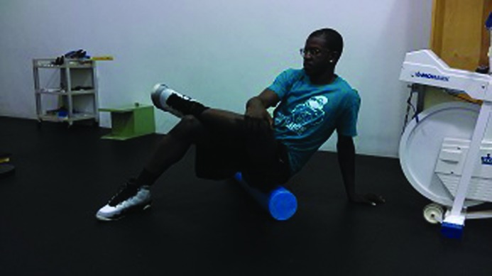
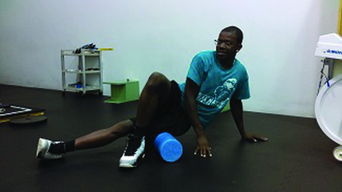
For the control session, the participant sat in a chair for three minutes prior to having the modified Ober test performed in the same manner as previously described for the post-test result. The procedure for the AFR and PFR sessions were the same as the control session with the exception of foam rolling being performed instead of resting for three minutes. For the AFR session, participants foam rolled over the gluteal muscle group for the hip that had the least amount of flexibility as determined during their first session. Participants sat on a 36 x 6 inch round, high-density ethylene vinyl acetate foam roller on the floor with their feet flat on the ground. The non-test leg was placed in a figure-four position, with the ankle placed just proximal to the opposite knee. The participant's hand on the test side was placed behind the participant on the floor for balance (Figure 1). Participants rolled the over gluteals, moving in a caudal-to-cephalad direction from the posterior superior iliac spine (PSIS) to the gluteal fold. Using a metronome to keep pace, participants foam rolled at a rate of 30 rolls per minute with rolling from the gluteal fold to the PSIS and back to the gluteal fold counting as one roll. Three sets of 30 seconds of foam rolling were completed with 30 seconds of rest between each set. For the PFR session, participants foam rolled over the ITB instead of the gluteal muscle group. During this session, the participant was in the side-lying position with the test leg bearing the weight of the foam roller. The ipsilateral hand was placed on the ground under the shoulder for support, and the contralateral leg was placed in front of the test leg with the foot flat on the floor for balance (Figure 2). Participants rolled in a caudal-to-cephalad direction from the greater trochanter to the lateral femoral condyle of the femur. Three sets of 30 seconds of foam rolling with 30 seconds of rest between sets were performed at the same pace as the AFR session. The post intervention Ober test was performed immediately after completing each FR session, and measured as previously described.
Figure 1.
Foam rolling the gluteal muscle group.
Figure 2.
Foam rolling for the iliotibial band.
Statistical Analysis
A two-way (session x time) Repeated Measures Analysis of Variance (RMANOVA) was used to assess statistical differences in ROM within and across each session. If the results of the RMANOVA were significant, paired t-tests using a Bonferroni adjustment was conducted as a post hoc analysis. Intrarater reliability was assessed across all three pretest measures using intaclass correlation coefficient (ICC) model 3,1 according to Shrout and Fleiss.28 All statistical analyses were performed using SPSS (version 22, IBM Corporation, Armonk, NY). The alpha level was set to 0.05. Results are reported as means ± SD.
RESULTS
Table 1 provides the hip adduction ROM measures from each session. AFR resulted in a 14% improvement in modified Ober measurement, whereas PFR saw a 1.1% improvement in modified Ober PROM. Controls had a 2% decrease in modified Ober testing. Results from the two-way RMANOVA revealed a significant interaction for session and time (F(2,25) = 25.202, p < 0.001,
= 0.502, 1 – β = 1.000). Preliminary analysis of a one way RMANOVA was performed on pre-test measurements across sessions. There was no significant difference across pre-test measurements (p > .05). The ICC for the pretest measurements was 0.815 (95% confidence interval = 0.680, 0.905) indicating a high level of reliability.28 Post hoc analysis showed that the post-test measurement for the AFR session was statistically significantly greater than the control (p < 0.001) or PFR post-test measurements (p < 0.001). There was no significant difference in post-test measurements between control and PFR (p = 0.188).
Table 1.
Pre-test and post-test hip adduction ROM, presented as Mean (SD). All measurements are in degrees. Negative numbers represent degrees below horizontal (0 °).
| Session | Pretest | Posttest |
|---|---|---|
| Control | −26.0 (5.1) | −25.5 (5.4) |
| AFR | −25.9 (5.3) | −29.6 (5.5)* |
| PFR | −25.8 (5.8) | −26.1 (5.8) |
AFR = Foam Rolling over the gluteal muscles, PFR = Foam Rolling over the Ilio-tibial band
Represents a statistically significant different from Control and PFR post-test measurements, p<0.05.
DISCUSSION
The results of this study suggest that foam rolling over the ITB directly has no immediate benefit in increasing hip adduction ROM, as measured by the modified Ober test. However, FR over the gluteal region resulted in an immediate, statistically significant increase in hip adduction ROM.
The clinical significance of these changes in ROM are less well defined. Generally, changes in outcomes that are considered important are determined by the patient or the clinician, so any changes that exceed these expectations are considered significant.29 No standardized value for improvement in hip adduction ROM is currently available for clinical purposes. However, some have suggested that using standard deviation (SD) within a study can be used to determine clinically significant changes. Wyrwich30 reported 2.3 times SD would ensure clinical significance. Using this measure, AFR needs an improvement of 4.1 ° to approach clinical significance (the current study saw a change of 3.5 °), whereas PFR needed 6.4 ° to ensure clinical significance (the current study only saw a change of 0.3 °).
The results of the effects of AFR are consistent with other findings on FR over muscle-fascia tissue in that a statistically significant increase in ROM was observed. However, AFR resulted in an improvement of 14.3% in Ober test ROM, more than the 6.2%,29 4.2%3 and 2%31 ROM improvements found in other studies. Further, previous studies have only looked at the effects of FR over tissue that contains both muscle and its surrounding fascia.1-3,31-35 This study has included the effects of FR over the ITB, a tissue composed primarily of connective tissue, devoid of any significant contractile component.
The current findings may be due to differences in pressure impulse, as the target tissue in the AFR group in the current study (the gluteal muscles) is much smaller in length than the target tissues in other studies,2,3,33,34 therefore in order to maintain pace with the metronome, subjects would have to roll slower than if rolling larger tissues at the same pace, resulting in a greater impulse moment on the tissue.
This study is consistent in design with previous studies on the effects of FR. Precautions were taken to isolate the target tissue of the gluteals or ITB. FR pacing was similar to other studies4,33,35-37 as well as in frequency and duration dosages.31,35,38 Therefore, the significant difference in ROM in the current study is likely due to other factors.
There are three theories proposed to explain observed changes in ROM after FR. Most prevalent is that myofascial adhesions develop over time, resulting in reduced ROM.40 Advocates of FR proport that FR is able to reduce fascial adhesions,41 thus improving ROM. Second, alterations in blood flow and vascularization within the fascia are shown to change as a result of FR, which may lead to reduced neural inhibition.42,45 Finally, there is a proposed neurological mechanism that involves the facilitation of muscle relaxation / inhibition, which would occur to a greater degree in myofascial tissue than fascia alone.15
Myofascial Adhesions
Fascia is made of connective tissue, mostly collagen and elastin.21 It does have Pancinian corpuscles and Ruffini nerve endings, suggesting it may play a role in proprioception. It also has free nerve endings and chemoreceptors, suggesting it can be a source of pain.13,44 Histologically, fascia is composed primarily of fibroblasts, which maintain the extracellular matrix (ECM).21,45 There is some speculation that sustained static positions cause a colloidal, congealing of the fluid within the fascial fibers, within the gel-matrix - described as ‘fuzz’ by Hedley.46 It is postulated that this stiffening or thickening (fuzzing) of the colloidal gel results in a restriction of ROM and altered lines of pull on muscle action and restricted motion, termed “fascial adhesions”.46
The results of the current study suggest that if any fascial adhesions existed in the sample pool, FR over myofascial tissue would improve ROM, whereas an acute bout of FR over fascia alone would not acutely change ROM. However, the long-term effects of FR may warrant further research. Since the population for this study were devoid of any lower limb injuries, so the presence of fascial adhesions seems unlikely. Different results may be found in a pathological population.
Reese and Bandy25 found normative Ober measurements of -18.9 ° (+/-7.6 °), and Hudson and Darthuy47 reported Ober to be 20.9 ° (+/-4.3 °) in healthy subjects. As the current sample had significantly more hip adduction ROM on Ober test, mean = -26.0 º (SD = 5.1), it is possible that the sample population lacked fascial adhesions, thus the possibility of detecting significant changes due to any effect on fascia was limited. The sample pool was composed primarily of kinesiology students, who may have an inheritably healthier profile than the general population, as they may be more active and health conscious due to the nature of their studies. In addition, we cannot rule out possible subtle differences in stabilization of the lateral hip during the modified Ober since we did not measure the angle created by the lateral hip and table. Regardless, the significant change in PROM present after AFR suggests that other mechanisms may need to be considered.
Alterations in Vascularity
There is some support for the suggestion that foam rolling can improve blood supply to an area by improving the elasticity of the arteries.42,43 Neo-vascularization in connective tissue has been postulated as a source of pain in patellar mal-tracking48 and some tendinopathies.49 This in turn may lead to guarding and pain inhibition, reducing ROM. Repetitive foam rolling may damage new vessels and nerves that form during the neo-vascularization phase of the formation of collagen as fascia tissue proliferates, as has been shown to be the case in patellar mal-tracking,48 leading to reduced pain and possibly less pain inhibition. As this study only exposed the area to FR for one session, the dosage required to elicit these proposed changes may have been inadequate to measure the effect of any alterations in vascularity or perfusion due to FR. Further, as injury to the lower limb was an exclusion criteria in this study, it is possible that the subject pool lacked any vascular adhesions for FR to act upon, explaining the lack of significant findings in the PFR group. Outcomes in a clinical population may yield different results.
Neural Plastic Changes
Recent evidence suggests that the changes in ROM observed as a result of FR are due to neural excitability and improved facilitation of muscle tissue.7,13 GTOs are located in connective tissue.50 GTOs respond to slow stretch – which is simulated by FR – by reducing their firing rate, thus reducing tonus in adjacent muscle. This “softening” may account for a perceived increase in joint ROM.13
Effects on Ruffini corpuscles have been suggested as a cause for the changes due to FR.13 These slow-adapting receptors could alter their neural transduction with a sustained stretching stimulus. With reduced transmission to the brain, there is less for the brain to perceive, resulting in less efferent activity to target muscles, and therefore an overall improved neural effect on tissue ROM.51 This in turn could result in improved neuromuscular activity. Fascia does contain some contractile capacity much the same way as smooth muscle does.52 While it is conceivable that reduced neural input to the smooth muscles in the ITB may result in increased ITB length, this was not shown in this study. Given the chronic exposure to abnormal forces required to form the adaptive changes found in the ITB in overuse injuries, it is unlikely that a single bout of FR would produce the stimulation necessary to promote cellular remodeling. However, the effects of repeated exposure to non-contractile tissues has not been explored in the literature and warrants future investigation.
Cavanaugh et al7 suggested that a neural inhibition response occurs after FR that may reduce pain perception. It is possible that due to the increased afferent neural environment of the myofascial tissue compared to the ITB, a reduction in inhibitory neural drive allows for improved ROM in the AFR population. Similar improvements in pain tolerances have been reported by Aboodarda et al.6 The Ober test uses passive ROM to determine end-range, rather than pain, subjects may have experienced an increase in pain as the limb approached end-range. After FR, improvements in pain inhibition may account for the observed changes.
Limitations to the study and Future Considerations
While the AFR session had a significant increase in hip adduction ROM, PFR did not have a similar effect. However, this subject population had good initial flexibility, mean = -26.0 º (SD = 5.1). It is possible that a population with lower initial Ober ROM would yield different outcomes. The traditional theory behind FR's mechanism of action on connective tissue is an improved compliance of fascia, however it is difficult to ascertain if such adhesions exist. Given the health status of the test population, it is unlikely that these adhesions were present in any subjects.
Indeed, only one study confirms fascial adhesions with imaging findings. Baumann et al53 correlated contrast-enhanced MRI findings of increased tissue thickness and signal abnormalities with clinical findings (loss of ROM, weakness, pain and skin thickening) and biopsy results to fascial adhesions. However, the subjects in their population had an autoimmune trigger to the adhesions. These findings likely do not apply to the general population.
It is possible that the intensity, cadence or duration of the sessions were insufficient to elicit detectable changes in fascial tissue. MacDonald et al3 examined up to 20 minutes of FR on ROM of the knee. However, the outcome (6% increase in ROM) was no better in ROM changes than shorter duration studies. Sullivan et al2 used a massage roller at a cadence of 120 bpm and found a 4.3% improvement in sit-and-reach. However, the sit-and-reach is an assessment of neural length and its validity of a hamstring assessment tool is less reliable than other outcome measures for the hamstring.54 Further, massage roller may not be the same as a FR in regards of force application or impulse. The current study used subjects' body weight as the intensity of the pressure elicited on the roller. As the pressure was not standardized otherwise, it is possible that those with greater body weight or less fat mass could elicit greater force over the tissue, generating greater effect from FR. Greater adipose dissipate some of the pressure exerted on the underlying tissues. Further, pain from FR over the ITB could cause some pain inhibition not seen when addressing the gluteals using FR. However, body weight is a common force used in other studies.3,33-35,38
This study only included one follow-up measurement on the Ober test, immediately post FR or control. Markovic55 found the effects of FR on ROM can last as long as 24 hours post rolling. While it is unlikely that any increases in ROM from PFR are likely to be observed later, the duration of AFR would be useful to know. Further, the effects of repeated FR could show different results. Knowing the duration of effect would be beneficial in determining protocol for repeated FR.
There are several models of foam rolling devices available on the market. This study used a smooth, 36 x 6-inch round, high-density ethylene vinyl acetate foam roller for all sessions. Other studies have found ROM improvements using textured foam rollers, roller massage machines and manual roller massagers, although with varying degrees of improvement. However, to date no study has examined the effectiveness of different modalities within groups.
The percentage change in the AFR group in this study is greater than those found in other studies. This may be due to the increased impulse of the FR over the tissue. The gluteal is smaller in longitudinal length than that of the muscle groups used in other studies. As such, in order to maintain a cadence of 30 rolls per minute, subjects would have rolled at a slower velocity to maintain cadence over the smaller distance, resulting in a greater overall contact time and possibly greater effects from the FR. This difference may be due to greater impulse over the tissue, the FR composition or a combination of the two.
Further study on the effects of repeated exposure to FR on both fascial and myofascial tissue is logical, especially in this region, given these findings. Additionally, the effective of various durations, cadences and repetitions of FR warrants investigation. Current dosages appear to be consistent within the literature, however, justification for these dosages appear to be lacking. Finally, comparison of the effects of foam rollers and rolling devices is lacking within the literature, and warrants further investigation.
CONCLUSION
The results of the current study suggest that foam rolling over the gluteal muscles is an effective means to significantly improve an immediate measure of passive hip adduction ROM. This is in accordance with other studies on the effects of FR on muscle tissue length. However, a single bout of foam rolling over the ITB did did not produce a significant increase in passive hip adduction ROM. It is unclear what effects repeated exposure to FR on the ITB may have on Ober test and ROM in general.
References
- 1.Mohr AR. Foam rolling can improve range of motion without effect on performance. J Strength Condit Res. 2013;37(3):812-821. [Google Scholar]
- 2.Sullivan KM Sivley DBJ Button DC et al. Roller-massager application to the hamstrings increases sit-and-reach range of motion within five to ten seconds without performance impairments. Int J Sports Phys Ther. 2013;8(3):1-9. [PMC free article] [PubMed] [Google Scholar]
- 3.MacDonald GZ Penney MDH Mullaley ME et al. An acute bout of myofascial release increases range of motion without a subsequent decrease in muscle activation or force. J Strength Cond Res. 2013;27(3):812–821. [DOI] [PubMed] [Google Scholar]
- 4.Pearcey EPG Bradbury-Squires DJ Kawamoto JE et al. Foam rolling for delayed-onset muscle soreness and recovery of dynamic performance measures. J Athl Train. 2015;50(1):5-13. [DOI] [PMC free article] [PubMed] [Google Scholar]
- 5.MacDonald GZ Button DC Drinkwater EJ et al. Foam rolling as a recovery tool after an intense bout of physical activity. Med Sci Sports Exerc. 2014;46(1):131-142. [DOI] [PubMed] [Google Scholar]
- 6.Aboodarda SJ Spence AJ Button DC. Pain pressure threshold of a muscle tender spot increases follow local and non-local rolling massage. BMC Musculoskelet Disord. 2015;16:265. [DOI] [PMC free article] [PubMed] [Google Scholar]
- 7.Cavanaugh MT Doweling A Young JD et al. An acute session of roller massage prolongs voluntary torque development and diminishes evoked pain. Euro J Appl Physiol. 2016;117(1):109-117. [DOI] [PubMed] [Google Scholar]
- 8.Green ST. Patellofemoral syndrome. J Bodyw Mov Ther. 2005;9(1):16-26. [Google Scholar]
- 9.Fredericson M Wolf C. Iliotibial band syndrome in runners – Innovations and treatment. Sports Med. 2005;35(5):451-459. [DOI] [PubMed] [Google Scholar]
- 10.Strauss EJ Nho SJ Kelly BT. Greater trochanteric pain syndrome. Sports Med Arthros. 2010;18(2):113-119. [DOI] [PubMed] [Google Scholar]
- 11.Sullivan KM Sivley DBJ Button DC et al. The immediate effects of self-myofascial release on female footballers. Sports Exerc Dynamics. 2013;38(10):12-17 [Google Scholar]
- 12.Chaudhry H Schleip R Ji Z et al. Three-dimensional mathematical model for deformation of human fasciae in manual therapy. J Am Osteopath Assoc. 2008;108:379-390. [DOI] [PubMed] [Google Scholar]
- 13.Schleip R. Fascial plasticity – a new neurobiological explanation. J Bodyw Mov Ther. 2003;7(1):11-19 and 7(2):104-116. [Google Scholar]
- 14.Curran PF Fiore RD Crisco JJ. A Comparison of the Pressure Exerted On Soft Tissue by 2 Myofascial Rollers. J Sport Rehabil. 2008;17:432-442 [DOI] [PubMed] [Google Scholar]
- 15.Cassar MP. Handbook of Clinical Massage: A Complete Guide for Students and Practitioners. 2nd ed 2004. London: Churchill Livingstone. [Google Scholar]
- 16.Schleip R. Fascial tonicity: from tissue contractures to hypermobility. Proceedings from the fascia research congress. 2007. Cambridge, MA. [Google Scholar]
- 17.Gray HFR. Gray's anatomy. 15th ed 1995. New York, NY: Barnes & Noble. [Google Scholar]
- 18.Marieb EN Mallatt J. Human Anatomy. 1997. Menlo Park, California: Benjamin / Cummings. [Google Scholar]
- 19.Evans P. The postural function of the ilitibial tract. Ann R Coll Surg. 1979;l61:271-280. [PMC free article] [PubMed] [Google Scholar]
- 20.Gose JC Schweizer P. Iliotibial band tightness. J Orthop Sports Phys Ther. 1989;4:399-407. [DOI] [PubMed] [Google Scholar]
- 21.Myer T. Anatomy Trains: Myofascial Meridians for Manual and Movement Therapists, 3e. 2014. London: Churchill-Livingstone. [Google Scholar]
- 22.Flack NAMS Nicholson HD Woodley SJ. A review of the anatomy of the hip abductor muscles, gluteus medius gluteus minimus and tensor fascia lata. Clinical Anat. 2012;25:697-708. [DOI] [PubMed] [Google Scholar]
- 23.Fairclough J Hayashi K Toumi K et al. The functional anatomy of the iliotibial band during flexion and extension of the knee: implications for understanding iliotibial band syndrome. J Anat. 2006;208:309-316. [DOI] [PMC free article] [PubMed] [Google Scholar]
- 24.Gottschalk F Kourosh S Leveau B. The functional anatomy of the tensor fascia latae and the gluteus medius and minimus. J Anat. 1989;166:179-789. [PMC free article] [PubMed] [Google Scholar]
- 25.Reese NB Bandy WD. The use of an inclinometer to measure flexibility of the iliotibial band using the Ober test and the Modified Ober test: Differences in magnitude and reliability of measurements. J Orthop Sports Phys Ther. 2003;33(6):326-330. [DOI] [PubMed] [Google Scholar]
- 26.Melchione W Sullivan M. The reliability of measurements obtained by use of an instrument designed to indirectly measure iliotibial band length. J Orthop Sports Phys Ther. 1993;18:511-515. [DOI] [PubMed] [Google Scholar]
- 27.Piva SR Fitzgerald K Irrgang J et al. Reliability of impairments associated with patellafemoral pain syndrome. BMC Musculoskelet Disord. 2006;7(33). [DOI] [PMC free article] [PubMed] [Google Scholar]
- 28.Shrout PE Fleiss JL. Intraclass correlations: uses in assessing rater reliability. Psychol Bull. 1979;86(2):420-428. [DOI] [PubMed] [Google Scholar]
- 29.Page P. Beyond statistical significance: clinical interpretation of rehabilitation research literature. Int J Sports Phys Ther. 2014;9(5):726-736. [PMC free article] [PubMed] [Google Scholar]
- 30.Wyrwich KW. Minimal important difference thresholds and the standard error of measurement: is there a connection? J Biopharm Stat. 2004;14(1):97-110. [DOI] [PubMed] [Google Scholar]
- 31.Škarabot J Beardsley C Štirm I. Comparing the effects of self-myofascial release with static stretching on ankle range-of-motion in adolescent athletes. Int J Sports Phys Ther. 2015;10(2):203-212. [PMC free article] [PubMed] [Google Scholar]
- 32.Vigotsky AD Lehman GJ Contreras B et al. Acute effects of anterior thigh foam rolling on hip angle #knee |angle, and rectus femoris length in the modified Thomas test. Peer J. 2015;3e:1281. [DOI] [PMC free article] [PubMed] [Google Scholar]
- 33.Mohr AR Long BC Goad CL. Effect of foam rolling and static stretching on passive hip-flexion range of motion. J Sport Rehabil. 2014;23(4):296-9. [DOI] [PubMed] [Google Scholar]
- 34.Bushell JE Dawson SM Webster MM. Clinical relevance of foam rolling on hip extension angle in a functional lunge position. J Strength Cond Res. 2015;29(9):2397-2403. [DOI] [PubMed] [Google Scholar]
- 35.Halperin I Aboodarda SJ Button DC et al. Roller massager improves range of motion of plantar flexor muscles without subsequent decreases in force parameters. Int J Sports Phys Ther. 2014;9(1):92-102. [PMC free article] [PubMed] [Google Scholar]
- 36.Jay K Sundstrup E Sondergaard SD, et al. Specific and cross over effects of massage for muscle soreness: randomized controlled trial. Int J Sports Phys Ther. 2014;9(1):82-91. [PMC free article] [PubMed] [Google Scholar]
- 37.Bradbury-Squires DJ Noftall JC Sullivan KM et al. Roller massager application to the quadriceps and knee-joint range of motion and neuromuscular efficiency during a lunge. J Athl Train. 2015;59(2):133-140. [DOI] [PMC free article] [PubMed] [Google Scholar]
- 38.Peacock CA Drein DD Antonio J et al. Comparing acute bouts of sagittal plane progression foam rolling vs. frontal plane progression foam rolling. J Strength Cond Res. 2015;29(8):2310-2315. [DOI] [PubMed] [Google Scholar]
- 39.Healey K Dorfman L Riebe D et al. The Effects of Foam Rolling on Myofascial Release and Performance. J Strength Cond Res. 2014;28(1):61-68. [DOI] [PubMed] [Google Scholar]
- 40.Rolf I. Rolfing: The Integration of Human Tissues. 1977. Santa Monica, CA: Dennis Landman Pub. [Google Scholar]
- 41.Barnes MF Myofascial Release: Morphologic change in connective tissue. J Bodyw Mov Theor. 1997;1(4):213-238. [Google Scholar]
- 42.Okamoto T Masuhara M Ikuta K. Acute effects of self-myofascial release using a foam roller on arterial function. J Strength Cond Res. 2014;28:69–73. [DOI] [PubMed] [Google Scholar]
- 43.Takanobu O Mitsuhiko M Komei I. Sensory innervation of the human thoracolumbar fascia: An immunohistochemical study. J Strength Cond Res. 2014;28(1):69-73.23575360 [Google Scholar]
- 44.Yahia L Rhalmi S Newman N et al. Sensory innervation of human thoracolumbar fascia. An immunohistochemical study. Acta Orthopaedica Scandinavia. 1992;63(2):195-7. [DOI] [PubMed] [Google Scholar]
- 45.Stecco C Gagey O Belloni A et al. Anatomy of the deep fascia of the upper limb. Second part: study of innervation. Morphologie. 2007;91(292):38-43. [DOI] [PubMed] [Google Scholar]
- 46.Hedley G. Demonstration of the Integrity of Human Superficial Fascia as an Autonomous Organ (Oral Presentation). First International Fascia Research Congress Cambridge, MA. November 23, 2007.
- 47.Hudson Z Darthuy E. Iliotibial band tightness and patellofemoral pain syndrome: A case-control study. Musculoskelet Sci Pract. 2009;14(2):147-151. [DOI] [PubMed] [Google Scholar]
- 48.Beidert RM Stauffer E Friederich NF. Occurrence of free nerve endings in the soft tissue of the knee joint. A histological investigation. Am J Sports Med. 1992;20(4):430-433. [DOI] [PubMed] [Google Scholar]
- 49.Malliaras P Purdam C Maffulli N Cook JL. Temporal sequence of gray-scale ultrasound changes and their relationship with neovascularity and pain in the patellar tendon. Br J Sports Med. 2010;44(13):944–947. [DOI] [PubMed] [Google Scholar]
- 50.Cottingham JT. Healing through touch – A history and review of the physiological evidence. 1985. Boulder CO: Rolf Institute Publications. [Google Scholar]
- 51.Butler DS Mosely GL. Explain Pain. 2013. Adelaide, Australia: NOI Group. [Google Scholar]
- 52.Schleip R Klingler W Lehmann-Horn F. Fascia is able to contract in a smooth muscle-like manner and thereby influence musculoskeletal mechanics. Proceedings of the 5th World Congress of Biomechanics, Munich. 2006.
- 53.Baumann F Brühlmann P Andreisek G et al. MRI for diagnosis and monitoring of patients with eosinophilic fasciitis. Am J Roentgenol. 2005;184:169–174 [DOI] [PubMed] [Google Scholar]
- 54.Davis SD Quinn RO Whiteman CT et al. Concurrent validity of four clinical tests used to measure hamstring flexibility. J Strength Cond Res. 2008;22(2):583-588. [DOI] [PubMed] [Google Scholar]
- 55.Markovic G. Acute effects of instrument assisted soft tissue mobilization vs. foam rolling on knee and hip range of motion in soccer players. Bodyw Mov Ther. 2015;19(4):690-696. [DOI] [PubMed] [Google Scholar]


