Abstract
Context:
Greater passive hip range of motion (ROM) has been associated with greater dynamic knee valgus and thus the potential for increased risk of anterior cruciate ligament injuries. Normative data for passive hip ROM by sex are lacking.
Objective:
To establish and compare passive hip ROM values by sex and sport and to quantify side-to-side differences in internal-rotation ROM (ROMIR), external-rotation ROM (ROMER), and total ROM (ROMTOT).
Design:
Cross-sectional study.
Setting:
Station-based, preparticipation screening.
Patients or Other Participants:
A total of 339 National Collegiate Athletic Association Division I athletes, consisting of 168 women (age = 19.2 ± 1.2 years, height = 169.0 ± 7.2 cm, mass = 65.3 ± 10.2 kg) and 171 men (age = 19.4 ± 1.3 years, height = 200.0 ± 8.6 cm, mass = 78.4 ± 12.0 kg) in 6 sports screened over 3 years: soccer (58 women, 67 men), tennis (20 women, 22 men), basketball (28 women, 22 men), softball or baseball (38 women, 31 men), cross-country (18 women, 19 men), and golf (6 women, 10 men).
Main Outcome Measure(s):
Passive hip ROM was measured with the athlete lying prone with the hip abducted to 20° to 30° and knee flexed to 90°. The leg was passively internally and externally rotated until the point of sacral movement. Three measures were averaged for each direction and leg and used for analysis. We compared ROMIR, ROMER, ROMTOT (ROMTOT = ROMIR + ROMER), and relative ROM (ROMREL = ROMIR − ROMER) between sexes and among sports using separate 2 × 6 repeated-measures analyses of variance.
Results:
Women had greater ROMIR (38.1° ± 8.2° versus 28.6° ± 8.4°; F1,327 = 91.74, P < .001), ROMTOT (72.1° ± 10.6° versus 64.4° ± 10.1°; F1,327 = 33.47, P < .001), and ROMREL (1.5° ± 16.0° versus −7.6° ± 16.5°; F1,327 = 37.05, P < .001) than men but similar ROMER (34.0° ± 12.2° versus 35.8° ± 11.5°; F1,327 = 1.65, P = .20) to men. Cross-country athletes exhibited greater ROMIR (37.0° ± 9.3° versus 30.9° ± 9.4° to 33.3° ± 9.5°; P = .001) and ROMREL (5.9° ± 18.3° versus −9.6° ± 16.9° to −2.7° ± 17.3°; P = .001) and less ROMER (25.7° ± 7.5° versus 35.0° ± 13.0° to 40.2° ± 12.0°; P < .001) than basketball, soccer, softball or baseball, and tennis athletes. They also displayed less ROMTOT (62.7° ± 8.1° versus 70.0° ± 9.1° to 72.9° ± 11.9°; P < .001) than basketball, softball or baseball, and tennis athletes.
Conclusions:
Women had greater ROMIR than men, resulting in greater ROMTOT and ROMREL. Researchers should examine the extent to which this greater bias toward ROMIR may explain women's greater tendency for dynamic knee valgus. With the exception of cross-country, ROM values were similar across sports. The clinical implications of these aberrant cross-country values require further study.
Key Words: anterior cruciate ligment injury, screening, risk factor, normative data
Key Points
Women had greater internal-rotation range of motion (ROM) than men regardless of sport, which resulted in women having greater total and relative ROM.
The greater bias toward internal-rotation ROM may help explain the greater potential for females to display greater dynamic knee valgus.
Cross-country athletes had greater internal-rotation ROM and less external-rotation ROM than athletes in every other sport studied except golf.
The appreciable bilateral differences in some athletes suggested that measurements on 1 limb did not necessarily represent the other limb.
Of the approximately 200 000 anterior cruciate ligament (ACL) injuries sustained each year in the United States, 70% occur via noncontact mechanisms.1,2 Dynamic knee valgus, comprising hip internal rotation (IR), hip adduction, knee abduction, and tibial external rotation (ER), is thought to contribute to noncontact ACL injury.3 This is particularly true in females, who are more likely to display greater dynamic knee valgus during inciting injury mechanisms than their male counterparts based on retrospective videographic evidence.3 However, less is known about factors precipitating dynamic knee valgus. Given that dynamic knee valgus is composed of hip-knee coupling in the frontal and transverse planes and movement occurs in a proximal-to-distal pattern,4 a lack of control at the hip may be key to a lack of control at the knee.
From this perspective, available passive hip range of motion (ROM) has been presented as a potential contributor to dynamic knee valgus.5,6 Whereas hip ROM can be influenced by active, or muscular, restraints, it is largely dictated by inert capsular constraints.7 Therefore, greater passive ROMIR may predispose an individual to move into more dynamic hip adduction and IR, leading to greater dynamic knee valgus. This may be especially apparent when greater ROMIR occurs in conjunction with a lack of dynamic hip strength and control. In separate studies,5,8 females with greater hip ROMIR and less ROMER moved into more dynamic knee valgus throughout landing tasks. Combining these 2 variables (ROMIR − ROMER) to represent an ROMIR bias, Howard et al6 showed that females who had greater ROMIR relative to ROMER, along with weak hip abductors and external rotators, were more likely to move into greater knee abduction during a single-legged landing (R2 = 0.47, P < .001). Whereas dynamic hip strength and control have long been considered a primary cause of dynamic knee valgus, these data suggest that excessive hip ROM may also play a role.
In addition to isolated ROMIR and ROMER values, total hip ROM (ROMTOT), which is the sum of ROMIR and ROMER, may also contribute to the risk of ACL injury.9 Individuals with greater ROMTOT also display other characteristics thought to increase ACL injury risk. For instance, greater ROMTOT has been associated with generalized joint laxity (r = 0.57),10 a prospectively identified ACL injury risk factor in females.11
Although passive hip ROM during functional activity has been empirically linked to dynamic knee valgus during functional activity6,12 and females have been reported to differ from males in passive hip ROM,10 literature quantifying normative ranges of motion by sex is lacking. This gap can be particularly problematic considering that females may exhibit different injury mechanisms than males.3 Previous reports of transverse-plane hip ROM values are presented in Table 1. Researchers who examined sex differences in ROMIR and ROMER have suggested that females possess more ROMIR than ROMER and more ROMIR than males in a healthy population10 and elite tennis players.16 However, as a result of the limited data on athletic populations, these sex comparisons may not be generalizable to individuals participating across a variety of sports. Furthermore, available ROM data by sport are limited to soccer and tennis athletes.8,15 Because of different lower extremity demands among sports, passive hip ROM may differ by sport. More importantly, given the higher rates of ACL injury in sports such as basketball and soccer2,17 and the evidence of a greater sex disparity in ACL injuries in these sports, understanding the sex- and sport-specific patterns of hip ROM may help explain these disparities. Therefore, the primary purpose of our study was to establish normative values for absolute and relative passive hip ROM (ROMIR, ROMER, ROMTOT, and relative ROM [ROMREL]) for each sex and various sports and to compare these values between sexes and among sports. We hypothesized that women would exhibit greater ROMIR, and thus greater ROMREL and ROMTOT, than men. With the higher rates of ACL injury in basketball and soccer players, we also hypothesized that women participating in these sports would have greater ROMIR,2,17 which would result in higher ROMREL values. Our secondary purpose was to quantify the side-to-side differences for ROMIR, ROMER, and ROMTOT to help clinicians determine whether 1 limb can represent both limbs during screening protocols and for return-to-play criteria.
Table 1.
Currently Available Hip Range-of-Motion Data
| Authors (Year) |
Population |
No. |
Range of Motion (Mean ± SD), ° |
|
| Internal Rotation |
External Rotation |
|||
| Nyland et al13 (2004) | Female, active | 18 | 43.3 ± 11 | Not applicable |
| Sigward et al8 (2008) | Female, soccer | 39 | 42.7 ± 9.9 | 38.9 ± 8.6 |
| Chiaia et al14 (2009) | Female, soccer | 26 | 32.5 ± 7.7 | 24.5 ± 6.3 |
| Howard et al6 (2011) | Male and female, active | 45 | 29 ± 11 | 35 ± 7 |
| Tainaka et al15 (2014) | Male and female, active | 123 | 50.2 ± 7.2a | 56.3 ± 6.8a |
| Young et al16 (2014) | Female, tennis | 125 | 38 ± 41 | 23 ± 24 |
| Fan et al10 (2014) | Male, healthy | 16 | 35.1 ± 4.7 | 41.9 ± 6.6 |
| Female, healthy | 16 | 42.7 ± 10.3 | 41.1 ± 6.4 | |
| Moreno-Pérez et al19 (2015) | Male, tennis | 81 | 30.0 ± 9.6 | 50.6 ± 8.0 |
| Female, tennis | 28 | 35.6 ± 8.2 | 49.3 ± 6.8 | |
Measured supine.
METHODS
We recruited 339 National Collegiate Athletic Association Division I varsity athletes from 1 institution over 3 years. Participants were 168 women (age = 19.2 ± 1.2 years, height = 169.0 ± 7.2 cm, mass = 65.3 ± 10.2 kg) and 171 men (age = 19.4 ± 1.3 years, height = 200.0 ± 8.6 cm, mass = 78.4 ± 12.0 kg). Female and male baseball, basketball, cross-country, golf, soccer, softball, and tennis athletes were included. The numbers of participants by sex, sport, and examiner are presented in Table 2. Athletes not cleared for full participation at the time of their preparticipation examinations were excluded from the study. All recruits provided written informed consent, and the study was approved by the Institutional Review Board at the University of North Carolina at Greensboro.
Table 2.
Number of Participants by Examiner, Sex, and Sport
| Participant |
Examiner 1 |
Examiner 2 |
Total |
| Women, No. | 75 | 93 | 168 |
| Soccer | 30 | 28 | 58 |
| Tennis | 11 | 9 | 20 |
| Basketball | 15 | 13 | 28 |
| Softball/baseball | 19 | 19 | 38 |
| Cross-country | 0 | 18 | 18 |
| Golf | 0 | 6 | 6 |
| Men, No. | 51 | 120 | 171 |
| Soccer | 30 | 37 | 67 |
| Tennis | 12 | 10 | 22 |
| Basketball | 9 | 13 | 22 |
| Softball/baseball | 0 | 31 | 31 |
| Cross-country | 0 | 19 | 19 |
| Golf | 0 | 10 | 10 |
| Total | 126 | 213 | 339 |
Procedures
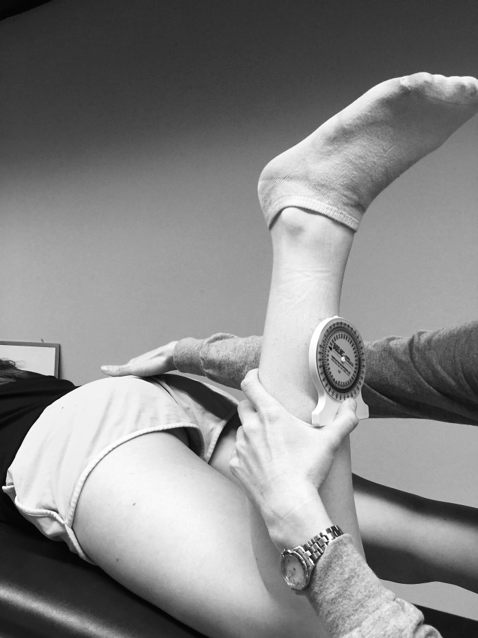
All data were collected using established measurement techniques18 in conjunction with the athletes' preparticipation examinations. Only data from the earliest year of data collection were included. We obtained all passive hip ROM measurements bilaterally using a 0° to 360° bubble inclinometer (Saunders Manufacturing, Readfield, ME) with 2° increments that was positioned on the anterior side of the tibia along the longitudinal tibial axis. Each participant was positioned prone with the hip in 0° of extension and 20° to 30° of abduction (Figure 1); sagittal-plane neutral hip positioning minimized muscular involvement. The knee was flexed to 90°. The leg was passively rotated internally and externally until initial sacral tilt as determined by the examiner's palpation. At this point, the transverse angle formed by true vertical and the tibial diaphysis were considered ROMER and ROMIR, respectively. We summed ROMER and ROMIR to calculate ROMTOT. To calculate ROMREL, we subtracted ROMER from ROMIR. This relative ROMIR variable retained the original degree unit and has been used in the literature to represent the amount of bias toward ROMIR.6 Three trials were obtained bilaterally for ROMIR and ROMER and averaged for analysis. Two examiners (J.A.H., A.N.) with good to excellent between-days reliability (intraclass correlation coefficient [2,3; standard error of measurement]) took all measurements (examiner 1: ROMIR = 0.97 [1.6°], ROMER = 0.85 [3.3°]; examiner 2: ROMIR = 0.87 [2.5°], ROMER = 0.83 [1.8°]). A single examiner (J.A.H. or A.N.) took all measurements within each year of testing. The numbers of men and women measured by each examiner were relatively balanced except for softball and baseball, as baseball was only measured for 1 year by examiner 2. The distribution of sports between examiners was also relatively balanced except for cross-country and golf, which were measured only by examiner 2 (Table 2).
Figure 1.
Positioning for all range-of-motion measures. The participant lay prone with the hip slightly abducted and the knee flexed. The examiner passively rotated the tibia while monitoring sacral tilt.
Statistics
Histograms were constructed for each variable of interest to inspect normality of distribution. Descriptive statistics (means and standard deviations) were also computed for each variable. Paired t tests confirmed no appreciable differences in bilateral ROMIR or ROMER. Therefore, four 2 × 6 (sex by sport) between-subjects analyses of variance were performed with average ROMIR, ROMER, ROMTOT, and ROMREL as dependent variables. Except for softball and baseball, all sports were represented by both male and female athletes. Given the similar lower extremity demands of softball and baseball, these sports were considered as 1 sport for comparisons between sexes and with the other sports. To adjust for all 4 comparisons using a Bonferroni correction, we set the a priori α level for the omnibus analysis-of-variance models at .0125. Tukey post hoc analyses (α ≤ .05) were applied to main effects as appropriate. We computed left-right limits of agreement and Bland-Altman plots to determine the suitability of using measures on 1 limb to represent both limbs. We used SPSS for all primary analyses (version 21; IBM Corp, Armonk, NY). Limits of agreement and Bland-Altman plots were computed using R (2015; R Foundation for Statistical Computing, Vienna, Austria).
RESULTS
Means and standard deviations for each variable, stratified by sex and sport, are presented in Table 3. Women exhibited greater ROMIR (38.1° ± 8.2° versus 28.6° ± 8.4°; F1,327 = 91.74, P < .001), ROMTOT (72.1° ± 10.6° versus 64.4° ± 10.1°; F1,327 = 33.47, P < .001), and ROMREL (1.5° ± 16.0° versus −7.6° ± 16.5°; F1,327 = 37.05, P < .001) than men. We observed similar ROMER between women (34.0° ± 12.2°) and men (35.8° ± 11.5°; F1,327 = 1.65, P = .20). Main effects by sport were present for ROMIR (F5,327 = 4.51, P = .001), ROMER (F5,327 = 9.47, P < .001), ROMTOT (F5,327 = 6.10, P < .001), and ROMREL (F5,327 = 11.51, P = .001). Follow-up post hoc analyses revealed that cross-country athletes exhibited greater ROMIR (37.0° ± 9.3° versus 30.9° ± 9.4° to 33.3° ± 9.5°), lesser ROMER (25.7° ± 7.5° versus 35.0° ± 13.0° to 40.2° ± 12.0°), and greater ROMREL (5.9° ± 18.3° versus −9.6° ± 16.9° to −2.7° ± 17.3°) than basketball, soccer, softball and baseball, and tennis athletes and less ROMTOT (62.7° ± 8.1° versus 70.0° ± 9.1° to 72.9° ± 11.9°) than basketball, softball and baseball, and tennis athletes. We observed no interactions for any variable by sex and sport (P range = .32–.82).
Table 3.
Hip Range of Motion by Sport and Sex
| Range of Motion (Mean ± SD), ° |
||||
| Internal Rotation |
External Rotation |
Total |
Relative Internal Rotation |
|
| Basketball | ||||
| Women | 37.9 ± 7.7 | 38.4 ± 11.2 | 76.2 ± 11.1 | −0.5 ± 14.0 |
| Men | 28.7 ± 9.5 | 39.9 ± 12.7 | 68.6 ± 11.8 | −11.1 ± 18.5 |
| Total | 33.3 ± 9.5 | 39.1 ± 11.8 | 72.9 ± 11.9 | −5.2 ± 16.8 |
| Cross-country | ||||
| Women | 41.4 ± 9.2 | 23.3 ± 7.1 | 64.7 ± 8.7 | 14.2 ± 15.9 |
| Men | 32.6 ± 7.2 | 28.5 ± 6.2 | 60.7 ± 7.3 | −1.9 ± 17.3 |
| Total | 37.0 ± 9.3a | 25.7 ± 7.5a | 62.7 ± 8.1b | 5.9 ± 18.3a |
| Golf | ||||
| Women | 41.7 ± 4.3 | 31.5 ± 7.6 | 73.2 ± 7.5 | 10.2 ± 8.4 |
| Men | 27.9 ± 7.7 | 34.3 ± 6.0 | 62.2 ± 5.6 | −7.5 ± 13.4 |
| Total | 34.8 ± 9.5 | 32.9 ± 6.6 | 66.3 ± 8.2 | −0.9 ± 14.5 |
| Soccer | ||||
| Women | 35.8 ± 7.5 | 34.9 ± 13.2 | 70.7 ± 10.9 | 1.4 ± 16.4 |
| Men | 26.2 ± 8.4 | 35.2 ± 12.9 | 61.4 ± 11.0 | −6.3 ± 17.4 |
| Total | 31.0 ± 9.3 | 35.0 ± 13.0 | 65.7 ± 11.9c | −2.7 ± 17.3 |
| Softball or baseball | ||||
| Women | 36.2 ± 8.4 | 38.7 ± 10.4 | 74.9 ± 8.2 | −2.5 ± 16.0 |
| Men | 29.4 ± 6.9 | 34.5 ± 6.4 | 63.9 ± 6.0 | −5.1 ± 10.8 |
| Total | 32.8 ± 8.5 | 36.6 ± 9.1 | 70.0 ± 9.1 | −3.6 ± 13.9 |
| Tennis | ||||
| Women | 35.3 ± 7.9 | 37.4 ± 11.8 | 72.7 ± 11.5 | −2.1 ± 14.1 |
| Men | 26.6 ± 8.9 | 43.0 ± 11.6 | 69.6 ± 10.3 | −16.5 ± 16.6 |
| Total | 30.9 ± 9.4 | 40.2 ± 12.0 | 71.1 ± 10.9 | −9.6 ± 16.9 |
| Total | ||||
| Women | 38.1 ± 8.2d | 34.0 ± 12.2 | 72.1 ± 10.6d | 1.5 ± 16.0d |
| Men | 28.6 ± 8.4d | 35.8 ± 11.5 | 64.4 ± 10.1d | −7.6 ± 16.5d |
Different from all groups except golf (P < .01).
Different from softball/baseball, basketball, and tennis (P < .01).
Different from basketball (P < .01).
Different from all other groups (P < .01).
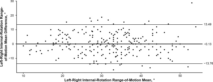
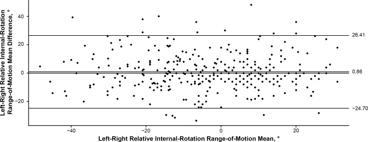
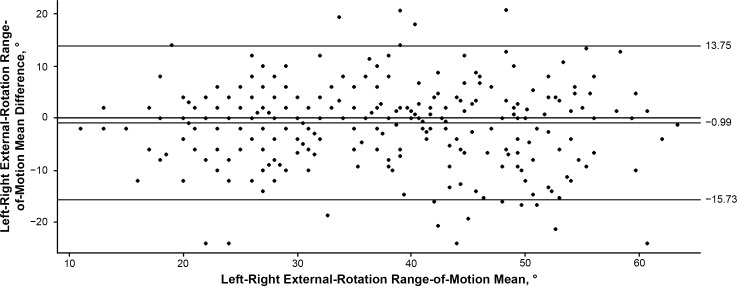
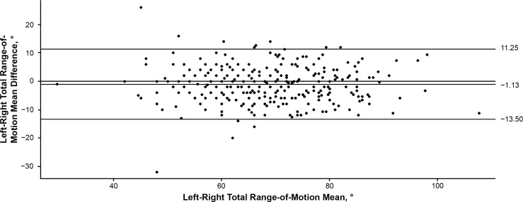
When we examined bilateral asymmetries, mean side-to-side differences (left-right) for ROMIR, ROMER, ROMTOT, and ROMREL were −0.13°, −0.99°, −1.13°, and 0.86°, respectively, indicating minimal systematic bias. We observed that 68% and 95% of values were within 6.8° and 13.4° of the mean for ROMIR, 7.4° and 14.5° for ROMER, 6.2° and 12.1° for ROMTOT, and 12.8° and 25.0° for ROMREL. Bland-Altman plots are presented in Figures 2 through 5.
Figure 2.
Bland-Altman plot comparing the left-right internal-rotation range of motion, representing the left-right mean difference and 95% confidence interval.
Figure 5.
Bland-Altman plot comparing the left-right relative internal-external rotation range of motion, representing left-right mean difference and 95% confidence interval.
Figure 3.
Bland-Altman plot comparing the left-right external-rotation range of motion, representing the left-right mean difference and 95% confidence interval.
Figure 4.
Bland-Altman plot comparing the left-right total range of motion, representing the left-right mean difference and 95% confidence interval.
DISCUSSION
Our primary findings that women exhibited greater ROMIR than and similar ROMER to men, resulting in greater ROMTOT and ROMREL, supported our hypothesis. Given that ROMER was similar between sexes, the observed sex differences in ROMTOT and ROMREL can be attributed to the greater ROMIR in women. Secondarily, cross-country athletes, regardless of sex, displayed greater ROMIR and lesser ROMER, resulting in increased ROMREL.
For women, the ROMIR and ROMER values were 38.1° ± 8.2° and 34.0° ± 12.2°, respectively, which agreed with previous work using similar methods (Table 1). We located only 1 published study19 in which the authors used a prone measurement technique in an all-male cohort and reported values of 30.0° ± 9.6° for ROMIR and 50.6° ± 8.0° for ROMER, which were considerably higher than the values of 28.6° ± 8.4° and 35.8° ± 11.5°, respectively, in our study. Differences in measurement collection may explain this; in the previous study,19 the ROM endpoint was designated as the examiner's perception of firm resistance, whereas in our study, the endpoint was signified by initial sacral tilt, which would likely occur before the perception of firm resistance.
Differences Between Sexes
Women displayed greater ROMIR than men (38.1° ± 8.2° versus 28.6° ± 8.4°). This could have clinical implications with respect to observed sex disparities in ACL injuries and patellofemoral pain syndrome.20,21 Evidence6 has suggested that an increase in ROMIR may be associated with a higher prevalence of dynamic knee valgus, especially in females. During activity, an athlete with more available ROMIR might be predisposed to exhibit greater dynamic IR, which is a component of dynamic knee valgus that is thought to increase the risk of ACL injury.22,23 Researchers24,25 have also postulated that excessive dynamic IR of the hip may be associated with chronic, repetitive loading of the patellofemoral joint, leading to patellofemoral pain, a condition more commonly observed in females.
Whereas sex differences in ROMTOT were affected by ROMIR, we are hesitant to discount the potential importance of ROMTOT. Increased ROMTOT, as a measure of ligamentous laxity, may have implications for lower extremity injury risk. Total ROM has been associated with generalized joint laxity,10 a prospectively identified ACL injury risk factor in females,11 and may place greater demands on the hip musculature to stabilize the joint during dynamic activity. Investigators should determine the contributions of ROMTOT to dynamic knee control and potential injury risk, whether these contributions are separate from those of ROMIR, and whether differences in ROMTOT may partially explain observed sex disparities in lower extremity injuries.
To address potential between-sexes effects due to examiner differences, we conducted secondary analyses within the cohort measured by examiner 2. The results were consistent, as females displayed greater ROMIR (39.3° ± 7.3° versus 29.8° ± 7.5°) and ROMTOT (65.7° ± 8.5° versus 59.9° ± 8.7°) than males. Therefore, we contend that the observed sex differences are true differences and not a result of interexaminer differences.
Differences Among Sports
Despite our hypothesis that passive hip ROM would differ among sports, the divergent values displayed in cross-country, a sport not known for ACL injuries, are surprising. Cross-country athletes had greater ROMIR (37.0° ± 9.3° versus 30.9° ± 9.4° to 33.3° ± 9.5°) and less ROMER (25.7° ± 7.5° versus 35.0° ± 13.0° to 40.2° ± 12.0°) than athletes in the other sports, resulting in less ROMTOT (62.7° ± 8.1° versus 70.0° ± 9.1° to 72.9° ± 11.9°) and greater ROMREL (5.9° ± 18.3° versus −9.6° ± 16.9° to −2.7° ± 17.3°). Given that the data on all cross-country athletes were obtained by the same examiner (Table 2), secondary analyses were conducted within those athletes measured by examiner 2 to confirm that differences were not due to the examiner. Similar trends were revealed; cross-country athletes had greater ROMIR than athletes in all other sports (37.0° ± 9.3° versus 32.5° ± 7.1° to 36.8° ± 8.2°) and less ROMER than athletes in all other sports except soccer (25.7° ± 7.1° versus 30.3° ± 6.3° to 32.9° ± 6.6°, soccer = 24.3° ± 6.0°). Within examiners, these differences resulted in cross-country athletes having lower ROMTOT than athletes in all other sports except soccer (62.7° ± 8.1° versus 63.6° ± 7.8° to 67.1° ± 10.2°, soccer = 58.2° ± 9.5°) and greater ROMREL than athletes in all other sports except soccer and basketball (5.9° ± 18.3° versus −0.9° ± 14.9° to 0.9° ± 11.1°, soccer = 9.0° ± 12.1°, basketball = 6.6° ± 10.6°). Whereas having multiple testers is frequently a reality in multiseason screening, it is not ideal and often introduces “noise” into the data. Despite this, our secondary analyses suggested that the observed differences are true and cannot be fully attributed to discrepancies between examiners.
Given the lack of cutting and lateral movement in cross-country, excessive dynamic hip IR may not lead to an increased ACL injury risk in this population, but it may help explain the increased prevalence of patellofemoral pain syndrome in runners. Ho et al26 postulated that excessive dynamic hip IR repetitively loads the patellofemoral joint, leading to an acute change in patellar composition. Excessive dynamic hip IR during running has also been repeatedly suggested25,27,28 to contribute to the higher incidence of patellofemoral pain observed in female runners.
Side-to-Side Limits of Agreement
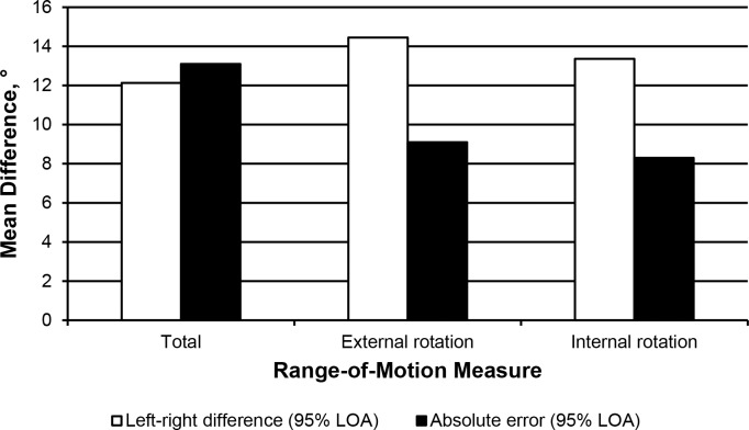
Calculating side-to-side differences via limits of agreement can aid clinicians in determining if 1 limb can be used as a suitable representation of both limbs. If passive hip ROM is equal bilaterally, the healthy limb could then serve as a baseline for the injured limb. In our sample, the average mean difference was close to zero, indicating little to no systematic bias present between left and right passive hip ROM. However, the observed magnitudes of left-right differences varied considerably. For ROMIR, ROMER, and ROMTOT, 32% of the sample had left-right ROM differences that exceeded 6.8°, 7.4°, and 6.2°, respectively, and 5% of the sample had left-right mean differences greater than 13.4°, 14.5°, and 12.1°. The observed left-right differences in ROMIR and ROMER were substantially greater than what we would expect simply because of test-retest measurement error (Figure 6), which suggests that, in some individuals, ROMIR and ROMER are appreciably different bilaterally, and therefore measurement of 1 limb may not adequately represent the other limb for screening purposes. In contrast to ROMIR and ROMER, left-right differences in ROMTOT were similar to the expected test-retest measurement error, indicating that measurement of the entire envelope of passive hip ROM on 1 side may appropriately represent the other side. Clinically, this may imply that some athletes exhibit a unilateral bias toward either ROMIR or ROMER while maintaining the same amount of ROMTOT, which was confirmed by the limits of agreement computed from ROMREL (Figure 5). One-third of the sample displayed side-to-side ROMREL differences exceeding 12.8°, and 5% exceeded bilateral differences of 25.0°, indicating that, in some athletes, the configuration of the passive hip ROM envelope may be markedly different bilaterally. This could result in a tendency for 1 limb to move into greater or lesser amounts of hip IR during movement. Howard et al6 used ROMREL as an estimate of femoral anteversion. Given that left-right differences in femoral anteversion have only been shown to exceed 6.9° in some individuals,29 more research is needed to determine the amount of ROMREL that can be explained by femoral anteversion and how much can be attributed to factors such as sport-specific adaptations.
Figure 6.
Comparison of absolute measurement error with the observed left-right differences. Abbreviation: LOA, limit of agreement.
Clinically, in some individuals, 1 limb may not be representative of the other with respect to ROMIR and ROMER. This may be especially important considering that potential left-right differences for ROMIR and ROMER account for a sizeable portion of the total available ROM. For example, two-thirds of our sample was within 6.8° of ROMIR bilaterally, and 95% was within 13.4°. For females, this could represent 19% to 37% of overall ROM, which may have important clinical implications. A bilateral difference of 5° of ROMIR may lead to more substantial consequences in an athlete with less ROMTOT than in an athlete with greater ROMTOT.
Our study had limitations. Whereas each examiner had established good to excellent intrarater reliability before data collection, we acknowledge that having 1 examiner obtain all measurements would have been preferable. Because the data were collected in nonconsecutive years, we were unable to establish intertester reliability. However, both examiners were experienced clinicians and were similarly trained in obtaining these measures using established laboratory measurement techniques. In addition, all data for any given year were collected by the same examiner, making it possible to compare means between sexes and among sports within each year and examiner to ensure data fidelity and that our primary findings held for both examiners. Another limitation was the accuracy of the bubble inclinometer used for collection, as we could record values only in 2° increments. However, the same instrument was used when assessing measurement error and our planned comparisons, and the observed differences well exceeded the measurement error. In addition, whereas these data were obtained from athletes medically cleared for full participation, athletes who had recently been injured or who had a history of lower extremity surgery were not excluded. Although we acknowledge that passive hip ROM may be affected by previous lower extremity surgery or injury, we maintain that our sample was representative of one typically found in athletics. Lastly, including normative hip-strength data would have rendered a more complete picture of how altered hip ROM couples with strength to influence lower extremity movement patterns. We did not collect these data, but they present an avenue for future research.
CONCLUSIONS
Women exhibited greater ROMIR than men regardless of sport, which resulted in women having greater ROMTOT and higher ROMREL. This may, in part, explain the increased potential for females to display greater dynamic knee valgus. Researchers should investigate the potential neuromuscular and biomechanical implications of these sex differences to determine the contributions of different ROM values to ACL injury risk and the extent to which passive hip ROM is modifiable or can be countered by dynamic hip strength and control and potentially serve as a target for injury-prevention strategies. Moreover, cross-country athletes displayed greater ROMIR and less ROMER than athletes in all other sports except golf. The causes of these divergent values and potential clinical implications warrant further investigation. Lastly, some athletes showed appreciable bilateral differences, suggesting that measurements on 1 limb do not necessarily represent the other limb. Reasons for these discrepancies, as well as the implications this may have on potential injury risk, require further study.
REFERENCES
- 1.Hewett TE, Shultz SJ, Griffin LY. Understanding and Preventing Noncontact ACL Injuries. Champaign, IL: Human Kinetics;; 2007. [Google Scholar]
- 2.Moses B, Orchard J, Orchard J. Systematic review: annual incidence of ACL injury and surgery in various populations. Res Sport Med. 2012;20(3–4):157–179. doi: 10.1080/15438627.2012.680633. [DOI] [PubMed] [Google Scholar]
- 3.Krosshaug T, Nakamae A, Boden BP, et al. Mechanisms of anterior cruciate ligament injury in basketball: video analysis of 39 cases. Am J Sports Med. 2007;35(3):359–367. doi: 10.1177/0363546506293899. [DOI] [PubMed] [Google Scholar]
- 4.Khamis S, Yizhar Z. Effect of feet hyperpronation on pelvic alignment in a standing position. Gait Posture. 2007;25(1):127–134. doi: 10.1016/j.gaitpost.2006.02.005. [DOI] [PubMed] [Google Scholar]
- 5.Nguyen A, Cone J, Stevens L, Schmitz R, Shultz S. Influence of hip internal rotation range of motion on hip and knee motions during landing [abstract] J Athl Train. 2009;44(suppl 3):S-68. [Google Scholar]
- 6.Howard JS, Fazio MA, Mattacola CG, Uhl TL, Jacobs CA. Structure, sex, and strength and knee and hip kinematics during landing. J Athl Train. 2011;46(4):376–385. doi: 10.4085/1062-6050-46.4.376. [DOI] [PMC free article] [PubMed] [Google Scholar]
- 7.Martin HD, Savage A, Braly BA, Palmer IJ, Beall DP, Kelly B. The function of the hip capsular ligaments: a quantitative report. Arthroscopy. 2008;24(2):188–195. doi: 10.1016/j.arthro.2007.08.024. [DOI] [PubMed] [Google Scholar]
- 8.Sigward SM, Ota S, Powers CM. Predictors of frontal plane knee excursion during a drop land in young female soccer players. J Orthop Sports Phys Ther. 2008;38(11):661–667. doi: 10.2519/jospt.2008.2695. [DOI] [PubMed] [Google Scholar]
- 9.Gomes JL, de Castro JV, Becker R. Decreased hip range of motion and noncontact injuries of the anterior cruciate ligament. Arthroscopy. 2008;24(9):1034–1037. doi: 10.1016/j.arthro.2008.05.012. [DOI] [PubMed] [Google Scholar]
- 10.Fan L, Copple TJ, Tritsch AJ, Shultz SJ. Clinical and instrumented measurements of hip laxity and their associations with knee laxity and general joint laxity. J Athl Train. 2014;49(5):590–598. doi: 10.4085/1062-6050-49.3.86. [DOI] [PMC free article] [PubMed] [Google Scholar]
- 11.Uhorchak JM, Scoville CR, Williams GN, Arciero RA, St Pierre P, Taylor DC. Risk factors associated with noncontact injury of the anterior cruciate ligament: a prospective four-year evaluation of 859 West Point cadets. Am J Sports Med. 2003;31(6):831–842. doi: 10.1177/03635465030310061801. [DOI] [PubMed] [Google Scholar]
- 12.Sigward SM, Cesar GM, Havens KL. Predictors of frontal plane knee moments during side-step cutting to 45 and 110 degrees in men and women: implications for anterior cruciate ligament injury. Clin J Sport Med. 2014;25(6):529–534. doi: 10.1097/JSM.0000000000000155. [DOI] [PMC free article] [PubMed] [Google Scholar]
- 13.Nyland J, Kuzemchek S, Parks M, Caborn DN. Femoral anteversion influences vastus medialis and gluteus medius EMG amplitude: composite hip abductor EMG amplitude ratios during isometric combined hip abduction-external rotation. J Electromyogr Kinesiol. 2004;14(2):255–261. doi: 10.1016/S1050-6411(03)00078-6. [DOI] [PubMed] [Google Scholar]
- 14.Chiaia TA, Maschi RA, Stuhr RM, et al. A musculoskeletal profile of elite female soccer players. HSS J. 2009;5(2):186–195. doi: 10.1007/s11420-009-9108-9. [DOI] [PMC free article] [PubMed] [Google Scholar]
- 15.Tainaka K, Takizawa T, Kobayashi H, Umimura M. Limited hip rotation and non-contact anterior cruciate ligament injury: a case-control study. Knee. 2014;21(1):86–90. doi: 10.1016/j.knee.2013.07.006. [DOI] [PubMed] [Google Scholar]
- 16.Young SW, Dakic J, Stroia K, Nguyen ML, Harris AHS, Safran MR. Hip range of motion and association with injury in female professional tennis players. Am J Sports Med. 2014;42(11):2654–2658. doi: 10.1177/0363546514548852. [DOI] [PubMed] [Google Scholar]
- 17.Arendt E, Dick R. Knee injury patterns among men and women in collegiate basketball and soccer: NCAA data and review of literature. Am J Sports Med. 1995;23(6):694–701. doi: 10.1177/036354659502300611. [DOI] [PubMed] [Google Scholar]
- 18.Nguyen AD, Zuk EF, Baellow AL, Pfile KR, DiStefano LJ, Boling MC. Longitudinal changes in hip strength and range of motion in female youth soccer players: implications for ACL injury, a pilot study. J Sport Rehabil. 2016;26(5):358–364. doi: 10.1123/jsr.2015-0197. [DOI] [PubMed] [Google Scholar]
- 19.Moreno-Pérez V, Ayala F, Fernandez-Fernandez J, Vera-Garcia FJ. Descriptive profile of hip range of motions in elite tennis players. Phys Ther Sport. 2015;19:43–48. doi: 10.1016/j.ptsp.2015.10.005. [DOI] [PubMed] [Google Scholar]
- 20.Witvrouw E, Lysens R, Bellemans J, Peers K, Vanderstraeten G. Open versus closed kinetic chain exercises for patellofemoral pain: a prospective, randomized study. Am J Sports Med. 2000;28(5):687–694. doi: 10.1177/03635465000280051201. [DOI] [PubMed] [Google Scholar]
- 21.Arendt EA, Agel J, Dick R. Anterior cruciate ligament injury patterns among collegiate men and women. J Athl Train. 1999;34(2):86–92. [PMC free article] [PubMed] [Google Scholar]
- 22.Sigward SM, Powers CM. Loading characteristics of females exhibiting excessive valgus moments during cutting. Clin Biomech (Bristol, Avon) 2007;22(7):827–833. doi: 10.1016/j.clinbiomech.2007.04.003. [DOI] [PubMed] [Google Scholar]
- 23.Ireland ML. Anterior cruciate ligament injury in female athletes: epidemiology. J Athl Train. 1999;34(2):150–154. [PMC free article] [PubMed] [Google Scholar]
- 24.Boling MC, Padua DA, Marshall SW, Guskiewicz K, Pyne S, Beutler A. A prospective investigation of biomechanical risk factors for patellofemoral pain syndrome: the Joint Undertaking to Monitor and Prevent ACLInjury (JUMP-ACL) cohort. Am J Sports Med. 2009;37(11):2108–2116. doi: 10.1177/0363546509337934. [DOI] [PMC free article] [PubMed] [Google Scholar]
- 25.Souza RB, Powers CM. Predictors of hip internal rotation during running: an evaluation of hip strength and femoral structure in women with and without patellofemoral pain. Am J Sports Med. 2009;37(3):579–587. doi: 10.1177/0363546508326711. [DOI] [PubMed] [Google Scholar]
- 26.Ho KY, Hu HH, Colletti PM, Powers CM. Running-induced patellofemoral pain fluctuates with changes in patella water content. Eur J Sport Sci. 2014;32(7):628–634. doi: 10.1080/17461391.2013.862872. [DOI] [PubMed] [Google Scholar]
- 27.Boling M, Padua D, Marshall S, Guskiewicz K, Pyne S, Beutler A. Biomechanical risk factors for the development of patellofemoral pain: the JUMP-ACL study. J Orthop Sport Phys Ther. 2010;40(3):A28–A29. [Google Scholar]
- 28.Powers CM. The influence of abnormal hip mechanics on knee injury: a biomechanical perspective. J Orthop Sports Phys Ther. 2010;40(2):42–51. doi: 10.2519/jospt.2010.3337. [DOI] [PubMed] [Google Scholar]
- 29.Shultz SJ, Nguyen AD. Bilateral asymmetries in clinical measures of lower-extremity anatomic characteristics. Clin J Sport Med. 2007;17(5):357–361. doi: 10.1097/JSM.0b013e31811df950. [DOI] [PubMed] [Google Scholar]


