Abstract
FlhF controls the number and position of the polar flagellar formation of Vibrio species. FlhF, is a paralog of FtsY, a GTPase acting in the Sec membrane transport system of bacteria, and localizes at the cell pole. Mutations in the conserved GTPase motif of FlhF lost polar localization capability and flagellar formation. Vibrio FlhF has not, until now, been purified as soluble protein. Here, we report that addition of MgCl2 and GTP or GDP at the step of cell lysis greatly improved the solubility of FlhF, allowing us to purify it in homogeneity. Purified FlhF showed GTPase activity only in the presence of FlhG. Of twelve FlhF GTPase motif mutants showing reduced function, eleven were recovered as precipitate after the cell disruption. The E440K substitution could be purified and showed no GTPase activity even in the presence of FlhG. Interestingly an FlhF substitution in the putative catalytic residue for GTP hydrolysis, R334A, allowed normal flagellar formation although GTPase activity of FlhF was completely abolished. Furthermore, size exclusion chromatography of purified FlhF revealed that it forms dimers in the presence of GTP but exists as monomer in the presence of GDP. We speculate that the GTP binding allows FlhF to dimerize and localize at the pole where it initiates flagellar formation, and the GDP-bound form diffuses as monomer.
Introduction
Many motile bacteria have filamentous protrusion organelles called flagella on the cell surface. It allows cells to swim in liquid by rotating their flagella like a screw. Among the bacterial species, the number and position of flagellar formation varies. For example, Escherichia coli and Bacillus subtilis have multiple, peritrichous flagella around the cell body while Campylobacter has a single flagellum at both poles, and Pseudomonas aeruginosa and Vibrio species have a single flagellum at one pole. FlhF and FlhG are proteins that control flagellar formation1–3. Overexpression of FlhF in a marine bacterium, Vibrio alginolyticus, which has a single polar flagellum, results in the synthesis of multiple polar flagella while the loss of FlhF results in the absence of a flagellum. Thus, FlhF positively controls flagellar number3. On the other hand, when FlhG is overexpressed, cells lack a flagellum whereas multiple flagella are produced at cell pole when FlhG is absent. Thus, FlhG negatively controls the number of flagella formed. FlhF is important for determining the position of flagellar formation at the cell pole. The polar localized FlhF promotes flagellar formation, and FlhG interferes with the polar localization of FlhF4,5. FlhF is localized at cell pole by itself, but FlhG localizes to the pole via the scaffolding membrane protein HubP and seems to inhibit the FlhF function directly6. The detailed mechanisms of flagellar formation controlled by FlhF and FlhG have not been clarified.
FlhG is a protein with an ATPase motif homologous to MinD, which is known as a factor controlling cell division in Escherichia coli. MinD constitutes the Min system together with MinC and MinE to control FtsZ ring formation at the cell division plane7. We made ATPase motif mutants in V. alginolyticus FlhG and found that the ATPase motif of FlhG plays an important role in the inhibition of FlhF function8. Furthermore, we have purified FlhG and measured its ATPase activity, showing that ATP-dependent polar localization of FlhG is crucial for its ability to regulate the number of polar flagella. We speculate that ATP hydrolysis by FlhG is required for the fine-tuning of its ability to regulate flagellar number at the pole. On the other hand, FlhF shows homology to FtsY and Ffh, which are GTPase that act to target membrane proteins of E. coli to the cytoplasmic membrane. Those membrane proteins have a hydrophobic Sec-dependent signal sequence to direct their insertion into the cytoplasmic membrane. Soon after the signal sequence is exposed from the ribosome during nascent protein synthesis, Ffh in the signal recognition particle (SRP) binds to the signal sequence. Then the SRP-nascent protein complex is bound to the SRP receptor FtsY and targeted to the Sec machinery9,10. FtsY and Ffh interact with each other by binding GTP to form an Ffh/FtsY complex and the ongoing polypeptide in the ribosome is guided to the Sec transport machinery. Both FtsY and Ffh have a GTPase motif, and the Ffh/FtsY complex dissociates by hydrolyzing GTP to become the GDP-bound form. The binding of FtsY and Ffh to GTP and the GTPase activity play an important role in a series of processes for targeting membrane proteins to membranes11,12.
FlhF has the conserved GTPase motifs I to IV which play an important role in GTP hydrolysis13. In Bacillus subtilis, the crystal structure of FlhF has been solved in a form of GTP-bound homodimer14. The GTPase activity of B. subtilis FlhF was detected and stimulated by FlhG11. In Campylobacter jejuni, Pseudomonas aeruginosa, Shewanella oneidensis and Shewanella putrefaciens, the GTPase activity was detected in FlhF15–18. In C. jejuni, the GTPase motif mutants that decrease the GTPase activity altered the flagellar number and the position of flagellar formation on the cell body15. On the other hand, in Vibrio cholerae, the mutation of the putative catalytic residue for GTP hydrolysis did not affect flagellar formation, whereas mutations that abolished GTP-binding ability did affect flagellar formation19. This suggests that GTP binding is essential for FlhF function but GTPase activity is dispensable. The phenotypes of the GTPase motif mutants might be different among the bacterial species.
In an earlier study20, we investigated eight V. alginolyticus FlhF mutants with amino acid substitutions in motifs I, III and IV (G299A, G304A, K305A, T306A, D377A, G380A, K437A, D439A, please see also Fig. S1). The T306A and D439A substitutions completely lost the ability to synthesize flagella. The G299A, G304A, K305A, D377A, and K437A mutants could form flagella but with a reduction in both flagellar number and polar localization of FlhF. The G380A mutant did not substantially decrease flagellar formation and was more strongly localized to the cell poles, but its motility was reduced. We also isolated flhF mutants by random mutagenesis to select for those with a polar localization deficient phenotype21. As a result, two point mutants (T436M and E440K) were isolated, both of which are located in GTPase motif IV. The T436M and E440K mutants completely lost the ability to form flagella, and their polar localization was greatly reduced as compared to the wild type. This strongly suggested that the GTPase motif was important for the polar localization of FlhF and the initiation of polar flagellation. Phenotypic analysis of V. alginolyticus FlhF has been extensively performed, however its biochemical characterization has been hampered because overproduced FlhF in E. coli cells precipitated after cell lysis. In this study, we tested various conditions to obtain soluble FlhF protein and establish a method for its purification, which would allow for the characterization of its biochemical properties. Based on the results obtained in this study, we present a model of the mechanism of flagellar regulation by FlhF.
Results
Purification of FlhF
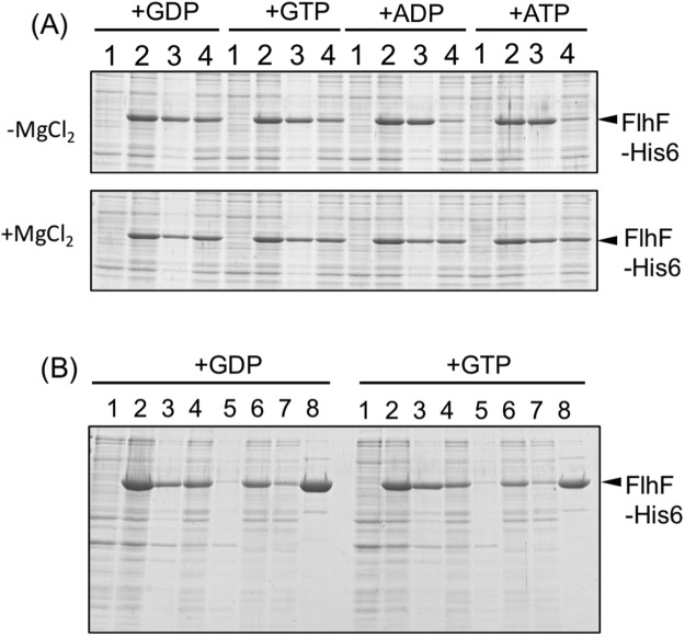
Purification of V. alginolyticus FlhF had been carried out using E. coli BL21 (DE3) transformed by the plasmid pET-FlhF, which overexpresses His6-FlhF from T7 promoter4. In that study, FlhF inclusion bodies were pelleted by low speed centrifugation followed by denaturation by treatment with urea and then affinity purified. However, when urea was removed FlhF again precipitated. In this study, we attached His6 tag to the C-terminus of V. alginolyticus FlhF resulting in a protein with a total length of 511 amino acids and a molecular weight of 57.4 kDa (Fig. S1). Plasmid expressed FlhF-His6 or FlhF showed similar motility in the flhF mutant strain (LPN1) strain (Fig. 1A), and flagellation was observed in both strains by a high-intensity dark-field microscope. This demonstrated that FlhF-His6 was functional in Vibrio cells. His-tagged FlhF was then produced in E. coli using a cold shock expression vector pCold IV, which allowed overexpression from the cspA promoter. Although we anticipated that low-temperature expression might improve protein stability, almost all of the FlhF protein precipitated out following low speed centrifugation after cells were broken by sonication in T-buffer [50 mM Tris-HCl (pH 8.0), 150 mM NaCl] (Fig. S2A). However, a small amount of FlhF protein remained in the supernatant after low-speed centrifugation that was capable of affinity purification. We investigated the conditions that FlhF could be recovered in the supernatant following low speed centrifugation. When 1 mM GTP was added to the T-buffer before sonication, more FlhF was recovered in the supernatant following low speed centrifugation (Fig. 1B, −MgCl2/+GTP). The intensity of the FlhF bands after the low speed centrifugation was measured, and its ratio (supernatant/pellet) increased in the presence of GTP (0.2 for −MgCl2/−GTP, but 0.3 for −MgCl2/+GTP). Therefore the stability of FlhF protein seemed to increase in the presence of GTP. We also tested the effect of Mg2+ since FtsY of E. coli as well as FlhF of Bacillus subtilis and Campylobacter were successfully purified by adding Mg2+ to buffer4,15,22. Indeed, addition of Mg2+ increased the recovery yield of His-tagged FlhF (Fig. 1B, +MgCl2/−GTP, sup/ppt ratio is 0.64). About half of the FlhF protein produced was recovered in the supernatant fraction when both GTP and MgCl2 were added to the T-buffer before sonication (Fig. 1B, +MgCl2/+GTP, sup/ppt ratio is 0.99). Therefore, cells were sonicated in a buffer containing 0.5 mM GTP and 10 mM MgCl2, and purification was performed in the presence of 10 mM MgCl2 (Fig. S2B). The purified His6-FlhF (hereafter, simply FlhF) protein aggregated when left at room temperature or at 4 °C for several hours. However, the aggregation was not observed if 10% (w/v) glycerol was added into the elution buffer. Since the addition of GTP to the buffer improved the purification of FlhF, we examined the effects of adding other nucleotides: GDP, ATP and ADP. We found that GDP was more effective for recovering soluble FlhF protein (Fig. 2A). Therefore, purification was carried out using buffer containing 0.5 mM GDP and 10 mM MgCl2 (Fig. 2B).
Figure 1.

Characterization of FlhF protein. (A) Cell motility. Cells of the flhF mutant (LPN1) harboring a plasmid, vector; pBAD33, FlhF; pAK322, or FlhF-His6; pTSK119, were inoculated into a soft agar plate containing 0.02% arabinose and incubated for 6 hours at 30 °C. (B) Cells of BL21(DE3) harboring a plasmid pTSK110 which has flhF-his6 in pColdIV vector were broken in the presence and absence of MgCl2 or GTP. The proteins were separated by SDS-PAGE and the gels were stained by CBB. The regions of interest were cropped from the gels. 1: the pre-induction cells, 2: the post-induction cells, 3: the low speed centrifugal precipitation, 4: the low speed supernatant.
Figure 2.
Purification of FlhF. (A) Cells of BL21(DE3) harboring a plasmid pTSK110 which has flhF-his6 in pCold IV vector were broken in the presence of GDP, GTP, ADP and ATP with (lower panel) or without (upper panel) MgCl2. The proteins were separated by SDS-PAGE and the gels were stained by CBB. The regions of interest were cropped from the gels. 1: the pre-induction cells, 2: the post-induction cells, 3: the low speed centrifugal precipitation, 4: the low speed supernatant. (B) Purification of FlhF by adding GTP or GDP. 1: the pre-induction cells, 2: the post-induction cells, 3: the low speed centrifugal precipitation, 4: the low speed supernatant, 5: the ultracentrifugation precipitation, 6: the post-ultracentrifugation supernatant, 7: the flow through fraction of Ni-NTA resin, 8: the eluted fraction of Ni-NTA resin. The proteins were separated by SDS-PAGE and the gels were stained by Coomassie brilliant blue (CBB).
A scale-up of FlhF purification was attempted from 100 ml of cell culture by the small-scale procedure described above. FlhF was recovered in the supernatant fraction following low-speed centrifugation and ultracentrifugation. However, when the ultracentrifuged supernatant was added to Ni-NTA resin and mixed by a rotator, the large amount of white precipitate was formed and FlhF was lost. Thus, to shorten the processing time, immediately after ultracentrifugation, the supernatant was applied to a disposable column packed with Ni-NTA resin, followed by immediate washing and elution with imidazole. As the result, almost all the FlhF protein adsorbed to the Ni-NTA resin without precipitation and was successfully eluted from the column (Fig. S3). From 100 ml of culture, 2.5 ml of purified FlhF protein solution was obtained at ca. 0.5 mg ml−1.
Measurement of the GTPase activity of FlhF
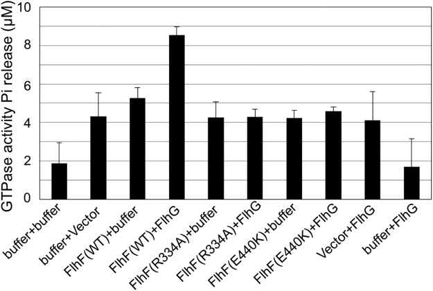
GTPase activity was measured using the purified FlhF protein in order to determine the amount of inorganic phosphate released by GTP hydrolysis. Because purified FlhF was slightly contaminated with other proteins, the background GTPase activity was measured using the sample that was prepared by the same purification procedure from cells containing only vector plasmid. 1 μM FlhF released 5.3 μM inorganic phosphate in 30 minutes, while the background sample released 4.3 μM inorganic phosphate (Fig. 3). This indicated the presence of significant background GTPase activity in addition to that from FlhF. However, when purified FlhF was mixed with purified FlhG at the same concentration, the released inorganic phosphate increased significantly to 8.5 μM (Fig. 3). When FlhG was added in the background sample or buffer, the amount of inorganic phosphate release did not increase. This shows that FlhG is necessary for stimulation of the FlhF GTPase activity.
Figure 3.
The GTPase activity of purified FlhF. The amount of inorganic phosphate release was measured in a mixture of 1.0 μM FlhF, 0.5 mM GTP, and 10 mM MgCl2 for 30 min. To examine the effect of FlhG on FlhF-GTPase, 1.0 μM FlhG was also added in the reaction. The sample solutions were prepared from BL21(DE3) cells harboring a plasmid pCold IV; Vector, pTSK110; FlhF(WT), pSK313; FlhF(R334A), or pSK311; FlhF(E440K), and of BL21(DE3)/pLysS harboring a plasmid pTrc-flhG; FlhG. The activity was measured by mixing of FlhG buffer containing FlhF and GTP buffer containing FlhG for three times with different purification batches. Error bars are standard deviation.
Purification of GTPase motif mutants
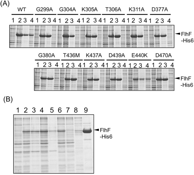
Our previous studies showed that GTPase motif mutants in FlhF resulted in defects in FlhF polar localization and flagellar formation20,21. Twelve mutants (G299A, G304 A, K305A, T306, K311A, D377A, G380A, T436M, K437A, D439A, E440K, and D470A) were examined. In an attempt to purify the twelve mutant proteins as described above for wild-type FlhF, we found that all but E440K precipitated out following the low speed centrifugation step (Fig. 4A). Although the expression level of E440K was lower than that of the wild type, we succeeded in purifying it to a concentration of ca. 0.3 mg ml−1 (Fig. 4B).
Figure 4.
Characterization and purification of the mutant FlhF proteins. (A) Cells of BL21(DE3) harboring a plasmid pTSK110(WT) or the derivative mutant plasmids carrying flhF-his6 in pCold IV vector were broken in the presence of GDP. The proteins were separated by SDS-PAGE and the gels were stained by CBB. The regions of interest were cropped from the gels. 1: the pre-induction cells, 2: the post-induction cells, 3: the low speed centrifugal precipitation, 4: the low speed supernatant. (B) BL21(DE3) cells harboring a pTSK110 derivative mutant plasmid (E440K) were broken in the presence of GDP. 1: the pre-induction cells, 2: the post-induction cells, 3: the low speed centrifugal precipitation, 4: the low speed supernatant, 5: the ultracentrifugation precipitation, 6: the post-ultracentrifugation supernatant, 7: the flow through fraction of Ni-NTA resin, 8: the washed fraction of Ni-NTA resin, 9: the eluted fraction of Ni-NTA resin. The proteins were separated by SDS-PAGE and the gels were stained by CBB.
The E440K mutant was obtained by a random mutagenesis21. We confirmed that the E440K mutant could not complement the motility defect of the LPN1 flhF null mutant cells. Motility was completely absent on soft agar medium and flagellar formation was not observed by high-intensity dark field microscope. The E440K mutant FlhF fused with GFP was expressed in the flhF null mutant background and observed by fluorescence microscopy. The polar localization ratio of wild-type FlhF-GFP was ~87%, whereas the localization ratio of FlhF (E440K)-GFP was ~33%. The GTPase activity of E440K mutant was the same as that from background vector which is only cell extracts (4.2 μM inorganic phosphate in 30 minutes, Fig. 3). This indicated that the E440K mutant lost GTPase activity.
Purification of FlhF mutant of putative GTPase catalytic residues
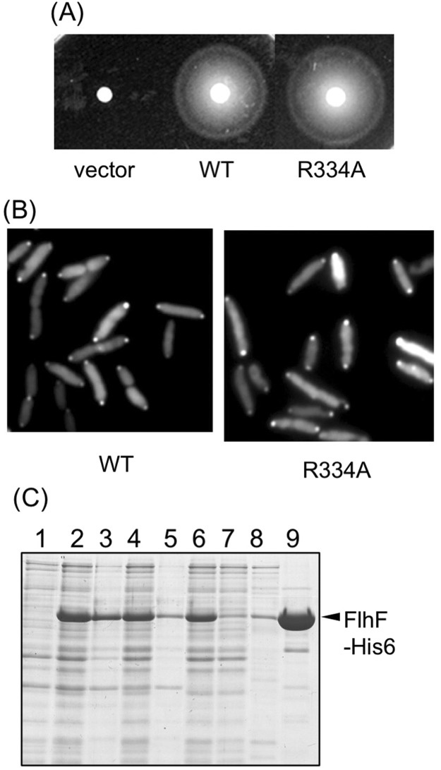
To further investigate the role of FlhF GTPase activity on its polar localization and flagellar formation, an R334A substitution was constructed in the putative catalytic residue for GTP hydrolysis. The corresponding residue of P. aeruginosa FlhF is R251 and its GTPase activity was lost by an R251G substitution16. When the R334A mutant was produced from our plasmid expression system in the flhF mutant strain LPN1, motility was comparable to wild type FlhF on soft agar medium (Fig. 5A). Its flagellar formation was also similar to that of wild type FlhF as observed by high-intensity dark field microscope. When R334A FlhF-GFP was expressed in the flhF mutant strain, fluorescent dots were observed at the cell pole in ca. 84% cells as was observed with wild-type FlhF-GFP (Fig. 5B). Overproduced R334A FlhF protein could be purified similar to the wild-type protein (Fig. 5C). The concentration of R334A FlhF protein was 0.42 mg ml−1. We used this protein to measure the GTPase activity and the inorganic phosphate was released at the background level even in the presence of FlhG (Fig. 3). The mutant R334A gave the similar polar localization phenotype as wild-type FlhF, but the GTPase activity of R334A was lost, suggesting that the GTPase activity is not necessary for the FlhF function.
Figure 5.
GTPase activity is dispensable for the FlhF function. (A) Cells of the flhF mutant (LPN1) harboring a plasmid, vector; pBAD33, WT; pAK322, or R334A; pSK111, were inoculated into a soft agar plate containing 0.02% arabinose and incubated for 6 hours at 30 °C. (B) Cells of the flhF mutant (LPN1) harboring a plasmid, WT; pAK325, or R334A; pSK211, were inoculated in VPG medium containing 0.02% arabinose and observed with a fluorescence microscope. (C) BL21(DE3) cells harboring a pTSK110 derivative mutant plasmid (R334) were broken in the presence of GDP. 1: the pre-induction cells, 2: the post-induction cells, 3: the low speed centrifugal precipitation, 4: the low speed supernatant, 5: the ultracentrifugation precipitation, 6: the post-ultracentrifugation supernatant, 7: the flow through fraction of Ni-NTA resin, 8: the washed fraction of Ni-NTA resin, 9: the eluted fraction of Ni-NTA resin. The proteins were separated by SDS-PAGE and the gels were stained by CBB.
Size estimation by gel filtration chromatography
It is known that FtsY and Ffh form a heterodimer upon binding GTP and the crystal structure of B. subtilis FlhF GTP-bound homodimer has been solved. Thus, we speculated that V. alginolyticus FlhF also forms a GTP-bound homodimer. To examine this, we performed gel filtration chromatography to estimate size changes of FlhF in the presence of either GTP or GDP (Fig. 6). FlhF eluted in fraction 20 in the presence of GTP, which corresponded to the size of the FlhF dimer (ca. 114 kDa). On the other hand, in the presence of GDP, FlhF eluted in fraction 22, which corresponded to the size of an FlhF monomer (ca. 57 kDa). These results demonstrated that V. alginolyticus FlhF forms a dimer in the presence of GTP and exists as a monomer in the presence of GDP.
Figure 6.
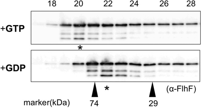
FlhF forms a dimer in the presence of GTP. FlhF was incubated in the presence of either 1 mM GTP or GDP and analyzed by gel filtration chromatography using buffer containing either 1 mM GTP or GDP. The upper panel shows the profile in the presence of GTP, and the lower panel shows it in the presence of GDP. Immunoblotting was performed for each fraction using the anti-FlhF antibody. The regions of interest were cropped from the immunoblotting. The peak fraction containing FlhF is indicated by the asterisk. The molecular size markers were carbonic anhydrase (29 kDa) and conalbumin (74 kDa).
Discussion
V. alginolyticus FlhF is a soluble protein, and is required to initiate flagellar formation and to regulate flagellar number in the presence of FlhG. FlhF and FlhG belong to the SIMIBI family (the nucleotide triphosphate binding protein family essential for protein targeting in a wide range of cellular processes). The SIMIBI proteins act as molecular switches with the NTP binding “on” state and the NDP binding “off” state23. V. alginolyticus FlhF aggregates easily, precipitating after low speed centrifugation following cell disruption. Thus, purification of Vibrio FlhF has proved challenging. In this study, FlhF was produced at low temperature using a cold shock expression vector and cells were disrupted in a buffer containing MgCl2 and GTP or GDP. by this method, soluble FlhF was recovered following low speed centrifugation. This may be due to stabilization of FlhF in the presence of Mg2+ or nucleotides. In Bacillus subtilis, the crystal structure of FlhF was solved bound with Mg2+ and nucleotides14. Therefore, we presume that V. alginolyticus FlhF binds to GTP or GDP with Mg2+ and the nucleotide binding state is stable. The GTP binding motif mutants, except for E440K, precipitated following low speed centrifugation. Mutations of the GTP binding motif probably affects the structure around the motif.
SIMIBI proteins form dimers in the NTP-bound form and play regulatory roles in various cellular processes such as cell division and protein transport23. FtsY and Ffh and MinD of E. coli belong to this family and form heterodimers in either a GTP-dependent manner for FtsY and Ffh or an ATP-dependent manner for MinD. Their crystal structures have been solved. In this study, analysis by gel filtration chromatography suggested that V. alginolyticus FlhF also forms a dimer in the presence of GTP and is stably present as a monomer in the presence of GDP. This may explain why more soluble FlhF could be recovered following low-speed centrifugation in disruption buffer containing GDP than buffer containing GTP. Higher ordered multimer formation is more likely to promote aggregation. We think that dimer formation is important for polar localization.
Until this study, V. alginolyticus FlhF had not been purified in its native form. We could detect GTPase activity, however, FlhF by itself only showed GTPase activity in the presence of FlhG. The putative catalytic residue mutant R334A lacked GTPase activity in the presence of FlhG though its intracellular localization and the formation of flagella were similar to wild type. These results suggest that the GTPase activity of FlhF is not necessary for the polar localization and the formation of flagella. Since the GTP-binding motif mutant E440K lost its GTPase activity and conferred an abnormal subcellular localization of FlhF and loss the ability to form flagella, the GTP binding seems to be important for polar localization of FlhF and flagellar formation. However, it has not been confirmed whether the purified wild-type FlhF actually binds GTP and whether the R334A mutant can bind GTP but the E440K mutant cannot.
It has been speculated that FlhF interacts to recruit the FliF protein to the cytoplasmic membrane to initiate the construction of the flagellar MS-ring structure, which is composed of FliF19. We have shown that FlhF localizes at the cell pole of rpoN-deficient strain of V. alginolyticus as well as in the flhDC-deficient strain of E. coli4. Therefore, we can conclude that FlhF by itself has an ability to localize at the cell pole, though FlhG needs HubP, which is a single transmembrane protein with an N-terminal periplasmic peptidoglycan-binding motif (LysM) and a large cytoplasmic domain24, to localize at the cell pole6. The GDP-bound form of FlhF diffuses around the cytoplasm as monomer and forms a dimer by binding with GTP to acquire the ability to localize the pole. How bacteria localize proteins to cell pole is not clear25–27. For example, chemoreceptors involved in chemotaxis of E. coli are located at cell poles28 and it has been recently shown that they become stably present at highly curved cell poles by forming multimers29. It has been shown that Caulobacter crescentus uses a multimeric pole-organizing factor (PopZ) that serves as a hub to concurrently achieve several polarizing functions and the polar accumulation of PopZ is central to its polarizing activity30. We speculate that a mechanism similar to that used by PopZ is employed by FlhF to localize to cell poles. Perhaps FlhF localizes at cell poles by forming a dimer, and the dimer might form multimers to retain polar localization. To initiate the flagellar assembly, it is inferred that FlhF assists in MS-ring formation at the poles.
It is noteworthy that FlhF is not essential for flagellation: very few cells of ΔflhF strain can still form a flagellum, and the flhF and flhG double mutant slightly increases the number of flagellated cells. When the additional defect of the membrane factor, SflA, is present in the ΔflhFG background, more than 50% of cells can generate multiple flagella at lateral positions31. The initiation of flagellar formation seems to be suppressed by SflA in the absence of FlhF. SflA is localized at cell poles, dependent on HubP32. HubP does not belong to the flagellar regulon and may be regulated independent from the flagellar genes. It was suggested that HubP is involved in the regulation of chromosome partitioning and chemoreceptor positioning in V. cholerae. It has been shown in V. cholerae and P. aeruginosa that FlhG (FleN) also functions as a transcriptional repressor of the polar flagellar genes2,33 and protein production of the polar flagellar genes increased in flhG deficient strain of V. alginolytius4. In FlhG (FleN) of P. aeruginosa, the reduction in activity of the master transcriptional regulator is dependent on ATP and its ATPase domain33,34.
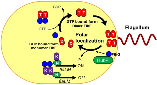
Based on the above evidence and previous reports, we present a model on how FlhF controls flagellar formation (Fig. 7). FlhG appears to negatively control flagellation by activation of FlhF GTPase activity. Since FlhG can exist throughout the cytoplasm and at the cell pole, it is possible that FlhG activates FlhF GTPase activity at both places to produce a GDP-bound or unbound form of FlhF that can no longer stay at the pole and is retained throughout the cytoplasm. As mentioned above, GTP hydrolysis activated by FlhG at the pole, but is not necessary to induce the release of FlhF from the pole. The GTP-bound form of FlhF by itself might be able to associate to the polar region and dissociate from the pole by the effects of FlhG whose polar localization is dependent on HubP. This is consistent with the fact that overexpression of FlhG in cells did not reduce the polar localization rate of FlhF and the hubP mutant gave the multi polar phenotype similar to the phenotype of the flhG mutant6. As suggested above, FlhF is likely to recruit the FliF to the pole and may also act on the formation of the FliF MS-ring. To unravel the mechanism of initiation for polar flagellar assembly, it will be necessary to investigate the dynamic interactions of proteins at the pole including FlhF, FlhG, FliF and HubP. For this purpose, it would be helpful to solve the crystal structure of V. alginolyticus FlhF though it has been determined in the homologous proteins, such as FlhF homolog of B. subtilis14 and FtsY or Ffh paralog of E. coli35.
Figure 7.
The model of FlhF polar localization in V. alginolyticus. FlhF diffuses into the cytoplasm as a GDP-bound monomer (shown as the red ellipse with “D”), but when it binds to GTP (shown as the red ellipse with “T”), it forms a dimer and becomes localized to the pole. Localized FlhF promotes flagellar formation at cell pole. FlhG diffuses in the cytoplasm and interacts with FlhF to inhibit polar localization of FlhF. Also, FlhG binds to FlaK (shown as the purple ellipse with “K”) to inhibit the early class of flagellar genes (flaLM), whose expression is dependent on FlaK and RpoN (shown as the green ellipse with “N”). At the poles, the GTPase activity of FlhF is activated by FlhG, which is localized to the pole by the HubP scaffold. FlhF is converted to the GDP bound form and dissociates from the cell pole. In the wild type V. alginolyticus, the amount of FlhF localized at the pole is controlled to a level resulting in biosynthesis of a single, polar flagellum.
Experimental procedures
Strains used, culture medium and culture conditions
The strains and the plasmids used in this study are shown in Table 1. E. coli cells were cultured at 37 °C in LB medium [1% (w/v) bactotryptone, 0.5% (w/v) yeast extract, and 0.5% (w/v) NaCl]. The gene under the control of the cold shock gene cspA promoter of E. coli was induced by the addition of IPTG at low temperature (16 °C). V. alginolyticus cells were cultured at 30 °C in the VC medium [0.5% (w/v) polypeptone, 0.5% (w/v) yeast extract, 0.4% (w/v) K2HPO4, 3% (w/v) NaCl, and 0.2% (w/v) glucose] or VPG medium [1% (w/v) polypeptone, 0.4% (w/v) K2HPO4, 3% (w/v) NaCl, and 0.5% (w/v) glycerol]. The gene under the control of the araBAD promoter was induced by addition of the appropriate amount of arabinose indicated. A solid and a soft agar medium were prepared by adding 1.25% and 0.25% agar, respectively. When necessary, the antibiotic chloramphenicol was used at 2.5 µg ml−1 for V. alginolyticus and 25 µg ml−1 for E. coli or the antibiotic ampicillin was added to a final concentration of 100 µg ml−1 for E. coli.
Table 1.
Bacterial strains and plasmids used in this study.
| Strain or plasmid | Genotype or description | Reference or source |
|---|---|---|
| V. alginolyticus | ||
| VIO5 | VIK4 (Rifr Pof+ Laf−) | 36 |
| LPN1 | VIO5 ΔflhF (Rifr Pof+ Laf−) | 4 |
| E. coli | ||
| DH5α | Recipient for DNA manipulation | |
| BL21(DE3) | Novagen | |
| BL21(DE3)/pLysS | ||
| Plasmids | ||
| pBAD33 | Cmr, PBAD | 37 |
| pTY57 | Cmr, PBAD, MCS from pBAD24 | 38 |
| pCold IV | Ampr, PcspA | Takara |
| pAK322 | flhF (wt) in pBAD33 | 3 |
| pAK325 | flhF-egfp (wt) in pBAD33 | 4 |
| pTSK110 | flhF-his6 in pCold IV | This study |
| pTSK119 | flhF-his6 in pTY57 | This study |
| pSK102 | flhF (E440K) in pBAD33 | 21 |
| pSK111 | flhF (R334A) in pBAD33 | This study |
| pSK202 | flhF-egfp (E440K) in pBAD33 | 21 |
| pSK211 | flhF-egfp (R334A) in pBAD33 | This study |
| pSK301 | flhF-his6(G299A) in pCold IV | This study |
| pSK302 | flhF-his6(G304A) in pCold IV | This study |
| pSK303 | flhF-his6(K305A) in pCold IV | This study |
| pSK304 | flhF-his6(T306A) in pCold IV | This study |
| pSK305 | flhF-his6(K311A) in pCold IV | This study |
| pSK306 | flhF-his6(D377A) in pCold IV | This study |
| pSK307 | flhF-his6(G380A) in pCold IV | This study |
| pSK308 | flhF-his6(T436M) in pCold IV | This study |
| pSK309 | flhF-his6(K437A) in pCold IV | This study |
| pSK310 | flhF-his6(D439A) in pCold IV | This study |
| pSK311 | flhF-his6(E440K) in pCold IV | This study |
| pSK312 | flhF-his6(D470A) in pCold IV | This study |
| pSK313 | flhF-his6(R334A) in pCold IV | This study |
| pTrc-flhG | his6-tev-flhG in pTrcHisB | 8 |
Ampr, ampicillin-resistant; Cmr, chloramphenicol-resistant; Rifr, Rifampicin-resistant; Pof+, possessing a polar flagellum; Laf−, lack of lateral flagella.
Transformation of V. alginolyticus by electroporation
Overnight cultures were added to 40 ml of VC medium at 100-fold dilution and grown at 30 °C until OD 660 = 0.8. The cells were collected by centrifugation (3,000 × g for 5 min), suspended in 10 ml of Osmotic solution [30 mM Tris-HCl(pH8.0), 20% (w/v) sucrose, 400 mM NaCl, 1 mM EDTA] and grown with shaking at 30 °C for 10 minutes or more. After centrifugation (3,000 × g for 5 min), cells were suspended in 10 ml of 20 mM MgSO4 and allowed to stand on ice for 10 minutes. After centrifugation (3,000 × g for 5 min), cells were suspended in 200 to 400 µl of 10% (w/v) glycerol, and dispensed in 40 µl aliquots. Electroporation was carried out using a Gene Pulser electroporation apparatus (Bio-Rad Laboratories) with a 0.2 cm cuvette under the conditions of resistance 200 Ω and voltage 1.4 kV. After that, the bacteria were recovered in 1 ml of VC medium, grown with shaking at 30 °C for 40 minutes, then inoculated into VC agar plate containing chloramphenicol and grown overnight at 30 °C.
Purification of FlhF
For the expression of FlhF, a system using E. coli BL21 (DE3) strain was used. FlhF was expressed from a plasmid (pTSK110) in which flhF derived from V. alginolyticus was cloned into the NdeI-XhoI site of a cold shock expression vector pCold IV. The His-tag is designed to be added to the C-terminus of FlhF and the final product is expected to be a protein with 511 amino acids and a molecular weight of 57.4 kDa.
For small scale purfications, 5 ml of induced cells were centrifuged (16, 000 × g, 1 min) and washed with FlhF buffer [20 mM Tris-HCl(pH8.0), 300 mM NaCl, 10 mM MgCl2, 10 mM KCl]. The cells were suspended in 1.2 ml of FlhF buffer containing 5 mM GTP or GDP and the complete protease inhibitor, EDTA-free (Roche). The complete protease inhibitor was prepared by dissolving 1 tablet in 1 ml of sterilized water and used in a 50-fold dilution. Cells were sonicated using a small probe (power = 5, 30 sec, 3 times, duty cycle 50%) and undisrupted cells were precipitated by centrifugation at low speed (6,100 x g for 5 min). The supernatant was fractionated by ultracentrifugation (154,000 × g for 30 min) using a TLA55 rotor and Optima MAX (BECKMAN COULTER). One third of the soluble fraction was added to 100 µg of Ni-NTA resin (Qiagen) equilibrated with FlhF buffer containing 5 mM imidazole and mixed for 1 h at 4 °C using a rotator. After centrifugation to remove the solution, the 1/3 of sample was added to the resin. After mixing for 1 h, the third sample was also added to the resin in the same procedure. The resin was twice washed by 1 ml of FlhF buffer containing 5 mM imidazole. Then, 1 ml of FlhF buffer containing 30 mM imidazole was added to wash the resin three times. Thereafter, 300 μl of FlhF buffer containing 300 mM of imidazole and 10% (w/v) glycerol was added and incubated at room temperature for 5 min. After the centrifugation, the supernatant was recovered. The concentration of the purified protein obtained was calculated from a calibration curve prepared by SDS-PAGE with known concentration of BSA and quantification using the Coomassie Brilliant Blue (CBB) stained bands using Image J.
For large-scale preparation, 100 ml of culture was centrifuged (22, 200 × g for 5 min) and the collected cells were washed with FlhF buffer. The cells were suspended in 50 ml of FlhF buffer containing 0.5 mM GDP and one tablet of complete protease inhibitor (EDTA-free, Roche). Cells were sonicated using large probe (power = 5, 60 sec, 6 times, duty cycle 50%) and unbroken cells were removed by low speed centrifugation (6,300 × g for 5 min) The obtained supernatant was fractionated by ultracentrifugation (157,000 × g for 30 min) with a P50AT2 rotor using himac CP80WX (HITACHI). The soluble fraction was poured into a 5 ml disposable column (Thermo Scientific) packed with 1.5 ml of Ni-NTA resin equilibrated with FlhF buffer containing 5 mM imidazole. After all the soluble fraction passed through the column, the resin was twice washed by 5 ml of FlhF buffer containing 5 mM imidazole and washed by 5 ml of FlhF buffer containing 30 mM imidazole 3 times, then the proteins were eluted by 2.5 ml of FlhF buffer containing 300 mM imidazole and 10% (w/v) glycerol. The 200 μl aliquots of the eluted sample were frozen by liquid nitrogen and stored at −80 °C.
Measurement of GTPase activity
GTPase activity was measured using PiColorLock™ Gold Phosphate Detection System (Innova Bioscience). For analysis of FlhF alone, 40 μl of purified FlhF, 40 μl of FlhG buffer [20 mM Tris-HCl (pH8.0), 300 mM NaCl, 10% (w/v) glycerol] and 40 μl of GTP buffer [20 mM Tris-HCl (pH8.0), 300 mM NaCl, 10 mM MgCl2, 1.5 mM GTP, 10% (w/v) glycerol] were mixed and reacted for 30 min at 25 °C. For analysis of FlhF with FlhG, 40 μl of purified FlhF, 40 μl of FlhG and 40 μl of GTP buffer were mixed and reacted for 30 min at 25 °C. Final concentration of FlhF and FlhG were 1 μM. To stop the hydrolysis reaction, 30 μl of reaction terminator was added and after 2 min, 12 μl of the stabilizer was added. After standing at 25 °C for 30 min, the absorbance at 595 nm was measured by a plate reader (DS Pharma Biomedical), and the concentration of inorganic phosphate was calculated from a calibration curve obtained by similarly measuring the known inorganic phosphate. The measurements were performed for 3 purified samples by 3 measurements for one sample, thus the average value of n = 9 with standard deviation was shown as the activity measurement value.
Purification of FlhG
His-tagged FlhG was purified as described previously8. BL21 (DE3)/pLysS cells, transformed with pTrc-flhG, were cultured in 1 L of SB medium at 37 °C. At an OD 660 of 0.3 to 0.4, IPTG was added to a final concentration of 0.4 mM. After incubation at 37 °C for 3 hours, the cells were collected by centrifugation (6,600 × g for 10 min) and frozen at −80 °C. Frozen cells were suspended in 50 ml of FlhG buffer containing 5 mM imidazole and 0.5 mM PMSF and disrupted by sonication using a large probe (power = 8, 60 sec, 5 times, duty cycle 50%) and unbroken cells were removed by low speed centrifugation (6,300 × g for 5 min). The supernatant was fractionated by ultracentrifugation (157,000 × g for 30 min) with the P50AT2 rotor using himac CP80WX (HITACHI). The soluble fraction was passed through a HiTrap TALON column (5 ml) connected to the AKTA PRIME system. After washing the column by FlhG buffer containing 20 mM imidazole at a flow rate of 1.0 ml/min, FlhG was eluted with FlhG buffer containing 65 mM imidazole at a flow rate of 1.0 ml min−1. The 100 μl aliquots of the eluted sample were frozen by liquid nitrogen and stored at −80 °C.
Detection of FlhF protein
The samples were mixed with one-fifth volume of SDS loading buffer [0.2 M Tris-HCl (pH 6.8), 37.5% (w/v) glycerol, 6% (w/v) SDS, and 0.004% (w/v) bromophenol blue] and one-tenth volume of 2-mercaptoethanol, and then boiled for 5 min. Proteins in the samples were separated by SDS-PAGE, and immunoblotting was conducted as described previously21, using the anti-FlhF antibody.
Sequencing
Reactions for DNA sequencing were performed by a dye terminator method using BigDye Terminator v3.1 Sequencing Standard Kit. The Nagoya University DNA sequencing core facility performed the DNA sequence analysis. Data were analyzed using GENETYX and 4 peaks software.
Site-specific mutagenesis using PCR
Routine DNA manipulations were carried out according to standard procedures. The mutations of flhF were introduced into plamids by the QuikChange site-directed mutagenesis method using a pair of primers for the desired mutation (Table S3). To amplify DNA, Pfu ultra polymerase was used, and the reaction cycle was 95 °C for 30 sec → 18 cycles of (95 °C for 1 min → 55 °C for 1 min → 68 °C for16 min) → 4 °C. Dpn I treatment was carried out to eliminate the template plasmid and introduced into E. coli DH5α. The plasmid was isolated from transformed cells and the nucleotide sequence of flhF was determined to confirm the mutation.
Detection of flagella in Vibrio cells
Overnight cultures were diluted 1:100 in VPG medium containing 0.02% (w/v) arabinose and 2.5 μg ml−1 chloramphenicol and further cultivated at 30 °C for 4 hours. The cultured cells were harvested and suspended in V buffer [50 mM Tris-HCl (pH7.5), 300 mM NaCl, 5 mM MgCl2]. Flagella were observed under a high-intensity dark-field microscope (Olympus model BHT) equipped with a 100-W mercury lamp (Ushio USH-102).
Observation of GFP fluoresence in cells
Vibrio cells were cultured overnight in VC medium at 30 °C. Overnight cultures were diluted 1:100 in a fresh VPG medium containing 0.02% (w/v) arabinose and 2.5 μg ml−1 chloramphenicol and cultivated further at 30 °C for 4 hours. The cells were collected by centrifugation (97,700 × g for 2 min), and suspended to V buffer at 1/4 volume of original culture. Fluorescence microscopic observations were carried out as described previously8.
Estimation of particle size by gel filtration chromatography
GTP or GDP was added to the purified FlhF protein to a final concentration of 1 mM, and the mixture was allowed to stand on ice for 10 minutes. A 100 μl portion of the protein solution was loaded on a SuperDex 75 Increase column (GE Healthcare) that had been equilibrated with buffer [20 mM Tris-HCl (pH 8.0), 300 mM NaCl, 10 mM MgCl2] containing 1 mM GTP or GDP, and eluted by the same buffer at a flow rate of 0.5 ml min−1. The elution was collected in 0.5 ml fractions and the elution position of the protein was confirmed by western blot.
Electronic supplementary material
Acknowledgements
We thank thank Hiroyuki Terashima for technical support and comments on the paper and also Kelly Hughes for critically reading the manuscript and editing the manuscript for English grammar. This work was supported in part by Grants-in-Aid for Scientific Research from the Japan Society for the Promotion of Science (16H04774 to S.K.).
Author Contributions
Sh.K. and Se.K. designed the experiments; Sh.K., Y.I. and A.M. performed the experiments; Sh.K., Y.I., A.M., Se.K. and M.H. analyzed the data; and Sh.K. M.H. and Se.K. wrote the manuscript.
Competing Interests
The authors declare no competing interests.
Footnotes
Publisher's note: Springer Nature remains neutral with regard to jurisdictional claims in published maps and institutional affiliations.
Electronic supplementary material
Supplementary information accompanies this paper at 10.1038/s41598-018-30531-5.
References
- 1.Balaban M, Hendrixson DR. Polar flagellar biosynthesis and a regulator of flagellar number influence spatial parameters of cell division in Campylobacter jejuni. PLoS path. 2011;7:e1002420. doi: 10.1371/journal.ppat.1002420. [DOI] [PMC free article] [PubMed] [Google Scholar]
- 2.Correa NE, Peng F, Klose KE. Roles of the regulatory proteins FlhF and FlhG in the Vibrio cholerae flagellar transcription hierarchy. J Bacteriol. 2005;187:6324–6332. doi: 10.1128/JB.187.18.6324-6332.2005. [DOI] [PMC free article] [PubMed] [Google Scholar]
- 3.Kusumoto A, et al. Regulation of polar flagellar number by the flhF and flhG genes in Vibrio alginolyticus. J Biochem (Tokyo) 2006;139:113–121. doi: 10.1093/jb/mvj010. [DOI] [PubMed] [Google Scholar]
- 4.Kusumoto A, et al. Collaboration of FlhF and FlhG to regulate polar-flagella number and localization in Vibrio alginolyticus. Microbiology. 2008;154:1390–1399. doi: 10.1099/mic.0.2007/012641-0. [DOI] [PubMed] [Google Scholar]
- 5.Pandza S, et al. The G-protein FlhF has a role in polar flagellar placement and general stress response induction in Pseudomonas putida. Mol Microbiol. 2000;36:414–423. doi: 10.1046/j.1365-2958.2000.01859.x. [DOI] [PubMed] [Google Scholar]
- 6.Takekawa N, Kwon S, Nishioka N, Kojima S, Homma M. HubP, a Polar Landmark Protein, Regulates Flagellar Number by Assisting in the Proper Polar Localization of FlhG in Vibrio alginolyticus. J Bacteriol. 2016;198:3091–3098. doi: 10.1128/JB.00462-16. [DOI] [PMC free article] [PubMed] [Google Scholar]
- 7.de Boer PA, Crossley RE, Hand AR, Rothfield LI. The MinD protein is a membrane ATPase required for the correct placement of the Escherichia coli division site. EMBO J. 1991;10:4371–4380. doi: 10.1002/j.1460-2075.1991.tb05015.x. [DOI] [PMC free article] [PubMed] [Google Scholar]
- 8.Ono H, Takashima A, Hirata H, Homma M, Kojima S. The MinD homolog FlhG regulates the synthesis of the single polar flagellum of Vibrio alginolyticus. Mol Microbiol. 2015;98:130–141. doi: 10.1111/mmi.13109. [DOI] [PubMed] [Google Scholar]
- 9.Herskovits AA, Bochkareva ES, Bibi E. New prospects in studying the bacterial signal recognition particle pathway. Mol Microbiol. 2000;38:927–939. doi: 10.1046/j.1365-2958.2000.02198.x. [DOI] [PubMed] [Google Scholar]
- 10.Pohlschroder M, Hartmann E, Hand NJ, Dilks K, Haddad A. Diversity and evolution of protein translocation. Annu Rev Microbiol. 2005;59:91–111. doi: 10.1146/annurev.micro.59.030804.121353. [DOI] [PubMed] [Google Scholar]
- 11.Bange G, et al. Structural basis for the molecular evolution of SRP-GTPase activation by protein. Nat Struct Mol Biol. 2011;18:1376–1380. doi: 10.1038/nsmb.2141. [DOI] [PubMed] [Google Scholar]
- 12.Cross BC, Sinning I, Luirink J, High S. Delivering proteins for export from the cytosol. Nat Rev Mol Cell Biol. 2009;10:255–264. doi: 10.1038/nrm2657. [DOI] [PubMed] [Google Scholar]
- 13.Kjeldgaard M, Nyborg J, Clark BF. The GTP binding motif: variations on a theme. FASEB J. 1996;10:1347–1368. doi: 10.1096/fasebj.10.12.8903506. [DOI] [PubMed] [Google Scholar]
- 14.Bange G, Petzold G, Wild K, Parlitz RO, Sinning I. The crystal structure of the third signal-recognition particle GTPase FlhF reveals a homodimer with bound GTP. Proc Natl Acad Sci USA. 2007;104:13621–13625. doi: 10.1073/pnas.0702570104. [DOI] [PMC free article] [PubMed] [Google Scholar]
- 15.Balaban M, Joslin SN, Hendrixson DR. FlhF and its GTPase activity are required for distinct processes in flagellar gene regulation and biosynthesis in Campylobacter jejuni. J Bacteriol. 2009;191:6602–6611. doi: 10.1128/JB.00884-09. [DOI] [PMC free article] [PubMed] [Google Scholar]
- 16.Schniederberend M, Abdurachim K, Murray TS, Kazmierczak BI. The GTPase activity of FlhF is dispensable for flagellar localization, but not motility, in Pseudomonas aeruginosa. J Bacteriol. 2013;195:1051–1060. doi: 10.1128/JB.02013-12. [DOI] [PMC free article] [PubMed] [Google Scholar]
- 17.Gao T, Shi M, Ju L, Gao H. Investigation into FlhFG reveals distinct features of FlhF in regulating flagellum polarity in Shewanella oneidensis. Mol Microbiol. 2015;98:571–585. doi: 10.1111/mmi.13141. [DOI] [PubMed] [Google Scholar]
- 18.Rossmann F, et al. The role of FlhF and HubP as polar landmark proteins in Shewanella putrefaciens CN-32. Mol Microbiol. 2015;98:727–742. doi: 10.1111/mmi.13152. [DOI] [PubMed] [Google Scholar]
- 19.Green JC, et al. Recruitment of the earliest component of the bacterial flagellum to the old cell division pole by a membrane-associated signal recognition particle family GTP-binding protein. J Mol Biol. 2009;391:679–690. doi: 10.1016/j.jmb.2009.05.075. [DOI] [PubMed] [Google Scholar]
- 20.Kusumoto A, Nishioka N, Kojima S, Homma M. Mutational analysis of the GTP-binding motif of FlhF which regulates the number and placement of the polar flagellum in Vibrio alginolyticus. J Biochem (Tokyo) 2009;146:643–650. doi: 10.1093/jb/mvp109. [DOI] [PubMed] [Google Scholar]
- 21.Kondo S, Homma M, Kojima S. Analysis of the GTPase motif of FlhF in the control of the number and location of polar flagella in Vibrio alginolyticus. Biophys Physicobiol. 2017;14:173–181. doi: 10.2142/biophysico.14.0_173. [DOI] [PMC free article] [PubMed] [Google Scholar]
- 22.Miller JD, Bernstein HD, Walter P. Interaction of E. coli Ffh/4.5S ribonucleoprotein and FtsY mimics that of mammalian signal recognition particle and its receptor. Nature. 1994;367:657–659. doi: 10.1038/367657a0. [DOI] [PubMed] [Google Scholar]
- 23.Bange G, Sinning I. SIMIBI twins in protein targeting and localization. Nat Struct Mol Biol. 2013;20:776–780. doi: 10.1038/nsmb.2605. [DOI] [PubMed] [Google Scholar]
- 24.Yamaichi Y, et al. A multidomain hub anchors the chromosome segregation and chemotactic machinery to the bacterial pole. Genes Dev. 2012;26:2348–2360. doi: 10.1101/gad.199869.112. [DOI] [PMC free article] [PubMed] [Google Scholar]
- 25.Laloux G, Jacobs-Wagner C. How do bacteria localize proteins to the cell pole? J Cell Sci. 2014;127:11–19. doi: 10.1242/jcs.138628. [DOI] [PMC free article] [PubMed] [Google Scholar]
- 26.Schuhmacher JS, Thormann KM, Bange G. How bacteria maintain location and number of flagella? FEMS Microbiol Rev. 2015;39:812–822. doi: 10.1093/femsre/fuv034. [DOI] [PubMed] [Google Scholar]
- 27.Kiekebusch D, Thanbichler M. Spatiotemporal organization of microbial cells by protein concentration gradients. Trends Microbiol. 2014;22:65–73. doi: 10.1016/j.tim.2013.11.005. [DOI] [PubMed] [Google Scholar]
- 28.Sourjik V, Armitage JP. Spatial organization in bacterial chemotaxis. EMBO J. 2010;29:2724–2733. doi: 10.1038/emboj.2010.178. [DOI] [PMC free article] [PubMed] [Google Scholar]
- 29.Strahl H, et al. Transmembrane protein sorting driven by membrane curvature. Nat Commun. 2015;6:8728. doi: 10.1038/ncomms9728. [DOI] [PMC free article] [PubMed] [Google Scholar]
- 30.Ebersbach G, Briegel A, Jensen GJ, Jacobs-Wagner C. A self-associating protein critical for chromosome attachment, division, and polar organization in Caulobacter. Cell. 2008;134:956–968. doi: 10.1016/j.cell.2008.07.016. [DOI] [PMC free article] [PubMed] [Google Scholar]
- 31.Kitaoka M, et al. A novel dnaJ family gene, sflA, encodes an inhibitor of flagellation in marine Vibrio species. J Bacteriol. 2013;195:816–822. doi: 10.1128/JB.01850-12. [DOI] [PMC free article] [PubMed] [Google Scholar]
- 32.Inaba S, Nishigaki T, Takekawa N, Kojima S, Homma M. Localization and domain characterization of the SflA regulator of flagellar formation in Vibrio alginolyticus. Genes Cells. 2017;22:619–627. doi: 10.1111/gtc.12501. [DOI] [PubMed] [Google Scholar]
- 33.Dasgupta N, Ramphal R. Interaction of the antiactivator FleN with the transcriptional activator FleQ regulates flagellar number in Pseudomonas aeruginosa. J Bacteriol. 2001;183:6636–6644. doi: 10.1128/JB.183.22.6636-6644.2001. [DOI] [PMC free article] [PubMed] [Google Scholar]
- 34.Baraquet C, Harwood CS. Cyclic diguanosine monophosphate represses bacterial flagella synthesis by interacting with the Walker A motif of the enhancer-binding protein FleQ. Proc Natl Acad Sci USA. 2013;110:18478–18483. doi: 10.1073/pnas.1318972110. [DOI] [PMC free article] [PubMed] [Google Scholar]
- 35.Doudna JA, Batey RT. Structural insights into the signal recognition particle. Ann Rev Biochem. 2004;73:539–557. doi: 10.1146/annurev.biochem.73.011303.074048. [DOI] [PubMed] [Google Scholar]
- 36.Okunishi I, Kawagishi I, Homma M. Cloning and characterization of motY, a gene coding for a component of the sodium-driven flagellar motor in Vibrio alginolyticus. J Bacteriol. 1996;178:2409–2415. doi: 10.1128/jb.178.8.2409-2415.1996. [DOI] [PMC free article] [PubMed] [Google Scholar]
- 37.Guzman LM, Belin D, Carson MJ, Beckwith J. Tight regulation, modulation, and high-level expression by vectors containing the arabinose pBAD promoter. J Bacteriol. 1995;177:4121–4130. doi: 10.1128/jb.177.14.4121-4130.1995. [DOI] [PMC free article] [PubMed] [Google Scholar]
- 38.Li N, Kojima S, Homma M. Characterization of the periplasmic region of PomB, a Na+-driven flagellar stator protein in Vibrio alginolyticus. J Bacteriol. 2011;193:3773–3784. doi: 10.1128/JB.00113-11. [DOI] [PMC free article] [PubMed] [Google Scholar]
Associated Data
This section collects any data citations, data availability statements, or supplementary materials included in this article.


