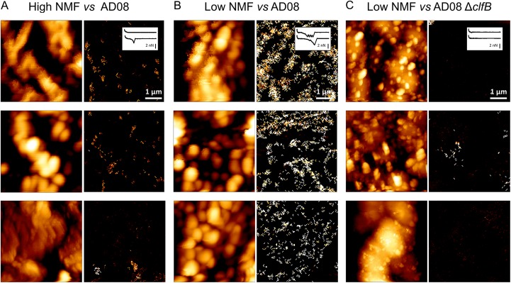
FIG 7 .
Nanoscale adhesion imaging shows that ClfB ligands are largely exposed on low-NMF corneocytes. (A and B) Simultaneous height (left) and adhesion (right) images of corneocytes recorded in PBS between different S. aureus AD08 cell probes and different high-NMF2 (A) or low-NMF1 (B) corneocytes. (C) Images obtained on different low-NMF1 corneocytes with different AD08 ΔclfB cell probes. The insets in the top right images show representative force curves. More images are presented in Fig. S5.

