Abstract
Ralstonia mannitolilytica is a Gram-negative soil bacteria. It is an emerging opportunistic pathogen in hospital-acquired infections. Maintenance hemodialysis patients at Manipal Hospital Outpatient Haemodialysis unit, Bengaluru, witnessed an outbreak of R. mannitolilytica infection between October 2016 and November 2016. Five patients were infected and one of them presented with infective endocarditis. All patients were successfully treated with antibiotic according to culture and sensitivity pattern. Immediately following the outbreak, environmental sampling was done. The culture from sterile water was positive for R. mannitolilytica growth. The Department of Infection Control ordered for discarding the whole batch of sterile water followed by water treatment with shock chlorination and room disinfection. Following implementation of the same, the outbreak of R. mannitolilytica infection was controlled. R. mannitolilytica infections are hospital acquired, affecting mainly immunocompromised patients. The disease onset and progression is rapid. Early identification of the organism and treatment with appropriate antibiotics is important.
Keywords: Hemodialysis, nosocomial infection, Ralstonia mannitolilytica
Introduction
Ralstonia mannitolilytica is a Gram-negative bacteria, widely present in the environment such as soil and plants.[1] It is a frequent contaminant of water supplies.[2] It rarely causes clinical infections, mainly affecting immunosuppressed individuals. It is a common source of hospital-acquired infections due to contamination of normal saline, sterile water, chlorhexidine, and medical equipment.[2] There have been outbreaks in a pediatric department in the USA due to oxygen equipment contamination.[1] No outbreaks of Ralstonia have been reported in India till date. Clinical isolation of Ralstonia is rare in India, while separation from blood culture and as a cause of infective endocarditis is even rarer. There are no typical symptoms of R. mannitolilytica infection, and there is a lack of sufficient experience with its diagnosis and treatment in India.
Case Series
At the Outpatient Haemodialysis Unit of Manipal Hospital, Bengaluru, between October 2016 and November 2016, we found 5 maintenance hemodialysis patients with a bloodstream infection of R. mannitolilytica. All of them had blood cultures drawn from either dialysis line or peripheral line positive for the organism. One of the patients presented with infective endocarditis.
A 61-year-old gentleman, diabetic and hypertensive with end-stage renal disease on maintenance hemodialysis for 12 years via arteriovenous fistula, presented with fever of 15–20 days duration, associated with headache, loss of appetite, and fatigue.
His physical examination revealed mild pedal edema, temperature of 102°F, pulse rate of 102/min, and BP of 110/70 mmHg. Cardiovascular system examination revealed high-pitched holosystolic murmur at the apex. Respiratory system examination revealed bilateral fine basal crepitations. There was no other remarkable finding.
Investigations revealed a total count of 13,100/mm3 with a neutrophilic predominance, hemoglobin of 9 g/dl, platelet count of 229,000/mm3, serum albumin of 2.2 g/dl, direct bilirubin of 0.3 mg/dl, and total bilirubin of 0.7 mg/dl. Serum glutamic pyruvic transaminase and serum glutamic oxaloacetic transaminase were within normal limits. Widal, Leptospira IgG and IgM, Weil Felix, dengue profile, peripheral smear for malaria parasite were negative.
Three blood culture samples (one aerobic and two anaerobic) were taken, one from dialysis line and two from peripheral line. The samples were submitted to the microbiology laboratory and cultured on blood, chocolate, and MacConkey agar. The culture from dialysis line was first to be flagged positive within 24 hours (BACTEC 9050 system). Gram stain revealed Gram-negative bacilli. Culture on MacConkey agar grew nonlactose fermenting, nonpigmented colonies that were catalase and oxidase positive. The organisms were motile. Acid was oxidatively produced from glucose, lactose, mannitol, and maltose. The organism was resistant to desferrioxamine and colistin. There was no acid production from ethylene glycol. It was also negative for nitrate and nitrite reduction tests. The isolate was differentiated biochemically from pseudomonas fluorescens, Pseudomonas Aeruginosa, and also from other Ralstonia species and identified as R. mannitolilytica. Disc diffusion was done for antibiotic sensitivity, which revealed sensitivity to quinolones, cefoperazone + sulbactam, imipenem, and meropenem.
He was empirically started on piperacillin-tazobactam (2.25 mg I.V. TID), but fever spikes persisted. Based on the blood culture report, antibiotic was changed to meropenem (500 mg I.V BID) and ofloxacin (200 mg PO BID) as per sensitivity pattern.
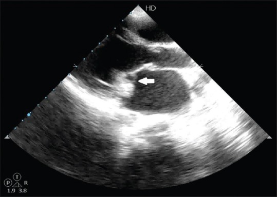
Two-dimensional echo showed 1 cm × 1.2 cm vegetation on the posterior mitral valve leaflet and moderate mitral regurgitation [Figure 1]. Chest X-ray was normal.
Figure 1.

Two-dimensional echo image showing vegetation on mitral valve
After 6 weeks of antibiotics, repeat blood culture was sterile and he improved symptomatically. He had normal appetite and was hemodynamically stable.
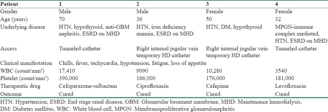
The clinical characteristics, blood culture sensitivity pattern, and outcome of the remaining 4 patients are depicted in Tables 1 and 2.
Table 1.
Clinical characteristics of the patients

Table 2.
Antibiotic sensitivity pattern of the patients

All the 4 patients had similar presentations with fever, chills, fatigue, tachycardia, and hypotension. Two of the patients had tunneled hemodialysis catheter. Two of the remaining patients had temporary hemodialysis internal jugular vein catheter, which was removed immediately. Total white blood cell count varied from leukopenia to leukocytosis. Antibiotic sensitivity pattern was similar with sensitivity to quinolones, carbapenems, and cefoperazone-sulbactam. All of them were cured with antibiotic treatment duration of 2 weeks. None of them required removal of the tunneled catheter.
Following the outbreak, environmental sampling was started. Samples were collected using sterile swabs in therapy room from furniture, electronic devices, hemodialysis equipment, tubings, and medicine trolley. Samples of liquid soap, chlorhexidine, were also cultured. In addition, sterile water and normal saline used for intravenous drug preparations were also cultured.
The sterile water culture was flagged positive for R. mannitolilytica. The whole batch of sterile water in use was discarded. Water treatment with shock chlorination and room disinfection was done by the Hospital Infection Control Committee, following which the outbreak subsided.
Rest of the cultures from chlorhexidine solution, liquid soap, and all environmental samples were negative.
Discussion
Very few cases of outbreaks of R. mannitolilytica have been reported till date in literature. The first reported outbreak occurred in the USA with 30 affected patients in 2005.[1] In 2013, Israel also reported infections in children due to infected vapotherm oxygen delivery systems.[3] Gröbner et al. reported 5 cases of catheter-related bloodstream infections in leukemia patients in Germany in 2007.[4] This is the first instance of reported outbreak of R. mannitolilytica infection in 5 maintenance hemodialysis patients with one patient presenting as infective endocarditis in India. To the best of our knowledge, the first report in India was by Mukhopadhyay et al. who presented a case of renal transplant with Ralstonia infection identified by routine culture and biochemical methods.[5]
There are no specific signs and symptoms of Ralstonia infection described. Most patients in our case series presented with signs of systemic inflammation such as fever, chills, fatigue, and loss of appetite.
Most of the isolates of R. mannitolilytica are overlooked as P. aeruginosa or Burkholderia species.[6] Hence, correct identification of species is important, especially in immunocompromised patients. Misidentification can lead to untreated infection and death of the patient. In our series, the antibiotic sensitivity patterns of Ralstonia infection were similar with sensitivity to quinolones, carbapenems, and cefoperazone-sulbactam, and all patients were cured with 2-week duration of therapy.
It is well-established fact that approximately only 1% of bacteria on earth can be readily cultivated in vitro, which is called the “great plate count anomaly.”[7,8] Because the majority of bacteria cannot be cultured, the pathogens are underestimated using routine culture methods. With the development of molecular techniques, which are culture independent, the pathogens are identified using polymerase chain reaction (PCR) amplification of housekeeping genes, especially 16S rRNA gene. Consequently, cloning is done for purification and sequencing for identification.[9,10] Molecular typing methods also play an important role in identifying fastidious, culture–negative, and slow-growing organisms. Pulsed-field gel electrophoresis is the gold standard in molecular typing for identification of Ralstonia species. Of late, PCR assay for the identification of R. mannitolilytica with 16S rRNA gene as target has been studied.[11,12] This has a sensitivity of 100% and a specificity of 99%. The incorporation of molecular methods routinely to detect microbes is limited by its cost-effectiveness. In addition, Ralstonia has been identified as a contaminant of DNA extraction kit or PCR reagents, which may be produce false-positive results.[12]
Various culture methods are studied to cultivate slow-growing organisms, such as filtration methods, density gradient centrifugation, and extinction dilution. Extended incubation times are necessary for the identification of these organisms. Addition of specific nutrient or chemical required for their growth and cocultivating helper strains hastens the identification of these slow-growing species.[13]
Conclusion
In our setup, this was the first case of infective endocarditis caused by R. mannitolilytica and also first outbreak at hemodialysis unit from infected sterile water. Early detection of the infection helps in initiating appropriate treatment according to culture and sensitivity. The use of molecular methods to detect pathogens may help in early identification of the organism. R. mannitolilytica infections require attention from physicians, microbiologists, and laboratory technicians to prevent undue calamity.
Declaration of patient consent
The authors certify that they have obtained all appropriate patient consent forms. In the form the patient(s) has/have given his/her/their consent for his/her/their images and other clinical information to be reported in the journal. The patients understand that their names and initials will not be published and due efforts will be made to conceal their identity, but anonymity cannot be guaranteed.
Financial support and sponsorship
Nil.
Conflicts of interest
There are no conflicts of interest.
References
- 1.Jhung MA, Sunenshine RH, Noble-Wang J, Coffin SE, St John K, Lewis FM, et al. A national outbreak of Ralstonia mannitolilytica associated with use of a contaminated oxygen-delivery device among pediatric patients. Pediatrics. 2007;119:1061–8. doi: 10.1542/peds.2006-3739. [DOI] [PubMed] [Google Scholar]
- 2.Dowsett E. Hospital infections caused by contaminated fluids. Lancet. 1972;1:1338. doi: 10.1016/s0140-6736(72)91064-1. [DOI] [PubMed] [Google Scholar]
- 3.Block C, Ergaz-Shaltiel Z, Valinsky L, Temper V, Hidalgo-Grass C, Minster N, et al. Déjà vu: Ralstonia mannitolilytica infection associated with a humidifying respiratory therapy device, Israel, June to July 2011. Euro Surveill. 2013;18:20471. [PubMed] [Google Scholar]
- 4.Gröbner S, Heeg P, Autenrieth IB, Schulte B. Monoclonal outbreak of catheter-related bacteraemia by Ralstonia mannitolilytica on two haemato-oncology wards. J Infect. 2007;55:539–44. doi: 10.1016/j.jinf.2007.07.021. [DOI] [PubMed] [Google Scholar]
- 5.Mukhopadhyay C, Bhargava A, Ayyagari A. Ralstonia mannitolilytica infection in renal transplant recipient: First report. Indian J Med Microbiol. 2003;21:284–6. [PubMed] [Google Scholar]
- 6.Pan HJ, Teng LJ, Tzeng MS, Chang SC, Ho SW, Luh KT, et al. Identification and typing of Pseudomonas pickettii during an episode of nosocomial outbreak. Zhonghua Min Guo Wei Sheng Wu Ji Mian Yi Xue Za Zhi. 1992;25:115–23. [PubMed] [Google Scholar]
- 7.Staley JT, Konopka A. Measurement of in situ activities of nonphotosynthetic microorganisms in aquatic and terrestrial habitats. Annu Rev Microbiol. 1985;39:321–46. doi: 10.1146/annurev.mi.39.100185.001541. [DOI] [PubMed] [Google Scholar]
- 8.Amann R, Fuchs BM, Behrens S. The identification of microorganisms by fluorescence in situ hybridisation. Curr Opin Biotechnol. 2001;12:231–6. doi: 10.1016/s0958-1669(00)00204-4. [DOI] [PubMed] [Google Scholar]
- 9.Giovannoni SJ, Britschgi TB, Moyer CL, Field KG. Genetic diversity in Sargasso Sea bacterioplankton. Nature. 1990;345:60–3. doi: 10.1038/345060a0. [DOI] [PubMed] [Google Scholar]
- 10.Pace NR. A molecular view of microbial diversity and the biosphere. Science. 1997;276:734–40. doi: 10.1126/science.276.5313.734. [DOI] [PubMed] [Google Scholar]
- 11.Coenye T, Vandamme P, LiPuma JJ. Infection by ralstonia species in cystic fibrosis patients: Identification of R.pickettii and R. mannitolilytica by polymerase chain reaction. Emerg Infect Dis. 2002;8:692–6. doi: 10.3201/eid0807.010472. [DOI] [PMC free article] [PubMed] [Google Scholar]
- 12.Prasad N, Singh K, Gupta A, Prasad KN. Isolation of bacterial DNA followed by sequencing and differing cytokine response in peritoneal dialysis effluent help in identifying bacteria in culture negative peritonitis. Nephrology (Carlton); Nov 16. doi: 10.1111/nep.12969. doi: 10.1111/nep.12969. Epub ahead of print. [DOI] [PubMed] [Google Scholar]
- 13.Vartoukian SR, Palmer RM, Wade WG. Strategies for culture of ‘unculturable’ bacteria. FEMS Microbiol Lett. 2010;309:1–7. doi: 10.1111/j.1574-6968.2010.02000.x. [DOI] [PubMed] [Google Scholar]


