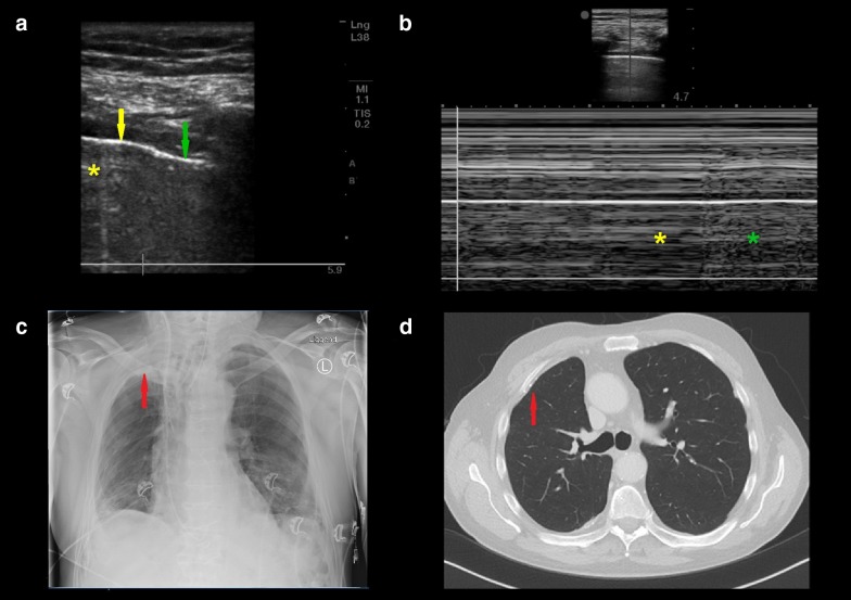
Given a reported 100% specificity for the diagnosis of pneumothorax, the lung point has been considered a sign that cannot be mimicked. However, we now present a unique case of the presence of lung point in the absence of pneumothorax in a 75-year-old man admitted after coronary artery bypass graft (Fig. 1).
Fig. 1.
a Lung point. The yellow arrow shows where lung sliding was absent. A single B-line, marked by the yellow asterisk, was present in the absence of lung sliding. The green arrow shows where lung sliding was present. b The M-mode of the lung point where the immobility of the affected lung alternates with the movement of the healthy lung which can be observed in the M-mode as stratosphere sign (yellow asterisk) alternating with seashore sign (green asterisk) respectively. c Post-operational chest X-ray showing pleural thickening. d Thoracic CT scan showing pre-existent asbestos-related pleural disease. The red arrows on the CT and X-ray indicate pleural thickening and calcification
Scanning at the right anterior third intercostal space showed absence of lung sliding (online video A), A-line and stratosphere sign indicating an A′-profile. Thus, a lung point was actively searched for and indeed found. Pneumothorax was ruled out by the presence of a B-line in the same view as the lung point (online video B), a post-operative chest X-ray and CT scan which indicated the pre-existence of asbestos-related pleural disease (Fig. 1a, c, d). We postulate that the healthy part of the lung moves unrestrictedly whereas the affected part is restricted, their transition resulting in a lung point. Another situation where lung point may be false positive is a ‘bleb’ point in bullous lung disease.
In conclusion, absent lung sliding and lung point can be observed in cases of pleural thickening and adhesion and may thus warrant revision of the perception that lung point is pathognomonic for pneumothorax.
Electronic supplementary material
Below is the link to the electronic supplementary material.
Author contributions
TSS, BH, PWGE and PRT contributed substantially to the study design and the writing of the manuscript.
Ethical approval
The commission for medical ethics (METc) of VUmc has approved this research: METC: 2016.053.
Informed consent
Written informed consent was given by the patient.
Conflicts of interest
The authors declare they have no conflict of interest relevant to this manuscript.
Contributor Information
Thei S. Steenvoorden, Email: t.steenvoorden@vumc.nl
Bashar Hilderink, Email: b.hilderink@vumc.nl.
Paul W. G. Elbers, Email: p.elbers@vumc.nl
Pieter R. Tuinman, Email: p.tuinman@vumc.nl
Associated Data
This section collects any data citations, data availability statements, or supplementary materials included in this article.


