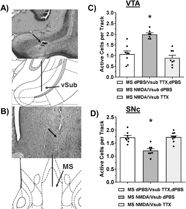
Fig. 2.
MS regulation of midbrain DA neurons requires the vSub. Representative histology and matching illustration for the target region of the (a) vSub and (b) MS. The line in the illustration denotes the cannula placement and the arrow in the histology indicates the ventral termination of the cannula track. TTX (1 µM, 0.5 µL, N = 7–10 rats per group) inactivation of the vSub prevented changes in the number of spontaneously active DA neurons in the (c) VTA (*MS NMDA/vSub dPBS significantly greater than both other groups, Tukey’s, P < 0.001) and (d) SNc (*MS NMDA/vSub dPBS significant decrease from both groups, Tukey’s, P < 0.001) compared to vehicle infusions (dPBS).

