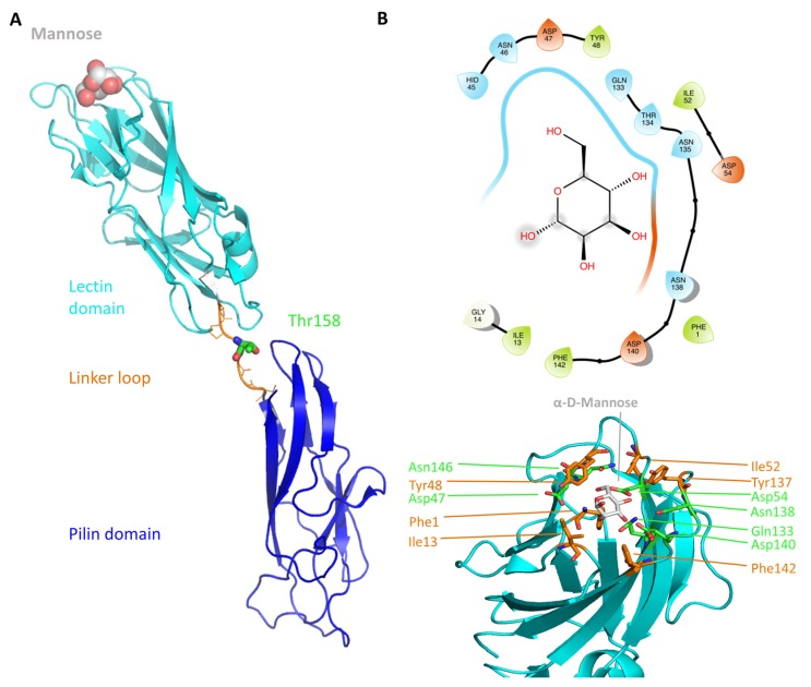
Figure 1.
The FimH structure and organization (A) An elongated linker (orange) connects the pilin (blue) and the lectin (cyan) domain of FimH (PDB code 1KLF [14]). The protein is shown in cartoon and the bound α-d-mannose molecule is depicted as atom-colored (grey for carbon) van-der-Waals spheres. Additionally, the position of T158 is shown as atom-colored sticks (green for carbon). (B) The mannose-binding site of the FimH lectin domain. On the top the 2D diagram of the binding site is depicted (prepared with Maestro using a cutoff of 5 Å) and on the bottom the 3D representation of the same site. The mannose molecule is highlighted in gray. The polar (green) binding site residues as well as the hydrophobic rim residues (orange) are additionally depicted. The 3D protein representations in this and the following figures were prepared using Pymol [29].

