Figure 1.
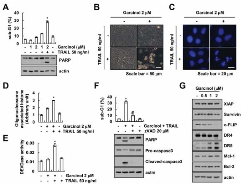
Garcinol sensitizes Caki cells to TRAIL-mediated apoptosis. (A–E) Caki cells were treated with garcinol (1–2 μM) and/or 50 ng/mL TRAIL for 24 h. Levels of apoptosis were assessed by flow cytometry, and western blot showing the PARP and actin (A). Morphology of cells was visualized with an optical microscope (B). DAPI staining detected condensation and fragmentation of nuclei (C). Detection of DNA fragmentation (D) and caspase activity (E). (F) Caki cells were treated with 2 μM garcinol and 50 ng/mL TRAIL for 24 h in the presence or absence of 20 μM z-VAD. Levels of apoptosis were assessed by flow cytometry, and western blot showing the PARP, pro-caspase-3, cleaved caspase-3 and actin. (G) Caki cells were treated with (0.5–2 μM) galcinol for 24 h. The related levels of proteins were detected by western blot using indicated antibody. * p < 0.01 compared to the control. # p < 0.01 compared to the garcinol plus TRAIL.

