Abstract
The western corn rootworm (WCR) is an important pest of corn and is well known for its ability to rapidly adapt to pest management strategies. Although RNA interference (RNAi) has proved to be a powerful tool for studying WCR biology, it has its limitations. Specifically, RNAi itself is transient (i.e. does not result in long-term Mendelian inheritance of the associated phenotype), and it requires knowing the DNA sequence of the target gene. The latter can be limiting if the phenotype of interest is controlled by poorly conserved, or even novel genes, because identifying useful targets would be challenging, if not impossible. Therefore, the number of tools in WCR's genomic toolbox should be expanded by the development of methods that could be used to create stable mutant strains and enable sequence-independent surveys of the WCR genome. Herein, we detail the methods used to collect and microinject precellular WCR embryos with nucleic acids. While the protocols described herein are aimed at the creation of transgenic WCR, CRISPR/Cas9-genome editing could also be performed using the same protocols, with the only difference being the composition of the solution injected into the embryos.
Keywords: Bioengineering, Issue 134, embryonic microinjection, functional genomics, germline transformation, transgenesis, CRISPR/Cas9 genome editing, western corn rootworm
Introduction
Western corn rootworm (WCR), Diabrotica virgifera virgifera, is an important pest of corn1. Interestingly, WCR appears to overcome control measures more rapidly than most agricultural pests since they not only adapt physiologically but also behaviorally2,3,4,5,6. Over the past decade, RNA interference (RNAi), a powerful functional genomic tool, has been investigated as a potential control method for WCR7,8, and has also been used as a means to study gene function9. However, while RNAi is frequently performed by microinjection of double-stranded RNA (dsRNA) in other species, injection-based RNAi is rare in WCR. In fact, there are only a few reports of RNAi via microinjection of dsRNA into WCR9. The reason is that WCR can attain high-levels of gene knockdown via ingestion of dsRNA7,10, even permitting the study of embryonic effects by feeding dsRNA to the mother11. While this method makes WCR an excellent subject for functional genomic analysis via RNAi, it has slowed progress on the development of methodologies for embryonic microinjection in this species.
Despite the power of RNAi, there are a few drawbacks. For example, not all genes respond equally to RNAi. This variability can make interpreting the results of a functional genomics assay more difficult. Also, RNAi is transient and does not generate heritable mutations. On the other hand, germline transformation, another functional genomic tool, can generate heritable mutations via insertion of a marked transposable element into the genome12,13. This makes germline transformation an excellent tool for creating mutant strains for use in long-term genetic studies. Transformation can also be used to rescue a mutation by delivering a functional copy of the gene to a mutant genome14. Moreover, germline transformation is the cornerstone of a wide variety of molecular genetic techniques. In addition to knocking out and/or rescuing gene function, germline transformation can be used for enhancer trapping15, gene trapping16, Gal4-based ectopic expression17, and genome-wide mutagenesis18.
More recently, tools that enable sophisticated genome editing have been developed. These tools include Transcription Activator-Like Effector Nucleases (TALEN) and the CRISPR/Cas9-nuclease system19,20. The advantage of these new tools is that they offer researchers the ability to induce a double-stranded DNA break at almost any location within almost any genome. Once induced, these breaks can be repaired through either non-homologous end joining, which can introduce deletions or insertions in the targeted gene, or through homology-directed repair, which in the presence of an engineered construct, can catalyze the replacement of entire genes or genetic regions21,22. However, these methods require microinjection of DNA, RNA and/or protein into very young embryos.
Importantly, unlike the dsRNAs used for RNAi, the nucleic acids and proteins used for germline transformation and genome editing cannot easily cross cell membranes. Therefore, microinjection of plasmid DNAs, mRNAs, and/or proteins must take place during the syncytial blastoderm stage (i.e. before the insect embryo cellularizes). This makes the timing of injections a critical factor. For example, in Drosophila melanogaster, embryos need to be injected within two hours after egg lay12. Therefore, development of a successful embryonic microinjection protocol for WCR must take into account the best conditions for female egg laying, as well as the best method for collecting sufficient quantities of precellular embryos.
An effort to bring a wide range of molecular genetic tools to bear on WCR biology requires developing methods for collecting and microinjecting precellular WCR embryos. Here we provide detailed instructions, along with tips and tricks, to help others use transformation-based techniques in WCR research. In addition to extending transgenic technologies to WCR for use in functional genomic studies, these techniques also enable powerful new pest control strategies such as gene drive23,24 to be tested in this economically important pest.
Protocol
1. Colony-level Rearing of WCR Adults
Obtain a sufficient quantity of WCR adults (500 - 1,000) from a reliable company or research laboratory (see Table of Materials for an example) and place in a 30 cm3 cage. NOTE: Use of a non-diapausing strain is highly recommended.
Prepare WCR artificial diet following manufacturer's protocol and pour a 1 cm-thick layer into a 38 oz container (see Table of Materials). After mixing, store unused diet at 4 °C for up to 2 months.
Put a Petri dish (100 x 15 mm) with adult diet (10 - 15 g) into a 30 cm3 cage. Add more diet when the food is low or dry.
Put a flask (300 mL) with water, covered with a cotton ball, which is used to hold in place a cotton roll (6" x 3/8"), to serve as a water source. Change the water when it runs low.
Maintain WCR at 26 °C with 60% humidity and a 14:10 light cycle in an insect rearing incubator.
2. Embryo Collection and Alignment
Make an egg collection chamber using 1% agar in water in a sterile 100 x 15 mm2 Petri dish. Store at 4 °C after the agar solidifies.
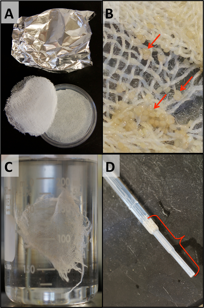
To collect newly laid eggs, place a single layer of filter paper, followed by 4 layers of cheesecloth (each cut to size) on the surface of the agar (Figure 1A). NOTE: Cheesecloth can be reused, after being washed in bleach and autoclaved, but it is not necessary to do this for the first use. Filter paper should not be reused and does not need to be sterilized.
Place egg collection plate into WCR cage as late in the day as possible (end of work day) and cover with a tinfoil tent. Leave overnight. NOTE: Having the lights on from 10 A.M. to 12 A.M. (14 h) delays egg laying, thus facilitating the collection of younger eggs for microinjection without requiring lab personnel to place collection chamber into cage late at night.
Remove the egg collection chamber around 8 or 9 A.M.
Use forceps to pick up 1 layer of cheesecloth at a time and place in a beaker (500 mL) of water. Wash eggs off of cheesecloth by gently stirring (Figure 1B and 1C).
Use a bulb pipette to transfer eggs to another beaker (500 mL) of water. Repeat 2 - 3x to clean the eggs.
Use the bulb pipette to transfer eggs onto filter paper (Figure 1D) while minimizing the amount of water carry-over.
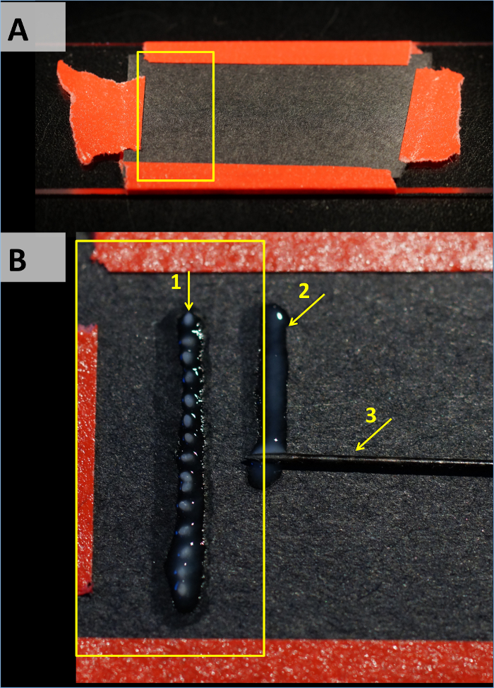
Cut black filter paper into strips as wide as a glass slide and tape firmly to the slide to make sure the paper lays flat (Figure 2A). NOTE: Black filter paper helps improve visibility under the microscope by reducing the amount of reflected light.
Apply non-toxic glue (see Table of Materials for an example) to the filter paper in fine lines (Figure 2B). Keep the glue lines thinner than the diameter of an egg to avoid getting the eggs coated in glue.
Use a fine brush to gently move WCR eggs one by one from their filter paper to the glue line. Try to lay the eggs on the glue before it dries out and keep at least one egg's distance between each egg.
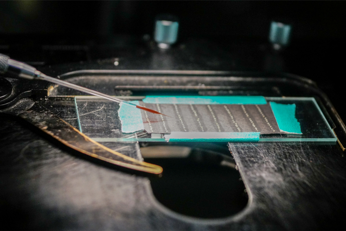
Maximize injection efficiency by placing multiple lines of eggs on one filter paper slide (Figure 3).
After all eggs are laid out on the filter paper, wait until all the glue dries before microinjection. NOTE: For germline transformation and for CRISPR/Cas9-mediated genome editing, inject all embryos by noon (12 P.M.) so that the oldest embryo is no more than 19 h old. However, for RNAi, there is no time limitation because, in WCR, dsRNA can move between cells. In fact, for RNAi, aging embryos might result in better survival rates.
3. Preparation of Plasmid DNAs and Injection Needles
Prepare high-quality supercoiled plasmid DNA using an endotoxin-free commercial kit (see Table of Materials for an example).
To prepare the injection solution, check the concentration of each plasmid and then combine helper plasmid (250 ng/µL final concentration), donor plasmid (750 ng/µL final concentration) and phenol red buffer (20% final concentration). Vortex the solution briefly and centrifuge for 3 min at maximum speed to precipitate particles that could clog the needle.
Pull borosilicate glass needles (O.D. 1 mm, I.D. 0.58 mm, 10 cm length) using a Flaming/Brown type micropipette puller. For storage, secure pulled needles on double-sided sticky tape in a Petri dish or other clear container. NOTE: For the micropipette puller system, the settings used were: Heat = 335, FIL = 4, VEL = 40, DEL = 200, PUL = 100.
Backfill the injection needle by using a micro-loader tip (see Table of Materials) to pipet 0.5 - 1.0 µL down inside the needle, near the tapered end. Remove bubbles from the tip of the needle if any occur.
Insert the injection needle into the needle holder.
Open the tip of the needle by gently breaking with a fine pair of forceps, or gently touching/dragging the tip on the coverslip or glass slide. NOTE: The goal is to have the smallest opening that still allows the injection mix to come out (smaller openings generally require higher injection pressure). Beveled needles don't require this step since they are already open.
4. Microinjection and Post-injection Care
Using a fine paintbrush, carefully wet the surface of 1 - 3 eggs with sterile water, insert the needle, and inject the solution. Inject each egg before the surface dries. Look for the appearance a small amount of red color inside eggs during injection to indicate the successful microinjection. Crush or remove any eggs that look damaged, empty, or otherwise cannot be injected. NOTE: Although injection pressure varies based on needle bore size, 10 - 13 psi is a good starting point.
After all eggs are injected, remove the filter paper from the glass slide.
Place the filter paper on the surface of a 1% agar dish (described above).
Use plastic paraffin film to seal the dish, and put in a 26 °C insect rearing incubator for 12 days. NOTE: The paraffin film will help prevent cross-contamination of mold between dishes. However, any other treatment to prevent mold and bacterial growth could reduce the survival of the injected embryos.
5. Culturing and Hatching of Embryos
Start checking embryos 12 days post-injection for newly-hatched larvae and continue daily for the next 2 weeks.
Transfer larvae, using a fine brush, to a rearing box with sprouted corn and soil. NOTE: Mold might grow in the agar dish, but larvae will still hatch in most cases. Look for larvae carefully within the mold to catch them all. Some larvae might move to the lid of the dish and might get stuck in the condensation.
Rear larvae at 26 °C following standard WCR rearing methods25.
Representative Results
An important consideration when developing this protocol was the stage of embryonic development at the time of microinjection. Specifically, germline transformation requires microinjection of DNAs prior to embryonic cellularization. This is because DNAs cannot easily cross cell membranes. Attempts to visualize the stages of WCR embryonic development using a nuclear stain were unsuccessful because dechorionation of WCR embryos essentially dissolves all of the protective membranes responsible for the integrity of the embryo. However, this line of inquiry was supplanted by another difficulty – acquiring sufficient quantities of embryos in a timely manner. In the field, WCR females lay their eggs in the soil, thus it should be no surprise that in the lab, females only lay eggs in the dark. This makes short daytime collections (2 - 4 h) difficult. To overcome this issue, a single overnight egg lay was performed and was found to yield large quantities of embryos. Although the process of collecting (Figure 1) and laying out (Figure 2) embryos for microinjection is somewhat laborious, if done quickly this protocol enables microinjection of embryos that are between 2 - 19 h old at the time of injection, and no older than 24 h if injections are performed later in the day. Comparison of development times between WCR and other species suggests that WCR embryos should still be precellular at 24 h post egg lay (see Discussion).
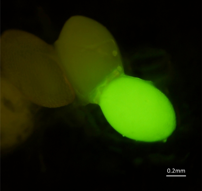
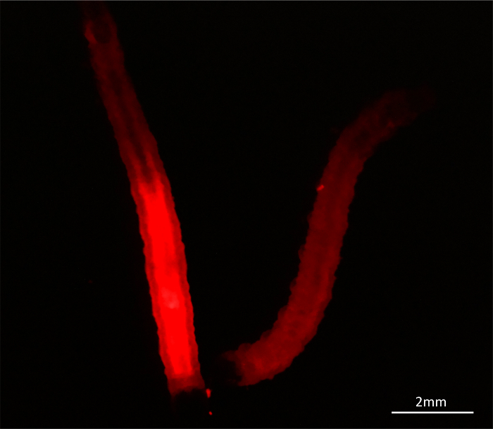
The first attempts at germline transformation of WCR involved co-injection (Figure 3) of helper (transposase source) and donor (transposon) plasmids into precellular embryos. The transformation marker for the donor plasmid was a green fluorescent protein (EGFP) gene driven by a constitutively active promoter from Tribolium castaneum (alpha-tubulin promoter26). Embryonic survival rates were low (Table 1), but 26 G0 adults were recovered. To determine if the donor element inserted into any germ cells, G1 embryos were screened for EGFP expression. Although fluorescent progeny were indeed identified (Figure 4), screening WCR embryos with the intense light source proved to be lethal27. The next round of germline transformation utilized a new donor plasmid, one possessing a red fluorescent protein (DsRed) gene driven by a constitutively active promoter from Drosophila melanogaster (heat shock protein 70 promoter28). While survival rates were much improved (Table 1), the surviving adults only produced a single DsRed-positive larva (Figure 5). However, these experiments were particularly informative in two regards. First, it was discovered that microinjection caused delays in development, resulting in an extended time-to-hatch. Rather than all embryos hatching within 2 weeks, injected embryos hatched between 12 and 26 days post microinjection, thus requiring an extended monitoring period to ensure all hatchlings were collected. Second, it was noticed that newly-hatched larvae were fairly active. They crawled onto the lid where they would get stuck, and sometimes escaped from the agar dish. These issues were overcome by sealing Petri dishes with a paraffin film and examining lids and agar carefully for hatchlings.
The final version of this injection protocol was tested by microinjecting WCR embryos with buffer alone, or with helper and eye-promotor (3xP329) marked donor DNA plasmids. As an additional control, another set of embryos were handled identically but were not microinjected. Four sets of embryos were injected with either injection solution. A total of 485 embryos were injected with buffer and of those, 148 hatched (hatch rate = 30.5%, Table 1). Of 1,450 embryos injected with plasmid DNAs, 289 hatched (rate = 26.8%). Interestingly, of the 470 embryos that underwent the same egg-handling conditions, but without microinjection, only 250 hatched (rate = 53.2%). That the survival rate of uninjected embryos is 20% higher than the survival rates for injected embryos is to be expected due to the unavoidable damage caused by microinjection. However, little difference is seen between buffer injection and DNA injection, which suggests the amount of DNA being added is acceptable for WCR (see Discussion). These results also show that survival rates improved over earlier efforts. Successful establishment of transgenic lines using this method has been reported elsewhere27.
Figure 1: Egg collection. A) Egg collection chamber. B) Zoomed-in view of a section of an egg chamber after 24-h egg lay. Note, arrows indicate the location of a few eggs. C) Washing eggs off cheesecloth. D) The final transfer of WCR eggs. Note, bracket indicates the region with densely packed WCR embryos (i.e. minimal water). Please click here to view a larger version of this figure.
Figure 2: Preparing an injection slide. A) Black filter paper taped tightly to a standard microscope slide. B) Zoomed-in view of a section of an injection slide during the preparation process. Note, arrow #1 indicates the location of a row of eggs, #2 partial line of glue and #3 the pin used to spread the glue. Please click here to view a larger version of this figure.
Figure 3: Injecting embryos. An injection slide with eight rows of eggs. Due to the angle of the injection needle, successive rows of embryos can be injected by simply moving the stage. Note the red dye in the injection needle greatly aids visualization of the injection. Please click here to view a larger version of this figure.
Figure 4: Transgenic G1 embryo. Embryos from a WCR adult that was co-injected with piggyBac-based helper and donor plasmids as an early embryo. The bright green embryo is positive for enhanced green fluorescent protein (EGFP) expression while the others are not. EGFP expression is driven by the Tribolium castaneum alpha-tubulin promoter26. Please click here to view a larger version of this figure.
Figure 5: Transgenic G1 larva. 3rd-instar larvae whose parent was co-injected with piggyBac-based helper and donor plasmids as an early embryo. The red glowing larva on the left is positive for DsRed expression, while the larva on the right is not. DsRed expression is driven by the Drosophila melanogaster heat shock 70 promoter28. Please click here to view a larger version of this figure.
| Experiment | Treatment | Eggs | Hatched | Hatch Rate |
| Exp. #1 | αtubEGFP-donor | 910 | 57 | 6.3% |
| Exp. #2 | hsp70DsRed-donor | 1580 | 211 | 13.4% |
| Exp. #3 | 3xP3EGFP-donor | 1450 | 389 | 26.8% |
| Buffer | 485 | 148 | 30.5% | |
| No-injection | 470 | 250 | 53.2% |
Table 1: Microinjection data. Hatch rates from three microinjection experiments on WCR embryos, showing the total number of eggs injected, the total number of larvae that successfully hatched, and the hatch rate.
Discussion
Although microinjection of WCR with dsRNAs for the purpose of RNAi has been reported9, this is the first protocol to establish best practices for microinjecting precellular WCR embryos, a critical process for conducting germline transformation and/or CRISPR/Cas9 genome editing in this species. Successful microinjection of WCR embryos is dependent on many factors, as is transformation efficiency. Discussed below are some of the major issues impacting the outcome of using this protocol for germline transformation in this beetle.
Obtaining precellular embryos As mentioned above, DNA does not cross cell membranes the way dsRNA can. This means that it is of critical importance that DNA, mRNA, and/or proteins be injected before cellularization occurs. Because the timing of injection is critical, it was important to determine a likely window for successful microinjection based on how long it takes the nondiapausing WCR strain30 to complete embryogenesis. This was calculated based on detailed studies of the timing of the syncytial blastoderm stage in another beetle, Tribolium castaneum. In Tribolium, cellularization occurs ~8 h post egg lay at 30 °C31. Since it takes ~3.5 days for Tribolium to complete embryogenesis at 30 °C, cellularization takes place a tenth of the way into the process. Since WCR embryos take nearly 2 weeks to develop, cellularization may not occur until the second day post egg lay. Therefore, it was assumed that the window for successful microinjection is at least 24 h long, which is helpful, since WCR require overnight egg laying to provide sufficient quantities of eggs for microinjection. While this method was used for successful transgenesis27, it is conceivable that results could be improved by injecting younger embryos, provided handling very early embryos doesn't cause them harm.
DNA quality and concentration It is not surprising that DNA concentration plays a pivotal role in successful transformation. It has long been thought that keeping the final concentration of plasmid DNA below 1 mg/mL was necessary to avoid potential toxicity to the injected embryos32. Although it is now unclear if the toxicity issues of the past were due to the DNA itself or were instead the result of contaminants carried over from the plasmid purification process, this rule of thumb has been steadfastly adhered to for two decades. However, we recently discovered that exceeding the 1 mg/mL limit was not only possible but also that doing so can achieve higher transformation rates, leading to speculation that the absence of egg toxicity may be due to advances in plasmid purification kits, resulting in fewer contaminants. Given the time required to rear WCR, any increase in transformation rates is helpful, even if it results in slightly higher mortality rates.
Softening egg surface before microinjection Unlike Tribolium, the outer-most surface (chorion) of a WCR embryo must be softened before microinjection. This is a critical step even when using quartz needles. Ideally, water should be applied to a single embryo immediately before injection, but can be applied to groups of 2 to 3 embryos at a time if microinjection can be accomplished before chorions reharden. Without this step, needles will generally fail to penetrate the chorion, thereby causing the injection attempt to fail, and potentially damaging the tip of the needle. Moreover, if the needle happens to successfully penetrate a dry chorion, the injection solution typically leaks back out due to the pressure inside the embryo being higher than ambient pressure. It is, however, easy to distinguish dry from wet (softened) embryos. Specifically, when dry, embryos have a distinct rigid, round shape, while the addition of water causes them to lose their spherical appearance. Softening not only allows the needle to easily penetrate the chorion but also reduces the pressure inside the embryo enough that the injection solution does not leak out.
Humidity and mold Injected embryos are held in a warm, moist environment throughout embryogenesis. These conditions not only provide the perfect environment for WCR development but also promote the growth of mold. During this two-week period, mold frequently grows on the agar dishes housing the WCR embryos. Unfortunately, the embryos require high humidity, and attempts to reduce it resulted in much lower hatch rates. Interestingly, the presence of mold generally had little impact on hatch rates, with most embryos hatching without problems. Moreover, antifungal treatments are harmful to WCR embryos, so the best way to reduce mold growth appears to be to avoid contamination; cleaning tools, containers, and microinjection system before use, and trying to keep the time spent microinjecting to a minimum since mold spores are prevalent in many laboratory environments.
Conclusions This protocol is intended to enable more researchers to employ transgenic technologies in their WCR research and will hopefully aid in the development of additional transgenics-based technologies for use in this insect. Sophisticated transgenics-based experiments conducted in Tribolium could potentially be adapted for WCR, including mutagenesis33 and CRISPR-based genome editing34. Because these techniques require microinjection of precellular embryos, this protocol will be useful not only for transposon-based functional genomic studies and/or CRISPR/Cas9-mediated genome editing but also for genetics-based pest management strategies35,36.
Disclosures
The authors have nothing to disclose.
Acknowledgments
This work was supported by a grant from the Monsanto Corn Rootworm Knowledge Research Program, grant number AG/1005 (to MDL and YC) and start-up funds to MDL from NCSU. FC was supported by grants from Monsanto's Corn Rootworm Knowledge Research Program (AG/1005) and the National Science Foundation, grant number MCB-1244772 (to MDL). The authors declare no competing interests. FC and MDL conceived and designed the experiments; FC, PW and SP performed the experiments; FC and MDL analyzed the results; and FC, NG and MDL wrote the manuscript. We thank Teresa O'Leary, William Klobasa, and Stephanie Gorski for their expert assistance in screening WCR. We also thank Dr. Wade French (USDA-ARS, North Central Agricultural Research Laboratory, Brookings, SD) for shipments of eggs to establish lab colonies and providing rearing protocols.
References
- Gray ME, Sappington TW, Miller NJ, Moeser J, Bohn MO. Adaptation and Invasiveness of Western Corn Rootworm: Intensifying Research on a Worsening Pest. Annual Review of Entomology. 2009;54:303–321. doi: 10.1146/annurev.ento.54.110807.090434. [DOI] [PubMed] [Google Scholar]
- Gassmann AJ, Petzold-Maxwell JL, Keweshan RS, Dunbar MW. Field-evolved resistance to Bt maize by western corn rootworm. PLoS One. 2011;6(7):e22629. doi: 10.1371/journal.pone.0022629. [DOI] [PMC free article] [PubMed] [Google Scholar]
- Gray ME, Levine E, Oloumi-Sadeghi H. Adaptation to crop rotation: western and northern corn rootworms respond uniquely to a cultural practice. Recent research developments in entomology. 1998;2:19–31. [Google Scholar]
- Meinke LJ, Siegfried BD, Wright RJ, Chandler LD. Adult Susceptibility of Nebraska Western Corn Rootworm (Coleoptera: Chrysomelidae) Populations to Selected Insecticides. Journal of Economic Entomology. 1998;91(3):594–600. [Google Scholar]
- Roselle R, Anderson L, Simpson R, Webb M. Annual report for 1959, cooperative extension work in entomology. Lincoln, NE: University of Nebraska Extension; 1959. [Google Scholar]
- Wright R, Meinke L, Siegfried B. Corn rootworm management and insecticide resistance management. Proceedings. 1996. pp. 45–53.
- Baum JA, et al. Control of coleopteran insect pests through RNA interference. Nat Biotechnol. 2007;25(11):1322–1326. doi: 10.1038/nbt1359. [DOI] [PubMed] [Google Scholar]
- Velez AM, et al. Parameters for Successful Parental RNAi as An Insect Pest Management Tool in Western Corn Rootworm, Diabrotica virgifera virgifera. Genes (Basel) 2016;8(1) doi: 10.3390/genes8010007. [DOI] [PMC free article] [PubMed] [Google Scholar]
- Alves AP, Lorenzen MD, Beeman RW, Foster JE, Siegfried BD. RNA interference as a method for target-site screening in the Western corn rootworm, Diabrotica virgifera virgifera. J Insect Sci. 2010;10:162. doi: 10.1673/031.010.14122. [DOI] [PMC free article] [PubMed] [Google Scholar]
- Rangasamy M, Siegfried BD. Validation of RNA interference in western corn rootworm Diabrotica virgifera virgifera LeConte (Coleoptera: Chrysomelidae) adults. Pest Management Science. 2012;68(4):587–591. doi: 10.1002/ps.2301. [DOI] [PubMed] [Google Scholar]
- Khajuria C, et al. Parental RNA interference of genes involved in embryonic development of the western corn rootworm, Diabrotica virgifera virgifera LeConte. Insect Biochem Mol Biol. 2015;63:54–62. doi: 10.1016/j.ibmb.2015.05.011. [DOI] [PubMed] [Google Scholar]
- Cooley L, Kelley R, Spradling A. Insertional mutagenesis of the Drosophila genome with single P elements. Science. 1988;239(4844):1121–1128. doi: 10.1126/science.2830671. [DOI] [PubMed] [Google Scholar]
- Robertson HM, et al. A stable genomic source of P element transposase in Drosophila melanogaster. Genetics. 1988;118(3):461–470. doi: 10.1093/genetics/118.3.461. [DOI] [PMC free article] [PubMed] [Google Scholar]
- Spradling AC, Rubin GM. Transposition of Cloned P Elements into Drosophila Germ Line Chromosomes. Science. 1982;218(4570):341–347. doi: 10.1126/science.6289435. [DOI] [PubMed] [Google Scholar]
- O'Kane CJ, Gehring WJ. Detection in situ of genomic regulatory elements in Drosophila. Proc Natl Acad Sci U S A. 1987;84(24):9123–9127. doi: 10.1073/pnas.84.24.9123. [DOI] [PMC free article] [PubMed] [Google Scholar]
- Lukacsovich T, et al. Dual-tagging gene trap of novel genes in Drosophila melanogaster. Genetics. 2001;157(2):727–742. doi: 10.1093/genetics/157.2.727. [DOI] [PMC free article] [PubMed] [Google Scholar]
- Brand AH, Perrimon N. Targeted Gene Expression as a Means of Altering Cell Fates and Generating Dominant Phenotypes. Development. 1993;118(2):401–415. doi: 10.1242/dev.118.2.401. [DOI] [PubMed] [Google Scholar]
- Horn C, Offen N, Nystedt S, Hacker U, Wimmer EA. piggyBac-based insertional mutagenesis and enhancer detection as a tool for functional insect genomics. Genetics. 2003;163(2):647–661. doi: 10.1093/genetics/163.2.647. [DOI] [PMC free article] [PubMed] [Google Scholar]
- Bassett AR, Tibbit C, Ponting CP, Liu JL. Highly efficient targeted mutagenesis of Drosophila with the CRISPR/Cas9 system. Cell Rep. 2013;4(1):220–228. doi: 10.1016/j.celrep.2013.06.020. [DOI] [PMC free article] [PubMed] [Google Scholar]
- Moscou MJ, Bogdanove AJ. A simple cipher governs DNA recognition by TAL effectors. Science. 2009;326(5959):1501. doi: 10.1126/science.1178817. [DOI] [PubMed] [Google Scholar]
- Bibikova M, Golic M, Golic KG, Carroll D. Targeted chromosomal cleavage and mutagenesis in Drosophila using zinc-finger nucleases. Genetics. 2002;161(3):1169–1175. doi: 10.1093/genetics/161.3.1169. [DOI] [PMC free article] [PubMed] [Google Scholar]
- Jeggo PA. DNA breakage and repair. Adv Genet. 1998;38:185–218. doi: 10.1016/s0065-2660(08)60144-3. [DOI] [PubMed] [Google Scholar]
- Gantz VM, Bier E. Genome editing. The mutagenic chain reaction: a method for converting heterozygous to homozygous mutations. Science. 2015;348(6233):442–444. doi: 10.1126/science.aaa5945. [DOI] [PMC free article] [PubMed] [Google Scholar]
- Windbichler N, et al. A synthetic homing endonuclease-based gene drive system in the human malaria mosquito. Nature. 2011;473(7346):212–215. doi: 10.1038/nature09937. [DOI] [PMC free article] [PubMed] [Google Scholar]
- Branson TF, Jackson JJ, Sutter GR. Improved Method for Rearing Diabrotica virgifera virgifera (Coleoptera, Chrysomelidae) Journal of Economic Entomology. 1988;81(1):410–414. [Google Scholar]
- Siebert KS, Lorenzen MD, Brown SJ, Park Y, Beeman RW. Tubulin superfamily genes in Tribolium castaneum and the use of a Tubulin promoter to drive transgene expression. Insect Biochem Mol Biol. 2008;38(8):749–755. doi: 10.1016/j.ibmb.2008.04.007. [DOI] [PubMed] [Google Scholar]
- Chu F, et al. Germline transformation of the western corn rootworm, Diabrotica virgifera virgifera. Insect Mol Biol. 2017. [DOI] [PubMed]
- Ingolia TD, Craig EA, McCarthy BJ. Sequence of three copies of the gene for the major Drosophila heat shock induced protein and their flanking regions. Cell. 1980;21(3):669–679. doi: 10.1016/0092-8674(80)90430-4. [DOI] [PubMed] [Google Scholar]
- Berghammer AJ, Klingler M, Wimmer EA. A universal marker for transgenic insects. Nature. 1999;402(6760):370–371. doi: 10.1038/46463. [DOI] [PubMed] [Google Scholar]
- Branson T. The selection of a non-diapause strain of Diabrotica virgifera (Coleoptera: Chrysomelidae) Entomologia Experimentalis et Applicata. 1976;19(2):148–154. [Google Scholar]
- Handel K, Grunfelder CG, Roth S, Sander K. Tribolium embryogenesis: a SEM study of cell shapes and movements from blastoderm to serosal closure. Dev Genes Evol. 2000;210(4):167–179. doi: 10.1007/s004270050301. [DOI] [PubMed] [Google Scholar]
- Handler AM, James AA. Insect transgenesis: methods and applications. CRC Press; 2000. [Google Scholar]
- Lorenzen MD, et al. piggyBac-based insertional mutagenesis in Tribolium castaneum using donor/helper hybrids. Insect Mol Biol. 2007;16(3):265–275. doi: 10.1111/j.1365-2583.2007.00727.x. [DOI] [PubMed] [Google Scholar]
- Gilles AF, Schinko JB, Averof M. Efficient CRISPR-mediated gene targeting and transgene replacement in the beetle Tribolium castaneum. Development. 2015;142(16):2832–2839. doi: 10.1242/dev.125054. [DOI] [PubMed] [Google Scholar]
- Phuc HK, et al. Late-acting dominant lethal genetic systems and mosquito control. BMC Biol. 2007;5:11. doi: 10.1186/1741-7007-5-11. [DOI] [PMC free article] [PubMed] [Google Scholar]
- Horn C, Wimmer EA. A transgene-based, embryo-specific lethality system for insect pest management. Nat Biotechnol. 2003;21(1):64–70. doi: 10.1038/nbt769. [DOI] [PubMed] [Google Scholar]


