Introduction
Increasingly, anthropologists have become involved with applied biomedical research and a variety of related activities in the health sciences. Wienker (1984) has recently reviewed some of the developments in what he has labeled biomedical anthropology, an area of physical anthropology that has emerged since the early 1970s. According to Wienker, the activity in biomedical anthropology has resulted quite naturally from the biocultural orientation of physical anthropology. He identifies the growth of applied anthropology in general, the changing academic market, and shifting resources for research as additional probable causes for interest in the area. It should be noted that a decade ago the late Albert Damon (1975) expressed his hope that an applied biological anthropology subarea would evolve among those physical anthropologists interested in medicine and bioengineering.
Medical genetics is a rapidly changing area of biomedical specialization to which both biologically and culturally oriented anthropologists can contribute. The relationship between medical genetics and physical anthropology has been explored in several recent reviews (Robinow 1982; O’Rourke and Petersen 1983; Ward and Meaney 1984). A potentially fruitful area for research by cultural anthropologists involves the problem of communication and the education process in genetic counseling, particularly when cross-cultural interaction is involved. These problems have not been investigated until recently by anthropologists. Two teams of anthropologists and medical geneticists have independently begun evaluation studies of genetic counseling services in two urban midwestern genetic counseling centers (Meier, Steltz-Lenarsky, and Hearst 1984; Steltz-Lenarsky, Mahnke, and Meier 1985; Ross, personal communication). Several recent case studies in genetic counseling (Applebaum and Firestein 1983) have explored the flow of information between genetic counselor and client in situations when the involved parties shared different cultural backgrounds. Evaluation studies of genetic counseling seem to be a potentially ripe area for research by cultural anthropologists, especially those medical anthropologists who have specialized in clinical settings (Shimkin and Golde 1983).
Clinical genetics, the subfield of medical genetics that involves the evaluation of patients and counseling of families with genetic diseases, is a particularly fertile area for the practice of Damon’s applied biological anthropology. Biological anthropologists with appropriate training have much to offer in the clinical evaluation of individuals with genetic disorders or with dysmorphic findings. Anthropometry, morphometric analysis and related techniques and approaches are important in at least two areas. First, measurements may be used in conjunction with other clinical findings to establish a diagnosis. Secondly, anthropometric and other clinical data may be utilized to delineate subgroupings of individuals with a heterogeneous genetic disorder. A five-year investigation of individuals with the Prader-Labhart-Willi syndrome (PLWS) by a team of investigators consisting of a cytogeneticist, clinicians, a biostatistician, and a biological anthropologist offers examples of applications in these two problem areas.
PLWS is a sporadic condition which was first described in 1956 (Prader, Labhart, and Willi 1956). It is characterized by decreased muscle tone (hypotonia) in early infancy, overweight of early childhood onset, mental deficiency, small genitalia, short stature, and small hands and feet. Diagnosis during infancy or even later is often difficult because features such as hypotonia, early feeding problems, and failure-to-thrive overlap with other conditions. In addition, the syndrome may represent a relatively heterogeneous condition, making it more difficult to diagnose. Interest in the etiology of the condition has been renewed by recent studies demonstrating a deletion on the long arm of chromosome 15 in 50–60% of the PLWS individuals observed (Ledbetter et al. 1981; Butler and Palmer 1983). The present paper presents certain of the results of an anthropometric survey and longitudinal study of the physical growth of a clinical population of PLWS individuals with and without the chromosome deletion and provides examples of the role of the applied biological anthropologist in these investigations.
Materials and Methods
Since early 1981, 38 individuals diagnosed with PLWS have been assessed anthropometrically as part of a cytogenetic and clinical survey of this syndrome by a team of investigators at the Indiana University School of Medicine. These individuals, 23 males and 15 females, ranged in age from two weeks to 39 years of age. It has been possible to obtain measurements on more than one occasion for 16 of the 38 individuals. The anthropometric evaluation of each individual consisted of measurements of weight, height, sitting height, and 23 other body measurements including four circumferences, skinfold thickness at seven sites, four head dimensions, and eight hand and foot measurements (Table 1). Certain of the measurements were also obtained on first degree relatives to allow an evaluation of the deviation of affected individuals from familial values. However, these data are not reported here. In addition to anthropometric data, medical histories, hand x-rays, hand and foot prints for analysis of dermatoglyphic patterns, and blood for chromosome tests were obtained. Results of the analyses of certain of these data have been reported previously (Butler et al. 1982a; Reed and Butler 1984; Butler, Meaney, and Kaler 1985). Of the 38 individuals, 20 (53 percent) were found to have the chromosome 15 deletion (Butler and Meaney 1985).
Table 1.
Anthropometric Measurements Obtained From Prader-Willi Syndrome Individuals.
| *Weight | CIRCUMFERENCES |
| *Height | Chest |
| *Sitting height | Waist |
| *Upper arm | |
| HAND | *Calf |
| *Total hand length | *Head |
| *Palm length | |
| *Middle finger length | SKINFOLDS |
| *Hand breadth | *Triceps |
| *Wrist breadth | *Subscapular |
| Forearm | |
| FOOT | Supra-iliac |
| *Ankle breadth | Abdomen |
| *Total foot length | Thigh |
| *Foot breadth | Medial calf |
| HEAD | |
| *Head length | |
| *Head breadth | |
| *Minimum frontal |
Measurements also obtained from first degree relatives
For some of the analyses of anthropometric and hand x-ray data, body measurements and measurements from the x-rays of the nineteen hand bones were converted to Z-scores to control for age and sex effects. Appropriate published standards (Garn et al. 1972; Poznanski et al. 1972; Frisancho 1974: Snyder et al. 1977; Farkas 1981) were used for the anthropometric variables. Measurement techniques and Z-score conversions for the nineteen hand bones are explained in Butler, Meaney, and Kaler (1985). The Z-score variables were then utilized in a number of analyses. For both the hand bone and anthropometric data, Z-score variables were plotted against age to determine whether there was any residual effect of age. Correlation values were computed for each of the comparisons between a Z-score variable and age. Forward stepwise discriminant analysis was used to make several group comparisons. Using the 19 Z-score variables for the hand bones plus age, the PLWS individuals were compared with a control group of 41 normal subjects. Discriminant analysis was also used to compare deletion and nondeletion subgroupings of PLWS individuals using the anthropometric and the hand bone Z-score variables. Finally, the raw measurements were plotted using smoothed curves based on standards obtained from published sources (Frisancho 1974; Snyder et al. 1977; Farkas 1981).
Results
Correlations between the anthropometric Z-score variables and age are negative for height, sitting height, hand and foot dimensions, and head circumference and length as shown in Table 2. A positive correlation is shown between the cephalic index and age. Table 3 displays correlations between age and each of the 19 Z-score variables for the hand bones. The values range from −0.42 (PC.009) for the third and fourth metacarpal to −0.75 (PC.00001) for the third distal phalanx. All are highly significant negative correlations. Based on these results, age was included as a potentially discriminating variable when group comparisons were made for the hand x-ray Z-score variables.
Table 2.
Anthropometric Z-Scores Correlated With Age In 38 Prader-Willi Syndrome Individuals.
| Anthropometric Variable | r |
| Weight | 0.16 |
| Height | −0.52*** |
| Sitting height | −0.34* |
| Hand length | −0.45** |
| Middle finger length | −0.54**** |
| Hand breadth | −0.39* |
| Foot length | −0.32 |
| Foot breadth | −0.38* |
| Head length | −0.42** |
| Head breadth | 0.15 |
| Cephalic index | 0.58**** |
| Frontal diameter (N = 14) | −0.25 |
| Head circumference | −0.44** |
| Arm circumference | 0.11 |
| Waist circumference (N = 33) | 0.17 |
| Calf circumference | 0.31 |
| Triceps skinfold (N = 30) | 0.13 |
| Subscapular skinfold (N = 30) | 0.19 |
P < .05
P < .02
P < .005
P < .001
Table 3.
Metacarpophalangeal Z-Scores Correlated With Age In 38 Prader-Willi Syndrome Individuals.
| Anthropometric Variable | r* |
| First metacarpal | −0.46 |
| Second metacarpal | −0.46 |
| Third metacarpal | −0.42 |
| Fourth metacarpal | −0.42 |
| Fifth metacarpal | −0.46 |
| First proximal phalanx | −0.59 |
| Second proximal phalanx | −0.63 |
| Third proximal phalanx | −0.54 |
| Fourth proximal phalanx | −0.54 |
| Fifth proximal phalanx | −0.62 |
| Second middle phalanx | −0.67 |
| Third middle phalanx | −0.55 |
| Fourth middle phalanx | −0.61 |
| Fifth middle phalanx | −0.60 |
| First distal phalanx | −0.55 |
| Second distal phalanx | −0.49 |
| Third distal phalanx | −0.75 |
| Fourth distal phalanx | −0.70 |
| Fifth distal phalanx | −0.51 |
All correlations are significant (P < .01)
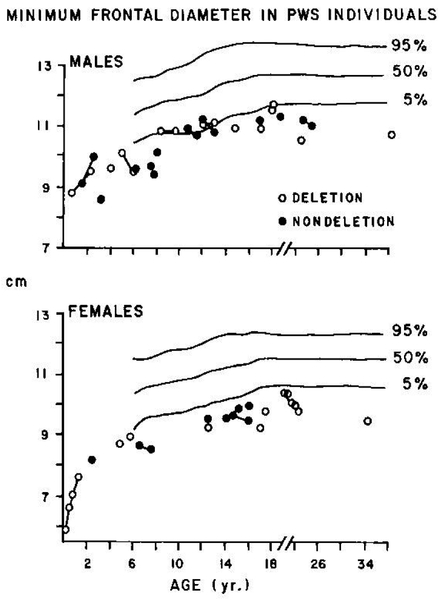
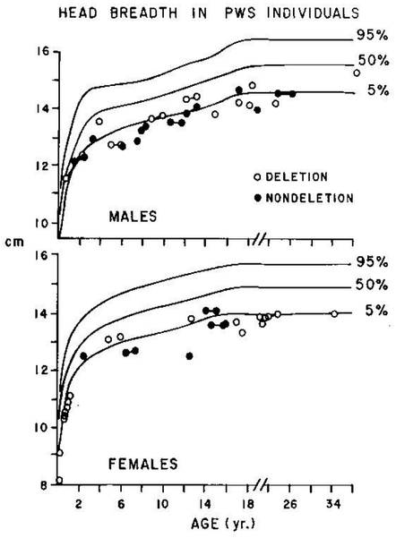
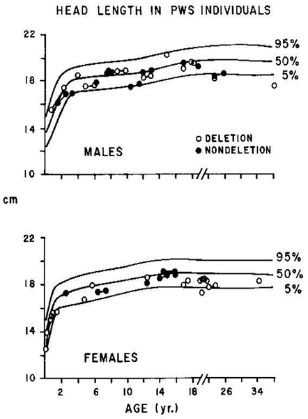
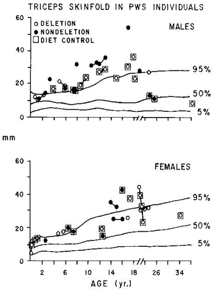
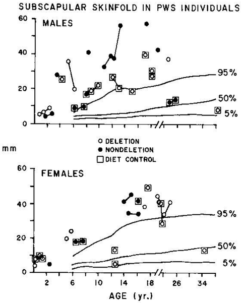
Figure 1 displays the raw values for minimum frontal diameter plotted against standards for this measurement based on published craniofacial data from a white Canadian population (Farkas 1981). Chromosome deletion and nondeletion individuals are shown by open and closed dots, respectively, in this and all subsequent figures. Most of the values fall well below the 5th percentile for both males and females. No differences between deletion and nondeletion individuals are observed. Similar results are observed for head breadth as shown in Figure 2. However, in the case of head breadth, the values do not fall as consistently below the 5th percentile as they do in the case of minimum frontal diameter. Head breadth measurements on PLWS individuals are consistently below the 50th percentile, with the majority of values approaching, if not on or below, the 5th percentile line. Length of the head does not seem to be affected in this syndrome, as seen in Figure 3. Almost all values fall within normal limits. Again, there are no differences observed between deletion and nondeletion individuals. Figures 4 and 5 display plots of the triceps and subscapular skinfolds for PLWS individuals. Those individuals on a relatively well-controlled diet are indicated with boxes. Strict dietary controls are necessary in this condition to control obesity. PLWS individuals are characteristically unable to exert self-control over food intake. In Figure 4 a rapid advance toward obesity is observed in a female with the deletion (see plot for female individuals less than 2 years of age—4 values plotted). This occurred over the first year of life while her weight progressed from a level less than the 5th percentile to the 50th percentile. A similar pattern is observed in four males whose triceps skinfolds were at or above the 95th percentile while their weights remained below the 50th percentile (see plots for two males, one with the deletion and one without the deletion, less than age 3 years, and two nondeletion males at 6 and 8 years of age). The triceps skinfold measurements for PLWS individuals consistently fall above the 95th percentile line after age 3 years with the exception of adult males whose dietary intakes have been controlled and several teenage and adult females, some of whose diets are under control. As can be seen in Figure 5, the results for subscapular skinfolds are similar.
Figure 1.
Minimum frontal diameter measurements in deletion and nondeletion PWS individuals plotted on standardized curves. Longitudinal measurements for an individual are represented with a line.
Figure 2.
Head breadth measurements in deletion and nondeletion PWS individuals plotted on standardized curves. Longitudinal measurements for an individual are represented with a tine.
Figure 3.
Head length measurements in deletion and nondeletion PWS individuals plotted on standardized curves. Longitudinal measurements for an individual are represented with a line.
Figure 4.
Triceps skinfold measurements in deletion and nondeletion PWS individuals plotted on standardized curves. Longitudinal measurements for an individual are presented with a line.
Figure 5.
Subscapular skinfold measurements in deletion and nondeletion PWS individuals plotted on standardized curves. Longitudinal measurements for an individual are represented with a line.
Discussion
Anthropometric differences between the deletion and nondeletion subgroups of PLWS individuals have been previously reported (Butler et al. 1982b). Further studies with increased sample sizes have failed to substantiate the original findings. However, we are encouraged to continue anthropometric studies of heterogeneity in PLWS, especially in light of molecular research in progress focusing on chromosome 15 to confirm the deletion. Collaboration between the cytogeneticist, molecular geneticist, and anthropometrist may eventually afford an opportunity to investigate major gene effects on obesity.
Meanwhile, anthropometric methods have proven useful as an additional clinical assessment in the early diagnosis of PLWS individuals. Butler and Meaney (1985) provide an update of a previous report (Butler et al. 1982a) on the metacarpophalangeal pattern profile and a discriminant function based on hand x-ray data as a tool in the diagnosis of PLWS. In this report a test of the original discriminant function reported by Butler et al. (1982a) was completed using hand x-ray data on 22 new PLWS individuals. Twenty of the 22 new individuals with PLWS (91%) were classified correctly as PLWS individuals, suggesting that the original function may have important application as a diagnostic tool in conjunction with other clinical findings.
Some clinicians have suggested that a narrow bi-frontal diameter should be considered a major feature of this syndrome, especially in younger individuals (Hall and Smith 1972). The data reported here support this notion, but also suggest that doli-chocephaly (a relatively narrow head) should be considered as an early diagnostic feature. A further description of the craniofacial data can be found in Meaney and Butler (1987).
The results of our longitudinal studies of PLWS individuals suggest that careful assessment of fatness by measurements of skinfold thickness may reveal a predisposition toward development of excessive fatness or obesity in these individuals before an abnormally high weight percentile is obtained. Thus an evaluation of early postnatal growth in weight and length in combination with assessment of fatness development may improve the chances of making a diagnosis in infants for which the clinician has observed such features as hypotonia, small genitalia, and early feeding problems. Earlier diagnosis of this condition might eventually improve the effectiveness of early dietary intervention to prevent obesity.
The negative relationships between age and anthropometric Z-scores suggest a deceleration of the growth of linear components of the body and of the hands and feet with increasing age in PLWS individuals relative to normal standards. Additional longitudinal studies will be required to establish the validity of these findings.
Conclusion
There can be little doubt that medical genetics offers many problems and challenges for the interested anthropologist. The flow is equally strong in the opposite direction as biological anthropologists with appropriate interests, training, and experience are already contributing in many research areas in the field. Much work remains to be done. A clear need exists for the collection of data on normal children for traits commonly used in the evaluation of dysmorphic conditions. It would be extremely worthwhile if the next national anthropometric survey included measurements of clinical relevance to medical genetics (e.g., cranial and facial measurements). More anthropometric studies of normal infants and preschool children would also be useful to the clinical geneticist for assessment of abnormal growth and dysmorphic features.
Cultural anthropologists with an interest in the evaluation of medical services may become major contributors to the evaluation of genetic services. Such topics as ethnic differences in knowledge and understanding of genetic inheritance and cultural and subcultural influences on reproductive decision-making in response to counseling have received scant attention in medical genetics. The need for such studies will become acute as the demand for genetic services broadens across all sectors of the population. In summary, then, many research niches remain unfilled while others continue to develop and flourish as some anthropologists follow in the footsteps of Damon, who worked so hard to build bridges between anthropology and medicine.
Acknowledgments.
This research was supported in part by Public Health Service grant PHS-5T32 GM07468 (Butler) and by Oral-Facial Genetics Training Grant DE 7043-05 (Meaney). Computer services were provided by the IUPUI computing facilities. An earlier version of this paper was presented in the symposium “Application of Anthropological Methods in Medical Genetics” on April 12, 1984, at the Fifty-Third Annual Meeting of the American Association of Physical Anthropologists in Philadelphia, Pennsylvania (Am. J. Phys. Anthropol. 63:111, 192–193).
REFERENCES CITED
- Applebaum EG, and Firestein SK 1983. A Genetic Counseling Casebook. New York: The Free Press. [Google Scholar]
- Butler MG, and Meaney FJ 1985. Metacarpophalangeal Pattern Profile Analysis in Prader-Willi Syndrome: A Follow-Up Report on 38 Cases. Clinical Genetics 28:27–30. [DOI] [PMC free article] [PubMed] [Google Scholar]
- Butler MG, and Palmer CG 1983. Parental Origin of Chromosome 15 Deletions in Prader-Willi Syndrome. Lancet 1:1285–1286. [DOI] [PMC free article] [PubMed] [Google Scholar]
- Butler MG, Meaney FJ, and Kaler SG 1985. Metacarpophalangeal Pattern Profile Analysis in Clinical Genetics: An Applied Anthropometric Method. American Journal of Physical Anthropology 70:195–201. [DOI] [PMC free article] [PubMed] [Google Scholar]
- Butler MG, Meaney FJ, and Palmer CG 1985. Clinical and Cytogenetic Survey of 39 Individuals With Prader-Labhart-Willi Syndrome. American Journal of Medical Genetics 23:793–809. [DOI] [PMC free article] [PubMed] [Google Scholar]
- Butler MG, Kaler SG, Yu PL, and Meaney FJ 1982a. Metacarpophalangeal Phalangeal Pattern Profile Analysis in Prader-Willi Syndrome. Clinical Genetics 22:315–320. [DOI] [PMC free article] [PubMed] [Google Scholar]
- Butler MG, Meaney FJ, Kaler SG, Yu PL, and Palmer CG 1982b. Clinical Differences Between Chromosome 15q Deletion and Nondeletion Prader-Willi Individuals. American Journal of Human Genetics 34:119A (abstr.). [Google Scholar]
- Damon A 1975. Biological Anthropology as an Applied Science In Physiological Anthropology. Damon A, ed. Pp. 360–367. New York: Oxford University Press. [Google Scholar]
- Farkas LG 1981. Anthropometry of the Head and Face in Medicine. New York: Elsevier North Holland. [Google Scholar]
- Frisancho AR 1974. Triceps Skinfold and Upper Arm Muscle Size Norms for Assessment of Nutritional Status. American Journal of Clinical Nutrition 27:1052–1058. [DOI] [PubMed] [Google Scholar]
- Garn SM, Hertzog KP, Poznanski AK, and Nagy JM 1972. Metacarpophalangeal Length in the Evaluation of Skeletal Malformation. Radiology 105:375–381. [DOI] [PubMed] [Google Scholar]
- Hall BD, and Smith DW 1972. Prader-Willi Syndrome. J. Pediatrics 81:286–293. [DOI] [PubMed] [Google Scholar]
- Ledbetter DH, Riccardi VM, Airhart SD, Strobel RJ, Keenan SB, and Crawford JD 1981. Deletions of Chromosome 15 as a Cause of the Prader-Willi Syndrome. New England Journal of Medicine 304:325–329. [DOI] [PubMed] [Google Scholar]
- Meaney FJ, and Butler MG 1987. Craniofacial Variation and Growth in the Prader-Labhart-Willi Syndrome. American Journal of Physical Anthropology 74:459–464. [DOI] [PMC free article] [PubMed] [Google Scholar]
- Meier RJ, Steltz-Lenarsky JS, and Hearst MT 1984. Anthropological Evaluation of Genetic Services: A Preliminary Report. American Journal of Physical Anthropology 63:193 (abstr.). [Google Scholar]
- O’Rourke DH, and Petersen GM 1983. Biological Anthropology and Genetic Disease Research: Introduction. American Journal of Physical Anthropology 62:1–2. [DOI] [PubMed] [Google Scholar]
- Poznanski AK, Garn SM, Gall JC, and Stern AM 1972. Metacarpophalangeal Pattern Profiles in the Evaluation of Skeletal Malformations. Radiology 104:1–11. [DOI] [PubMed] [Google Scholar]
- Prader A, Labhart A, and Willi H 1956. Ein syndrom von adipositas, klienwuchs, kryptorchimus und oligophrenie nach myatonieartigen Zustand im Neugeborenenalter. Schweizerische Medizinische Woehenschrift 86:1260–1261. [Google Scholar]
- Reed T, and Butler MG 1984. Dermatoglyphic Features in Prader-Willi Syndrome With Respect to Chromosomal Findings. Clinical Genetics 25:341–346. [DOI] [PMC free article] [PubMed] [Google Scholar]
- Robinow M 1982. Clinical Applications of Physical Anthropology. Yearbook of Physical Anthropology 25:169–179. [Google Scholar]
- Shimkin DB, and Golde P, eds. 1983. Clinical Anthropology: A New Approach to American Health Problems? Lanham, MD: United Press of America. [Google Scholar]
- Snyder RG, Schneiden LW, Owings CL, Reynolds HM, Gollomb DH, and Schork MA 1977. Anthropometry of Infants, Children, and Youths to Age 18 for Product Safety Design SP-450. Warrendale, PA: Society of Automobile Engineers, Inc. [Google Scholar]
- Steltz-Lenarsky JS, Mahnke GN, and Meier RJ 1985. Anthropological Evaluation of Genetic Services: Preliminary Findings. American Journal of Physical Anthropology 66:234 (abstr.). [Google Scholar]
- Ward RE, and Meaney FJ 1984. Anthropometry and Numerical Taxonomy in Clinical Genetics: An Example of Applied Biological Anthropology. American Journal of Physical Anthropology 64:147–154. [DOI] [PubMed] [Google Scholar]
- Wienker CW 1984. The Emergence of Biomedical Anthropology and its Implications for the Future. American Journal of Physical Anthropology 64:141–146. [DOI] [PubMed] [Google Scholar]


