

Abstract
Worldwide, 2–10% of human population is infested with pubic lice which are mostly found in the hair of pubic area, occasionally under the armpits, in the beard or mustache and on the eyebrows and eyelashes. They are usually transmitted through sexual contact or through toilet seats and bedding material. A twenty-year-old boy suffering from severe itching in the genital region during night hours submitted the lice for their identification in Department of Veterinary Parasitology, College of Veterinary Science and Animal Husbandry, Mhow. These lice were identified as Phthirus pubis.
Keywords: Human louse, Madhya Pradesh (India), Phthirus pubis
Introduction
Amongst 3000 species of lice, Pediculus humanus and Phthirus pubis (pubic lice) prefer the human beings. According to Light et al. (2008), P. humanus has two morphotypes, viz. P. humanus morphotype capitis (head louse) and P. humanus morphotype corporis (body louse). Head, body and pubic lice live on the head, clothing and pubic area, respectively. Based on archeological studies, Reinhart and Buikstra (2003) reported that pubic lice are infesting human beings since thousands of years and Linnaeus was the first to describe it as a Pediculus pubis in 1758, and later it was studied by several workers (Denny 1842; Nuttall 1918; Ferris 1951). These are usually transmitted through sexual contact and hence infestation is higher in 15–45 year-old sexually active population (Anderson and Chaney 2009; Mimouni et al. 2002). However in heavy infestations, they are also found in hair of axilla, chest, eyebrows and eyelashes (Manjunatha et al. 2006) and its occurrence is generally associated with poor hygiene and overcrowding. It causes itching in pubic region (Chosidow 2000). Generally, lice infestation is diagnosed by identification of adult lice and nits on the hair of different body regions (Faber 1996).
Materials and methods
A twenty-year-old boy suffering from severe itching in the genital region during night hours submitted the lice for their identification. Approval of the Institutional Animal Ethical Committee (IAEC) was obtained for processing of the lice. Further, consent for participation in the study was also obtained from the patient. The lice collected in 70% alcohol were boiled in 10% potassium hydroxide and subsequently dehydrated in ascending grades of alcohol (30, 50, 70, 90% and absolute alcohol) by keeping the specimen for 20 min in each grade of alcohol. For clearing, the specimens were transferred to a cavity block containing carboxylol for 20 min. After clearing, the specimens were mounted on the slide by using phenol balsam and were identified based on the morphological features described by Kettle (1995).
Results and discussion
Based on the location (pubic region) and morphological features, the lice were identified as Phthirus pubis. The lice were 1.25–2 mm long and were found to be smaller than Pediculus humanus. They were having an oval body which was wider than long resembling a crab. The head was distinctly smaller than the body with simple eyes and long antennae. All the legs were terminated with a tarsal claw. Claws on the 2nd and 3rd pairs of legs were huge as compared to 1st pair of legs. Thumb of tibia (extension of tibia) was evident which helps the lice to grasp the flattened hairs of pubic region of man. Four pairs of tubercles were stick out on each side of the abdomen (Fig. 1).
Fig. 1.
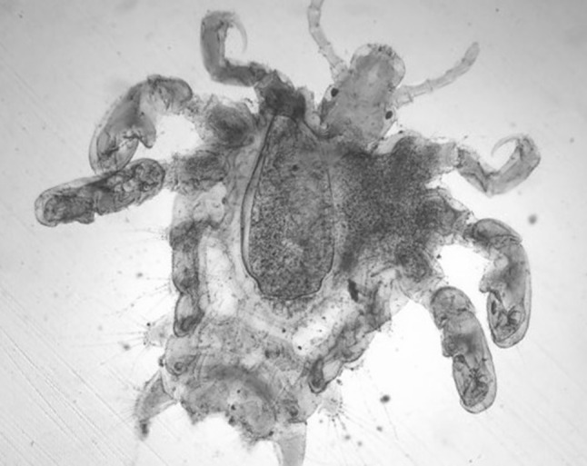
Photomicrograph of Phthirus pubis showing crab like oval body (100×)
Length of the louse (1.25–2 mm) and crab like body observed in the present study is in consonance with the findings of Kiran et al. (2012) who reported P. pubis as a cause of Phthiriasis palpebrarum with blepheroconjuctivitis in a 70-year-old woman from Chennai. Ashraf et al. (2014) treated unilateral phthiriasis palpebrarum caused due to P. pubis in a 28-year-old man from Aligarh. Palanisamy et al. (2015) confirmed P. pubis as a cause of itching in the pubic area in a 25-year-old married male from Puducherry with a history of extramarital contact. Singh et al. (2016) evinced pain and irritation in both the eyes due to P. pubis in a 41-year-old female from Delhi. Phthirus pubis as a causative agent for various conditions, viz. axillary pruritus in a 62 year-old HIV infected man from United States, blepharitis in a 42-year-old woman from Turkey and Phthiriasis palpebrarum in a 63-year-old woman from China has been reported by So and Carlos (2014), Sundu et al. (2015) and Wu et al. 2017, respectively. Although P. pubis prefers the pubic area it is also recorded from armpits, eyebrows, eyelashes, and beard (Couch et al. 1982).
The scanty literature pertaining to this louse from India could be due to under reporting of the cases due to the social stigma as lice commonly infest hair of pubic and perianal regions. Although, less reports are available in comparison with head lice or body lice, study of its epidemiology becomes mandatory because of its correlation with the occurrence of sexually transmitted diseases.
Acknowledgements
The authors are highly thankful to the Dean, College of Veterinary Science and Animal Husbandry, Mhow for providing the necessary facilities to carry out the research work.
Authors contribution
M. Shakya processed parasitic specimens for its correct identification. A.K. Jayraw helped in identification of the specimen and drafted the article. M. Singh revised the article and reviewed critically.
Compliance with ethical standards
Conflict of interest
Authors would hereby like to declare that there is no conflict of interests that could possibly arise.
References
- Anderson AL, Chaney E. Pubic lice (Pthirus pubis): history, biology and treatment vs. knowledge and beliefs of US college students. Int J Environ Res Public Health. 2009;6:592–600. doi: 10.3390/ijerph6020592. [DOI] [PMC free article] [PubMed] [Google Scholar]
- Ashraf M, Waris A, Kumar A, Akhtar N. A case of unilateral phthiriasis palpebrarum infestation involving the left eye. BMJ Case Rep. 2014 doi: 10.1136/bcr-2013-203307. [DOI] [PMC free article] [PubMed] [Google Scholar]
- Chosidow O. Scabies and pediculosis. Lancet. 2000;355:819–826. doi: 10.1016/S0140-6736(99)09458-1. [DOI] [PubMed] [Google Scholar]
- Couch JM, Green WR, Hirst LW, de la Cruz ZC. Diagnosing and treating Phthirus pubis palpebrarum. Surv Ophthalmol. 1982;26:219–225. doi: 10.1016/0039-6257(82)90082-0. [DOI] [PubMed] [Google Scholar]
- Denny H (1842) Monographia anoplurorum brittaniae. London
- Faber BM. The diagnosis and treatment of scabies and pubic lice. Prim Care Update OB/GYNS. 1996;3:20–24. doi: 10.1016/1068-607X(95)00054-M. [DOI] [Google Scholar]
- Ferris GF. The sucking lice. Mem Pac Coast Ent Soc. 1951;1:280–282. [Google Scholar]
- Kettle DS. Medical and veterinary entomolgy. Wallingford: CAB International; 1995. [Google Scholar]
- Kiran B, Kareem SA, Illamani V, Chitralekha S. Case of Phthiriasis palpebrarum with blepheroconjunctivitis. Indian J Med Microbiol. 2012;30:354–356. doi: 10.4103/0255-0857.99504. [DOI] [PubMed] [Google Scholar]
- Light JS, Toups MA, Reed DL. What’s in a name: the taxonomic status of human head and body lice. Mol Phylogenet Evolut. 2008;47:1203–1216. doi: 10.1016/j.ympev.2008.03.014. [DOI] [PubMed] [Google Scholar]
- Manjunatha NP, Jayamanne GR, Desai SP, Moss TR, Lalik J, Woodland A. Pediculosis pubis: presentation to ophthalmologist as pthriasis palpebrarum associated with corneal epithelial keratitis. Int J STD AIDS. 2006;17:424–426. doi: 10.1258/095646206777323445. [DOI] [PubMed] [Google Scholar]
- Mimouni D, Ankol OE, Gdalevich M, Grotto I, Davidovitch N, Zangvil E. Seasonality trends of Pediculosis capitis and Phthirus pubis in a young adult population: follow-up of 20 years. J Eur Acad Dermatol Venereol. 2002;16:257–259. doi: 10.1046/j.1468-3083.2002.00457.x. [DOI] [PubMed] [Google Scholar]
- Nuttall GHF. The biology of phthirus pubis. Parasitol. 1918;10:383–405. doi: 10.1017/S0031182000003954. [DOI] [Google Scholar]
- Palanisamy AP, Kanakaram KK, Vadivel S, Kothandapany S. Crab louse. Indian Dermatol Online. 2015;6:375. doi: 10.4103/2229-5178.164475. [DOI] [PMC free article] [PubMed] [Google Scholar]
- Reinhart KJ, Buikstra J. Louse infestation of the Chiribaya culture, southern Peru: variation in prevalence by age and sex. Mem Inst Oswaldo Cruz. 2003;98:173–179. doi: 10.1590/S0074-02762003000900026. [DOI] [PubMed] [Google Scholar]
- Singh A, Tripathy K, Gupta N, Kale P, Verma N, Mirdha BR. Phthirus pubis in the eye. Indian J Med Microbiol. 2016;34:405–406. doi: 10.4103/0255-0857.188384. [DOI] [PubMed] [Google Scholar]
- So JM, Carlos CA. Phthirus pubis as a cause of axillary pruritus. IDCases. 2014;1:55. doi: 10.1016/j.idcr.2014.07.001. [DOI] [PMC free article] [PubMed] [Google Scholar]
- Sundu C, Dinc E, Kurtulus UC, Yildirim O. Common blepharitis related to Phthiriasis Palpebrarum: argon laser phototherapy. Turk Parazitol Derg. 2015;39:252–254. doi: 10.5152/tpd.2015.3861. [DOI] [PubMed] [Google Scholar]
- Wu NA, Zang H, Sun FY. Phthiriasis palpebrarum: a case of eyelash infestation with Pthirus pubis. Exp Ther Med. 2017;13:2000–2002. doi: 10.3892/etm.2017.4187. [DOI] [PMC free article] [PubMed] [Google Scholar]