

Abstract
Anemia is a common problem in the neonatal period. Presenting symptoms may suggest numerous possible diagnoses ranging from anemia seen as a normal part of development to anemia due to critical pathology. An illustrative case is presented to highlight the appropriate evaluation of the neonate with significant anemia. Several important features of the evaluation of neonatal anemia are highlighted. The constellation of signs and symptoms that occur in conjunction with the anemia are critical for the evaluation. The evaluation should be performed in a step-wise process that starts by eliminating common causes of anemia. Manual review of the peripheral blood smear with a hematologist can be helpful.
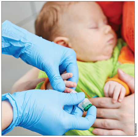
A 3-month-old baby presented for evaluation of congenital anemia. She was born at 35 weeks gestational age to a 34-year-old via cesarean delivery because of nonreassuring fetal heart tracings during preterm labor. Prior to the initiation of preterm labor the pregnancy had been uncomplicated. The mother received regular prenatal care and all serologies were negative. One-minute and 5-minute APGAR (appearance, pulse, grimace, activity, respiration) scores were 8 and 8, respectively. These APGAR scores reflected cyanosis and requirement for blow-by oxygen therapy in the first 10 minutes of life. In addition, the patient was noted to be pale with mild jaundice at delivery. This was corroborated by her complete blood count that revealed a hemoglobin (Hgb) of 6.1 g/dL. Aside from pallor, the infant had an unremarkable physical examination with a birth weight of 2,350 g (51%-75% for gestational age). Specifically, she had no hepatosplenomegaly, petechiae, or bruising, and her limbs were normal without radial defects.
Evaluation of her anemia at that time also revealed 189 × 103/mcL nucleated erythrocytes. Lactate dehydrogenase (LDH), haptoglobin, and bilirubin were not evaluated. Her blood type was found to be A+ and direct Coombs testing was negative. At 8 hours of life, the bilirubin was 12.6 mg/dL with a reticulocyte percentage of 32%. Examination of the peripheral smear revealed marked elevation in the number of schistocytes. Urine studies at the time revealed hemoglobinuria. Urgent treatment was initiated with phototherapy and exchange transfusion. After the exchange transfusion the patient’s bilirubin was 7.2 mg/dL with Hgb of 16.9 g/dL.
During the week after the exchange, the patient remained in the neonatal intensive care unit (NICU) and her bilirubin initially trended upward, but peaked at 14 mg/dL and improved with phototherapy. The patient’s Hgb slowly decreased to 10.9 mg/dL at time of discharge (9 days of life). Prior to discharge she was noted to have a normal hemoglobin electrophoresis.
In the weeks after her discharge from the NICU her Hgb level drifted down and she required multiple transfusions during her first months of life (Table 1).
TABLE 1.
Laboratory Dataa
| Measured Parameters | Reference Range | Neonatal Intensive Care Unit Discharge (DOL 9) | First Primary Care Physician Visit (DOL 19) | DOL 31 | DOL 33 | DOL 38 | DOL 52 | DOL 59 | DOL 76 |
|---|---|---|---|---|---|---|---|---|---|
| Hemoglobin (g/dL) | 10.3-13.2 | 10.9 | 8.2 | 7.4b | 10.4 | 8.3 | 7.1b | 8.9 | 6.8b |
| White cell count (× 103/uL) | 5.5-16.8 | 6.6 | 6.4 | 7.3 | 6.6 | 8.2 | 5.7 | 7.1 | |
| Platelet count (× 103/uL) | 150-450 | 162 | 390 | 146 | 282 | 388 | 147 | 195 | 438 |
| Reticulocyte count (%) | 0.6-1.7 | 3.04 | 1.49 | 1.71 | 2.73 | 1.08 | 2.69 | ||
| Total bilirubin (mg/dL) | 0.1-1.0 | 7.5 | 3.3 | ||||||
| Direct bilirubin (mg/dL) | 0.0-0.3 | 0.65 | |||||||
| MCV (fL) | 69-85 | 88 | 85 |
Abbreviation: DOL, day of life; MCV, mean corpuscular volume.
Neonatal intensive care unit through age 3 months.
Packed red blood cells transfusion.
EVALUATION OF HEMOLYTIC ANEMIA IN THE NEONATE
At initial presentation, the patient’s constellation of signs and symptoms were consistent with a hemolytic anemia based on the presence of anemia with reticulocytosis, marked unconjugated hyperbilirubinemia, elevated LDH, and decreased haptoglobin. Hemolytic anemia is due to the rupture of erythrocytes, releasing the contents of the red blood cells and most notably free hemoglobin. Haptoglobin binds to free hemoglobin to potentiate its excretion and therefore a decrease in haptoglobin is seen with even a small amount of hemolysis. The remaining free hemoglobin undergoes a series of enzymatic reactions that ultimately produces bilirubin.1 Hemolytic anemia is also commonly associated with a reticulocytosis as the healthy bone marrow increases erythropoiesis in an attempt to compensate for the anemia.
The differential diagnosis for hemolytic anemia in the newborn period includes alloimmunity, hemoglobinopathies, erythrocyte membrane defects, and enzyme deficiencies (Table 2). Thoughtful laboratory evaluation of the patient and mother can help determine the etiology of hemolytic anemia. However, it is important to also monitor closely for clinical jaundice and have a low threshold to test and treat for hyperbilirubinemia to avoid kernicterus.
TABLE 2.
Hemolytic Anemia in the Neonate
| Antibody Mediated | Hemoglobinopathies | Erythrocyte Membrane Defects | Enzyme Deficiencies |
|---|---|---|---|
| ABO incompatibility/Rh incompatibility | Sickle cell disease | Hereditary spherocytosis | G6PD deficiency |
| Transplacental transfer of maternal anti-red blood cell antibody | Alpha thalassemia | Hereditary elliptocytosis | Pyruvate kinase deficiency |
Evaluation of hemolytic anemia in the neonatal period should include comparing blood types of both the mother and the neonate, Coombs testing, hemoglobin electrophoresis, and evaluation of the peripheral blood smear. The presence of mismatched ABO or Rh red blood cell antigens can be a clue that maternal antibodies directed against neonatal red blood cells are the culprit and can be confirmed by a positive direct or indirect Coombs test that evaluates for the presence of such antibodies. Equally important in this assessment is a hemoglobin electrophoresis to identify hemoglobinopathies that may predispose patients to hemolytic disease. It should be noted that some hemoglobinopathies can be missed on the newborn screen depending on the type of assay performed. Deletion of a single complete beta-globin gene locus, although exceptionally rare, can lead to significant hemolysis during the neonatal period without abnormalities on hemoglobin electrophoresis.2,3 Another hemoglobinopathy associated with critically ill infants and hemolytic anemia is alpha thalassemia with deletion of all four alpha globin genes, also called hydrops fetalis. These fetuses are often lost due to spontaneous miscarriages in-utero. If they do survive to gestation, they often present with other fetal anomalies in addition to hemolysis, such as genitourinary anomalies, limb deformities, microcephaly, hypoplasia of the lungs, and cardiac defects.4–7
Examination of the peripheral blood smear is also important. With significant hemolysis, fragmented red blood cells, also called schistocytes, are often present. However, close attention should be paid to the presence of spherocytes or elliptocytosis. Hereditary spherocytosis and elliptocytosis, the most commonly inherited membrane defects, can present with hemolytic anemia. If there is clinical suspicion of a membrane defect, a supportive diagnostic test is the osmotic fragility test. This test exposes a patient’s blood to decreasing concentrations of sodium chloride, starting with isotonic normal saline, thereby causing increasing hypotonic stress. The degree of hemolysis in each tube is then plotted against tonicity.8 Normal biconcave disc-shaped erythrocytes have an increased surface-area-to-volume ratio and can avoid lysis in increasingly hypotonic fluid. In contrast, spherocytes have a lower surface-area-to-volume ratio, and lyse more easily in hypotonic fluids. A diagnosis of hereditary spherocytosis is supported when increased hemolysis is observed at a given sodium chloride concentration during the osmotic fragility test compared to control erythrocytes. It is important to note that spherocytes are not always due to hereditary spherocytosis, but can be present when there are other causes of hemolysis, which will lead to positive osmotic fragility testing. Therefore, a positive family history is also important to make the diagnosis of hereditary spherocytosis. Flow cytometry analysis can also be used to make this diagnosis by examining red blood cells for decreased expression of the transmembrane protein band 3.9
The final consideration in the differential diagnosis of a neonate with hemolysis is a genetically acquired enzyme defect, or an enzymopathy. The diagnosis of an enzymopathy requires either decreased activity of the specific enzyme or the identification of a mutation in the gene of interest. Individual enzyme levels can be ordered, or alternatively there are panels available for a broader diagnostic evaluation. Care should be taken in interpreting enzyme levels. A relative reticulocytosis can lead to falsely normal levels, as reticulocytes have higher levels of most enzymes, which then decrease as the red cell ages. Glucose-6-phosphate dehydrogenase (G6PD) is the most common enzyme defect causing hereditary hemolytic anemia. G6PD deficiency is inherited in an X-lined recessive pattern, so it is more likely in the male gender, except in rare cases when there is skewed lyonization or X-inactivation.
PATIENT DIAGNOSIS
Our patient had a normal hemoglobin electrophoresis, negative direct and indirect Coombs tests, and a smear that revealed polychromasia and schistocytes but no spherocytes or elliptocytes. Enzyme levels were not useful because the patient was transfusion dependent. Ultimately, gene sequencing was sent for a panel of red cell enzymes and membrane proteins. This testing revealed that she was compound heterozygous for two different mutations in the pyruvate kinase (PK) gene.
DISCUSSION
Genetics
PK is a key component of the glycolysis pathway that is active as a homotetramer. The role of the enzyme in vivo is to transfer a phosphate group from phosphoenolpyruvate to adenosine diphosphate, forming one molecule of pyruvate and one molecule of adenosine triphosphate (ATP). The subunit for pyruvate kinase is encoded by the PKLR gene located on chromosome 1. Mutations in the PK gene are the most frequent genetic defect found in the glycolytic pathway. At least 133 different mutations have been identified, the majority of which are point mutations. Insertions and frameshift mutations are found less commonly. PK mutations are distributed worldwide, although they are most commonly encountered in Northern European populations. There is a strong regional association with individual mutations.
People who are heterozygous carriers of PK mutations are clinically unaffected. The frequency of heterozygous gene mutation in the PKLR gene ranges between 0.6% and 6%.10 With only one mutated gene, the normal allele provides sufficient enzyme activity for cells to survive normally. Therefore, clinical pyruvate kinase deficiency (PKD) is inherited in an autosomal recessive fashion and manifestations of the disease occur in patients who are homozygous or compound heterozygous for mutations. Patients with homozygous null mutations, with a complete lack of functional protein, display intrauterine growth retardation, severe anemia at birth, and sometimes intrauterine or perinatal death.11
Pathophysiology
PK catalyzes the final reaction in the glycolytic pathway. The role of PK is of paramount importance in erythrocyte metabolism because mature erythrocytes lack mitochondria and are dependent on glycolysis for the production of ATP.12 As the PK-deficient erythrocytes age, they become ATP deplete, and develop abnormal potassium (K+) homeostasis.13 Ultimately, the PK-deficient cells become K+ depleted, leading to loss of intracellular water. This cellular dehydration leads to rigid cell walls and premature cell lysis.14 Hemolysis can be exacerbated by the hypoxic and acidic environment of the spleen as the cells become rigid in the pathway described above and then lyse while attempting to navigate the fenestrations between the splenic cord and sinuses with difficulty.15
Despite ongoing hemolysis and baseline low hemoglobin, patients with PKD tend to tolerate lower levels of hemoglobin without evidence of tissue hypoxia. This is thought to be due to accumulation of 2,3-diphosphoglycerate (2,3-DPG) upstream of the PK enzymatic reaction. Up to a 3-fold increase in the intracellular concentration of 2,3-DPG results in patients with PKD.10 Because 2,3-DPG shifts the oxygen dissociation curve rightward, there is improved oxygen delivery to tissues despite anemia.16
Clinical Manifestations
PKD is characterized by lifelong hemolysis that ranges in severity from mild or fully compensated anemia to life-threatening hemolysis that can present as early as the neonatal period and require exchange transfusion.17 The incidence is estimated to be 51 cases per 1 million persons among whites.18 Incidences in other races have not been characterized.
Hematologic features include ongoing jaundice and associated anemia. As the erythrocytes break down, patients have unconjugated hyperbilirubinemia, decreased haptoglobin, and elevated LDH. Schistocytes are present on the peripheral smear. In addition, most patients with PKD exhibit splenomegaly. The hematologic findings are nonspecific and therefore the diagnosis is reliant on family history, genetic sequencing, and enzyme levels.
Treatment
Current treatment is largely supportive in nature. Patients with severe disease require regular transfusions in the early years. Most often, the Hgb stabilizes in the school-aged child at 6 to 8 g/dL and transfusions are only necessary if hemolysis is exacerbated by infection, pregnancy, or other triggers. As discussed above, patients with PKD have improved tissue oxygen delivery due to increased intracellular 2,3-DPG and therefore tolerate a lower threshold for transfusion. As with all patients who receive chronic transfusions, patients with PKD are at risk for iron overload and may require chelation therapy.
In patients with severe disease, splenectomy can greatly diminish hemolysis, eliminating the need for chronic transfusion and increasing the Hgb by 1-3 g/dL.19 Most physicians try to wait until patients are at least 5-years-old, ensuring a fully developed immune system and time for adequate immunizations against encapsulated bacteria prior to splenectomy.
The only curative therapy that currently exists is hematopoietic stem cell transplantation. However, the risks of allogeneic transplant, including infections and graft-versus-host disease, generally outweigh the benefits for this particular nonmalignant disease.20 Gene transfer is currently being studied as an alternative approach, and successful gene transfer of the PKLR gene into human cell lines and mouse models has occurred.21,22 Future studies are needed to determine if gene transfer will be a successful therapy for patients with PKD.
PATIENT FOLLOW UP
The patient in the illustrative case is now age 21 months, and is growing and developing normally. She initially required monthly transfusions until age 6 months. Her hemolysis has decreased as she has grown, so that she is now able to maintain a hemoglobin of >7 g/dL with transfusions every 6 weeks. Her ferritin has been monitored regularly to assess for iron overload and remains <600 ng/mL.
Footnotes
Disclosure: The authors have no relevant financial relationships to disclose.
Contributor Information
Michele L. Nassin, Comer Children’s Hospital, Pritzker School of Medicine, University of Chicago.
Gabrielle Lapping-Carr, Section of Pediatric Hematology/Oncology/Stem Cell Transplant, Comer Children’s Hospital, Pritzker School of Medicine, University of Chicago.
Jill L. O. de Jong, Section of Pediatric Hematology/Oncology/Stem Cell Transplant, Comer Children’s Hospital; Pritzker School of Medicine, University of Chicago.
REFERENCES
- 1.Ryter SW, Alam J, Choi AM. Heme oxygenase-1/carbon monoxide: from basic science to therapeutic applications. Physiol Rev 2006;86:583–650. [DOI] [PubMed] [Google Scholar]
- 2.Rooks H, Bergounioux J, Game J, et al. Heterogeneity of the epsilon gamma delta beta-thalassaemias: characterization of three novel English deletions. Br J Haematol 2005; 128(5)722–729. [DOI] [PubMed] [Google Scholar]
- 3.Rooks H, Clark B, Best S, et al. A novel 506 kb deletion causing εγδβ thalassemia. Blood Cells Mol Dis 2012;49:121–127. [DOI] [PubMed] [Google Scholar]
- 4.Singer ST, Styles L, Bojanowski J, Quirolo K, Foote D, Vichinsky EP. Changing outcomes of homozygous alpha-thalassemia: cautious optimism. J Pediatr Hematol Oncol 2000;22:539–542. [DOI] [PubMed] [Google Scholar]
- 5.Bain BJ. Haemoglobinopathy Diagnosis. 2nd ed. Malden, MA: Blackwell; 2006. [Google Scholar]
- 6.Adam MP, Chueh J, El-Sayed YY, et al. Vascular-type disruptive defects in fetuses with homozygous alpha-thalassemia: report of two cases and a review of the literature. Prenat Diagn 2005;25:1088–1096. [DOI] [PubMed] [Google Scholar]
- 7.Dame C, Albers N, Hasan C, et al. Homozygous alpha-thalassemia and hypospadias--common aetiology or incidental association? Long-term survival of Hb Bart’s hydrops syndrome leads to new aspects for counseling of alpha-thalassemia traits. Euro J Pediatr 1999;158:217–220. [DOI] [PubMed] [Google Scholar]
- 8.Parapart A, Lorenz P, et al. The osmotic resistance (fragility) of human red cells. J Clin Invest 1947;26(4):636–640. [PubMed] [Google Scholar]
- 9.Stoya G, Baumann E, Junker U, Herman J, Linss W. Flow cytometric analysis of band 3 protein of human erythrocytes. Acta Histochem 1997;99(1):29–36. [DOI] [PubMed] [Google Scholar]
- 10.Zanella A, Bianchi P. Red cell pyruvate kinase deficiency: from genetics to clinical manifestations. Baillieres Best Pract Res Clin Haematol 1999;13(1):57–81. [DOI] [PubMed] [Google Scholar]
- 11.Zanella A, Bianchi P, Fermo E. Pyruvate kinase deficiency. Haematologica. 2007;92:721–723. [DOI] [PubMed] [Google Scholar]
- 12.Zanella A, Fermo E, Bianchi P, Valentini G. Red cell pyruvate kinase deficiency: molecular and clinical aspects. Br J Haematol 2005;130:11–25 [DOI] [PubMed] [Google Scholar]
- 13.Oski F, Nathan D, Sidel V, Diamond L. Extreme hemolysis and red-cell distortion in erythrocyte pyruvate kinase deficiency. I. Morphology, erythokinetics and family enzyme studies. N Engl J Med 1964;270:1023–1030. [DOI] [PubMed] [Google Scholar]
- 14.Nathan DG, Shohet SB. Erythrocyte ion transport defects and hemolytic anemia: “hydrocytosis” and “desicytosis.” Semin Hematol 1970;7(4):381–408. [PubMed] [Google Scholar]
- 15.Orkin SH, Nathan DG, Ginsburg D, et al. Nathan and Oski’s Hematology of Infancy and Childhood. 7th ed. Philadelphia, PA: Saunders; 2009. [Google Scholar]
- 16.Oski FA, Marshall BE, Cohen PJ, Sugerman HJ, Miller LD. The role of the left-shifted or right-shifted oxygen-hemoglobin equilibrium curve. Ann Intern Med 1971;74(1):44–46. [DOI] [PubMed] [Google Scholar]
- 17.Matthay KK, Mentzer WC. Erythrocyte enzymopathies in the newborn. Clin Haematol 1981; 10(1):31–55. [PubMed] [Google Scholar]
- 18.Beutler E, Gelbart T. Estimating the prevalence of pyruvate kinase deficiency from the gene frequence in the general white population. Blood 2000;95(11):3585–3588. [PubMed] [Google Scholar]
- 19.Hamblin TJ. Pyruvate-kinase (PK) deficiency In Dacie J, ed. The Hemolytic Anemias. 3rd ed. New York, NY: Churchill Livingstone; 1985:284–320. [Google Scholar]
- 20.Tanphaichitr VS, Suvatte V, Issaragrisil S, et al. Successful bone marrow transplantation in a child with red blood cell pyruvate kinase deficiency. Bone Marrow Transplant 2000;26:689–690. [DOI] [PubMed] [Google Scholar]
- 21.Tani K, Yoshikubo T, Ikebuchi K, et al. Retrovirus-mediated gene transfer of human pyruvate kinase (PK) cDNA into murine hematopoietic cells: implications for gene therapy of human PK deficiency. Blood. 1994;83(8):2305–2310. [PubMed] [Google Scholar]
- 22.Kanno H, Utsugisawa T, Aizawa S, et al. Transgenic rescue of hemolytic anemia due to red blood cell pyruvate kinase deficiency. Haematologica. 2007; 92:731–737. [DOI] [PubMed] [Google Scholar]