ABSTRACT
Daily torpor is used by small mammals to reduce daily energy expenditure in response to energetic challenges. Optimizing the timing of daily torpor allows mammals to maximize its energetic benefits and, accordingly, torpor typically occurs in the late night and early morning in most species. However, the regulatory mechanisms underlying such temporal regulation have not been elucidated. Direct control by the circadian clock and indirect control through the timing of food intake have both been suggested as possible mechanisms. Here, feeding cycles outside of the circadian range and brain-specific mutations of circadian clock genes (Vgat-Cre+ CK1δfl/fl εfl/+; Vgat-Cre+ Bmal1fl/fl) were used to separate the roles of the circadian clock and food timing in controlling the timing of daily torpor in mice. These experiments revealed that the timing of daily torpor is transiently inhibited by feeding, while the circadian clock is the major determinant of the timing of torpor. Torpor never occurred during the early part of the circadian active phase, but was preferentially initiated late in the subjective night. Food intake disrupted torpor in the first 4–6 h after feeding by preventing or interrupting torpor bouts. Following interruption, re-initiation of torpor was unlikely until after the next circadian active phase. Overall, these results demonstrate that feeding transiently inhibits torpor while the central circadian clock gates the timing of daily torpor in response to energetic challenges by restricting the initiation of torpor to a specific circadian phase.
KEY WORDS: Body temperature, Circadian rhythm, Clock mutant, Energetic savings, Metabolism, Suprachiasmatic nucleus
Highlighted Article: Daily torpor in energetically challenged mice is transiently inhibited by feeding while the circadian clock gates the expression of torpor, thereby restricting torpor from occurring during the early subjective night.
INTRODUCTION
Ensuring that energy expenditure does not exceed available energy stores is an essential and often challenging requirement for organisms. The thermoregulatory costs associated with maintaining body temperature represent the greatest energetic demands placed on small mammals (Speakman, 1997). Energy saving strategies are used by small mammals to cope with energetic challenges, with reductions in metabolic rate and the ensuing fall in body temperature (daily torpor) being an effective adaptation to reduce energetic requirements. Daily torpor in mice is characterized by a drop in body temperature from ∼37°C to 20–25°C for a few hours, resulting in a reduction in energy expenditure of 50–90% during the torpor bout (Geiser, 2004).
Although daily torpor is often employed as a strategy to cope with unexpected energetic challenges, the time of day at which it occurs appears to be tightly regulated. It has long been known that the circadian clock plays an important role in the timing of daily torpor (Kirsch et al., 1991; Lynch et al., 1980; Ruf et al., 1989). Daily torpor in endotherms is typically initiated in the late night/early morning (Hut et al., 2012; Körtner and Geiser, 2000), even in the absence of environmental lighting cycles (Kirsch et al., 1991; Lynch et al., 1980; Ruf et al., 1989). In laboratory mice, daily torpor is typically induced by restricting food intake to a single small meal a day, with torpor bouts ending and body temperature returning to ∼37°C a few hours before meals. The ability of the circadian clock to control the timing of daily torpor in the absence of feeding was shown in mice by Bechtold and colleagues (2012). In the absence of a lighting cycle and without access to food for 48 h, mice initiated daily torpor early in the subjective morning and terminated it in the late subjective morning of each day (Bechtold et al., 2012). An interaction between regulation by circadian control and food timing was illustrated in Siberian hamsters receiving food either in the morning or in the evening, resulting in significant differences in torpor timing (∼6 h), but not by the 12 h expected if food intake alone controlled the timing of torpor (Paul et al., 2004). Another experimental approach has been to expose food-deprived mice to a sudden drop in ambient temperature, resulting in a nearly instantaneous onset of torpor except when the ambient temperature reduction coincided with the start of the active phase (Oelkrug et al., 2011). When activity onset and the reduction in ambient temperature coincided, torpor was delayed by several hours (Oelkrug et al., 2011). These examples illustrate a role for both the circadian clock and the timing of food intake in regulating the timing of torpor, but how these processes interact at different times of day remains poorly understood.
Daily rhythms in physiology and behavior are controlled by endogenous circadian clocks (Dibner et al., 2010; Mohawk et al., 2012). At the molecular level, mammalian circadian clocks consist of transcription–translation feedback loops in which positive limb proteins (BMAL1, CLOCK and NPAS2) drive the expression of negative limb proteins (PERIOD1–3 and CRYPTOCHROME1–2), which subsequently inhibit the activity of the positive limb (Takahashi, 2017). This molecular feedback loop has a period of ∼24 h. The period length is set by the degradation rate of negative limb proteins, with casein kinase 1 delta and epsilon (CK1δ/ε) being important for setting the speed of the clock by controlling the stability of PERIOD proteins (Etchegaray et al., 2009; Meng et al., 2008; van der Vinne et al., 2018). The molecular components of the circadian clockwork are widely expressed, and cell-autonomous circadian clocks are present in nearly all cells of the body. These peripheral clocks are synchronized through timing cues controlled by the master clock in the suprachiasmatic nucleus (SCN) (Dibner et al., 2010). The SCN is typically synchronized (entrained) to the external lighting cycle through input from the eyes, but, in the absence of light, SCN-driven neuronal, hormonal and behavioral rhythms persist and maintain synchrony among tissues. While light is the strongest and most prominent entraining cue, circadian clocks can also be entrained by other external timing cues such as timed food availability (Stephan, 2002).
Considering the existing evidence indicating that both the circadian clock and food timing influence the timing of daily torpor, we designed experiments to test the hypothesis that the circadian clock and the timing of food intake have distinct and separable influences on the timing of torpor. To compare the relative importance of the circadian clock and the timing of food intake at all possible phases, feeding schedules with different periods (T-cycles) and brain-specific mutation of genes regulating circadian rhythms in mice (Vgat-Cre+ CK1δfl/fl εfl/+; Vgat-Cre+ Bmal1fl/fl) were used to systematically alter the phase relationship between circadian and feeding cycles. This approach revealed that the timing of daily torpor during caloric restriction is regulated by a combination of two separable events: (1) torpor is inhibited for several hours after the provision of food, and (2) the circadian clock restricts entry into daily torpor. Torpor never occurred during the circadian active phase, but was preferentially initiated shortly after it. In addition to the circadian regulation, feeding also prevents torpor in the first hours after a meal.
MATERIALS AND METHODS
Animals
All animal procedures were reviewed and approved by the Institutional Animal Care and Use Committee of the University of Massachusetts Medical School (Protocol A-1315) or Williams College (Protocol SS-N-14).
Six female wild-type C57BL/6J mice were purchased from Jackson Laboratories. Vgat-Cre+ CK1δfl/fl εfl/+ mice (5 male, 4 female) (van der Vinne et al., 2018), Vgat-Cre+ Bmal1fl/fl mice (5 male, 5 female) and No-Cre Bmal1fl/fl mice (5 male, 7 female) (Weaver et al., 2018) were generated by crossing previously described genotypes (Etchegaray et al., 2009; Storch et al., 2007; Vong et al., 2011). Genotyping was performed using ear or tail biopsies by PCR as described previously (Etchegaray et al., 2009; Weaver et al., 2018).
Experimental procedures
Mice were implanted in the peritoneal cavity with a telemetry transmitter to measure locomotor activity and core body temperature (TA-F10, Data Sciences International, St Paul, MN, USA) as described previously (Chu and Swoap, 2012). All transmitters were calibrated before implantation. Post-surgery, mice were maintained on a 12 h light:12 h dark cycle for at least 10 days before the start of the experiment. Throughout the experiment, mice were individually housed in constant darkness at 23±1°C in cages without a running wheel, with water available ad libitum. Body temperature and general locomotor activity were recorded through telemetry in 1 min bins throughout the experiment.
Daily torpor was induced in mice by restricting food intake to a single meal per feeding cycle (D12450B, 3.82 kcal g−1, Research Diets Inc., New Brunswick, NJ, USA), with meals being placed on top of the wire lid of the cage by the experimenter at the appropriate time while minimizing sound and movement to avoid disturbing the mice. Meal size was adjusted to compensate for the different intervals between meals and keep the average multi-day food intake constant, resulting in meal sizes of 2.20 g (24 h T-cycle), 1.83 g (20 h T-cycle) and 2.57 g (28 h T-cycle), with the first meal provided at the time of lights off. Mice were exposed to different sequences of feeding cycles depending on genotype (wild-type mice: 10 cycles at 24 h, 12 cycles at 20 h, 10 cycles at 28 h; Vgat-Cre+ CK1δfl/fl εfl/+ mice: 19 cycles at 24 h; Vgat-Cre+ Bmal1fl/fl mice and No-Cre Bmal1fl/fl mice: 10 cycles at 24 h, 15 cycles at 20 h). Because of technical difficulties, the sixth feeding bout (day 9–10) for mice in the last experiment (Vgat-Cre+ Bmal1fl/fl mice and No-Cre Bmal1fl/fl mice) was delayed by ∼20 h while simultaneously the ambient temperature dropped to ∼16°C , and the computer failed to collect data on day 21–22.
Data analyses
Torpor bouts were defined as the time intervals during which body temperature consistently dropped (1 min sampling interval), resulting in a body temperature reduction of at least 2°C from the start of the decrease in temperature, as well as all time points during which body temperature decreased while it was below 34°C. To visualize the timing of torpor in relation to the circadian active phase, torpor bouts are depicted in actograms by a blue bar. The circadian active phase and feeding-induced thermogenic response are visualized by black bars with the height of the bar representing the body temperature above 36.5°C. The timing of general locomotor activity correlated strongly with time intervals during which body temperature was high. A body temperature >36.5°C was chosen to visualize the circadian active phase and feeding response because the amplitude of the general locomotor activity rhythm was influenced by the phase relationship between the circadian and feeding cycles. Body temperature traces and actograms of all individual mice are presented in Dataset 1. The analysis assessing the timing of the first torpor bout in each feeding cycle (Fig. S2) used a more stringent criterion for the initiation of torpor, whereby body temperature had to decrease consistently (1 min sampling interval), resulting in a body temperature reduction of at least 4°C.
Circadian time (period and phase) was determined for all wild-type and Vgat-Cre+ CK1δfl/fl εfl/+ mice by eye-fitting a line through the onsets of the active phase as defined by body temperature (e.g. Fig. S1). Based on visual inspection of all actograms, feeding bouts were assumed to be 4 h in duration while the circadian active phase was assumed to last 6 h, with the resting phase covering all other time intervals (Fig. S1). The actual length of the circadian active phase was often <6 h, thus resulting in an underestimation of the effect described below and in Fig. S3A. Profiles depicting the percentage of time spent in torpor at different times were determined for 2 h intervals of the circadian and feeding cycles by averaging the time spent in torpor over multiday intervals. By definition, a circadian cycle is 24 circadian hours with CT 12 representing the onset of activity. In wild-type mice, average daily profiles of the percentage of time spent in torpor were calculated during the 24 h T-cycle (day 9–15), 20 h T-cycle (day 16–25) and 28 h T-cycle (day 26–37). Profiles of Vgat-Cre+ CK1δfl/fl εfl/+ mice were based on body temperature data from day 4–19. Because of the technical difficulties experienced on day 10 of the experiment using Vgat-Cre+ Bmal1fl/fl and No-Cre Bmal1fl/fl mice, the 24 h T-cycle profiles were determined using only 3 days of data (day 12–14) while the 20 h T-cycle profiles were based on day 15–27.
Statistical comparisons between the time spent in torpor during resting phase intervals preceded by either the circadian active phase or feeding were made using restricted maximum likelihood mixed models (α=0.05) in SAS JMP 7.0 for MS Windows. Animal ID was included as a random independent variable to correct for the differences in absolute torpor levels between animals. Data are presented as means±s.e.m. Temperature traces and actograms for each individual animal as well as raw data files supporting this article can be found in Dataset 1.
RESULTS
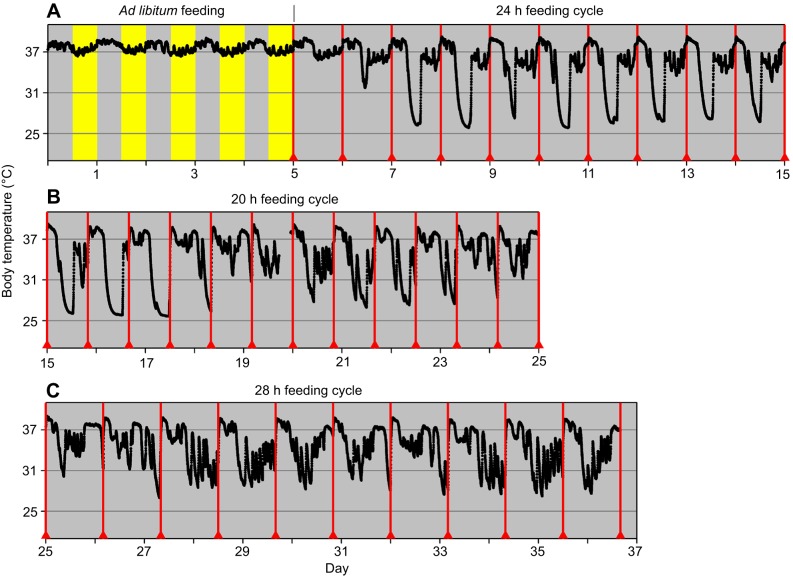
Daily torpor was induced in wild-type mice housed in the absence of a light–dark cycle by restricting daily food intake to a 2.2 g meal (∼70% of ad libitum food intake) that was initially provided to the mice at intervals of 24 h (24 h T-cycle). After 2–5 days, this resulted in daily torpor bouts starting a few hours after feeding, with body temperature dropping to 26–28°C (Fig. 1A; data of all individual mice is presented in Dataset 1). To dissect the role of the circadian clock from the timing of food intake in regulating the timing of daily torpor, the feeding interval was subsequently shortened to 20 h with the meal size being reduced proportionately to 1.8 g (20 h T-cycle). As expected (Stephan, 2002), the circadian clock was unable to entrain to this short feeding cycle, resulting in discordance between the timing of feeding and the circadian timing of behavior. Daily torpor bouts were delayed successively relative to the feeding time, with torpor bouts being abruptly terminated upon food provision (Fig. 1B). Daily torpor was absent on days where torpor was expected to start shortly after feeding. A similar pattern emerged in the final 12 days of the experiment, where 2.5 g food was provided every 28 h (28 h T-cycle; Fig. 1C). The timing of daily torpor advanced relative to the time of feeding, with torpor being absent when feeding occurred shortly before the expected torpor bout.
Fig. 1.
Feeding at the wrong time disrupts torpor. Body temperature recording of a wild-type mouse exposed to food restriction by providing (A) a single 2.2 g meal every 24 h (day 6–15), (B) a 1.8 g meal every 20 h (day 16–25) and (C) a 2.5 g meal every 28 h (day 26–37). Non-24 h feeding cycles revealed that feeding terminates torpor bouts and prohibits the initiation of torpor bouts when it occurs shortly before the expected onset of torpor. Mice were fed ad libitum on day 1–5 and thereafter food was restricted to a single meal per feeding cycle. Red triangles and lines indicate meal timing. The yellow and gray background represents lights on and off, respectively.
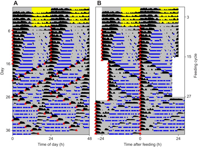
The interaction between food timing and the circadian clock in regulating the timing of daily torpor was assessed by comparing actograms in which body temperature profiles were plotted with different periods. In these actograms, the timing of torpor was plotted with either a 24 h period (∼circadian period; Fig. 2A) or a period matching the duration of the feeding cycle (Fig. 2B). Comparison of the timing of torpor between the two time scales showed that daily torpor occurred predominantly in the hours following the circadian active phase, while torpor rarely resumed after it had been interrupted by feeding. In order to quantify these differences, the rest phase was subdivided into portions preceded by feeding or preceded by the circadian active phase (Fig. S1). Mice spent significantly more time in torpor during portions of the resting phase preceded by the circadian active phase, compared with portions preceded by feeding (F4,20=101.8, P<0.0001; Figs 2 and 3A). The importance of the circadian clock in regulating the timing of daily torpor was further assessed by comparing the circadian time with the time after feeding of the first torpor bout during each feeding cycle (Fig. S2). The initial torpor bout typically started within a few hours of feeding (<½ feeding cycle) when feeding occurred during the circadian active phase (CT 12–24). In contrast, when feeding occurred during the circadian rest phase (CT 0–12), the first torpor bout was delayed until after the circadian active phase.
Fig. 2.
Timing of torpor depends on circadian clock and food timing. Double-plotted actograms of body temperature of a wild-type mouse exposed to timed food restriction. The same data are plotted with a period of 24 h (A) as well as with the feeding cycle period (24, 20 and 28 h; B). The actograms reveal that the timing of torpor is under circadian control. Body temperature is depicted in black with the height of the bar representing the body temperature above 36.5°C. Torpor is indicated by blue bars. Red triangles indicate meal timing. The yellow and gray background represents lights on and off, respectively.
Fig. 3.
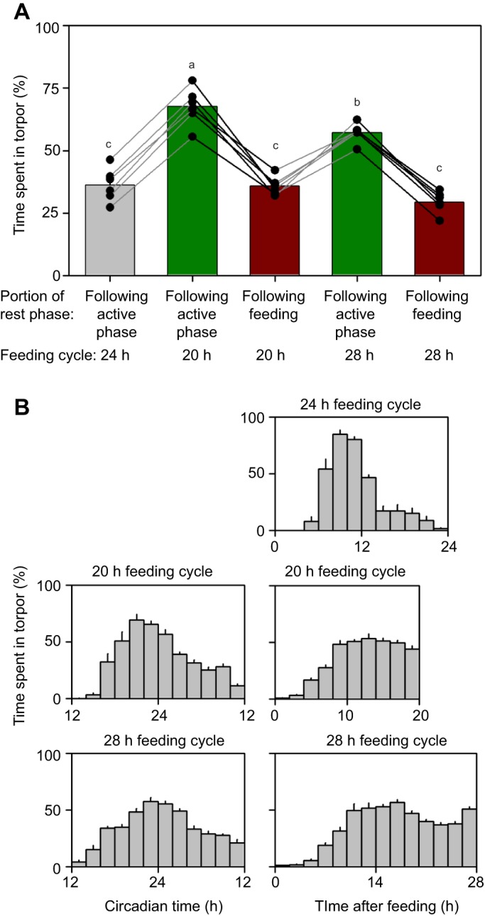
Timing of torpor is under circadian control and inhibited by recent feeding. (A) Wild-type mice exposed to feeding cycles with different periods (24, 20, 28 h T-cycles) spent a significantly larger proportion of time in torpor during intervals preceded by the circadian active phase (green) than following feeding bouts (red). These colors correspond to the subdivision as indicated in Fig. S1. Connected data points indicate measurements of the same animal. Bars with different letters are significantly different. (B) The portion of time spent in torpor at all phases of the circadian (left) and feeding cycle (right) in mice exposed to feeding cycles with different periods (top: 24 h; middle: 20 h; bottom: 28 h). By definition, a circadian cycle lasts 24 circadian hours with CT 12 indicating the onset of activity. Data are binned in 2 h intervals. Because of behavioral entrainment to 24 h feeding cycles, the circadian profile of torpor occurrences during this interval is identical to the profile defined by the time after feeding and is therefore not shown. Bar graphs are means+s.e.m.
To further separate the interacting influences of the circadian clock and feeding on the timing of torpor, daily profiles representing the average time spent in torpor at different phases over multiple cycles of the circadian and feeding cycles were generated (Fig. 3B). Under 24 h feeding T-cycles, torpor usually occurred during the second half of the subjective night (6–14 h after feeding) with torpor seldom occurring in the first 4–6 h after or ∼10 h before feeding. During periods of discordance between the circadian and feeding cycles (e.g. when food was provided at intervals of 20 or 28 h), the circadian regulation of torpor timing was still present, with torpor occurring mostly in the second half of the subjective night and at lower levels throughout the rest of the circadian cycle (Fig. 3B). Regulation by feeding was limited to suppression of daily torpor during the first 4–6 h after feeding, with intermediate levels of torpor throughout the rest of the feeding cycle. Overall, discordance between circadian and food timing revealed that the timing of daily torpor was closely linked to endogenously generated circadian rhythms, with feeding only influencing torpor by inhibiting it in the first hours after feeding.
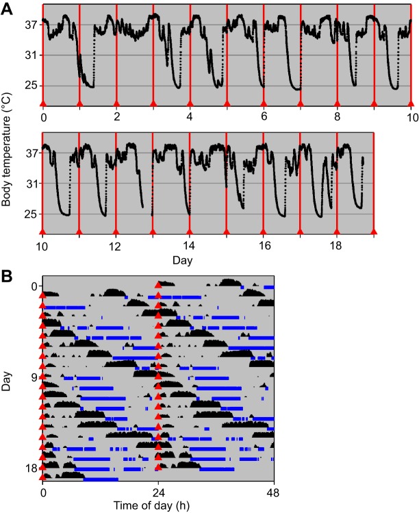
To confirm the relative importance of the circadian clock vis-à-vis the timing of food intake in regulating the timing of daily torpor, a second experiment was performed in which discordance between the feeding cycle and circadian timing was generated by lengthening the period of the central circadian clock. Mutation of three of four conditional alleles of CK1δ and CK1ε in GABAergic neurons slows the central clock in the SCN that regulates behavioral rhythms (Vgat-Cre+ CK1δfl/fl εfl/+; van der Vinne et al., 2018). When housed in constant darkness, these mice express circadian rhythms in locomotor activity with a period of ∼27.4 h (van der Vinne et al., 2018). Restriction of food intake to a single meal of 2.2 g every 24 h resulted in discordance between circadian and food timing as the circadian clock of these mutant mice was unable to entrain to the 24 h feeding cycle (Fig. 4B). Similar to what was observed in wild-type mice exposed to discordant 20 or 28 h feeding T-cycles, torpor bouts in long-period mice were abruptly curtailed by feeding, and torpor bouts were skipped on days when torpor was expected to start shortly after the time of feeding (Fig. 4A). Visual inspection of actograms indicated that torpor mostly occurred following the circadian active phase (Fig. 4B; Dataset 1). Torpor occurred significantly more during portions of the rest phase preceded by the circadian active phase, than during portions preceded by feeding (F1,8=27.84, P=0.0007; Fig. 4B; Fig. S3A). Daily profiles of the time spent in torpor at different phases in the circadian or feeding cycles (Fig. S3B) confirmed the conclusion from our study in wild-type mice, indicating that the timing of daily torpor was under circadian control.
Fig. 4.
Timing of torpor in a long-period (Vgat-Cre+ CK1δfl/fl εfl/+) mouse. (A) Body temperature recording of a long-period mouse exposed to food restriction by providing a single 2.2 g meal every 24 h. Discordance between the long circadian period and the 24 h feeding cycle resulted in termination of torpor when feeding occurred during torpor bouts and torpor was not initiated when feeding occurred shortly before its expected onset. (B) Double-plotted actogram of body temperature of a long-period mouse exposed to a feeding cycle with a 24 h period in constant darkness. Torpor occurs in the hours following the circadian active phase unless disrupted by feeding. Body temperature is depicted in black with the height of the bar representing the body temperature above 36.5°C. Torpor is indicated by blue bars. Red triangles indicate meal timing.
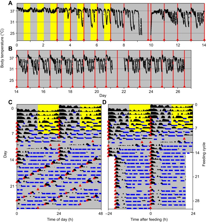
The two studies using the discordance paradigms presented above indicate that the circadian clock has primacy over feeding in the regulation of the timing of daily torpor. The assessment of the role of food timing in the regulation of torpor might thus be confounded by the presence of circadian regulation. To study the isolated role of food timing in controlling the timing of torpor, we next disrupted endogenous circadian rhythmicity by disrupting the essential clock gene Bmal1 in GABAergic neurons including the SCN, while leaving clocks in peripheral tissues effectively wild-type (Vgat-Cre+ Bmal1fl/fl; Weaver et al., 2018). Food was provided at 24 h (2.2 g) or 20 h (1.8 g) intervals (Fig. 5). In the absence of circadian regulation and environmental light–dark cycles, torpor showed a consistent temporal organization relative to feeding, with torpor being suppressed in the first 6 h after feeding and a high likelihood of torpor during the rest of the feeding cycle (Fig. 5; Fig. S4 and Dataset 1). This pattern was observed irrespective of the interval between meals (Fig. 5). The resulting temporal profile (Fig. S4) was similar to that observed in endogenously rhythmic mice challenged by a discordant feeding cycle in the first two experiments.
Fig. 5.
Timing of torpor bouts in mice without a functional central clock (Vgat-Cre+ Bmal1fl/fl). (A,B) Body temperature recording of a mouse without a functional central clock exposed to food restriction by providing a single 2.2 g meal every 24 h (day 3–14; A) followed by a 1.8 g meal every 20 h (day 15–27; B). The absence of a functional central clock did not prevent torpor. Because of technical difficulties, the meal on day 9 was delayed by ∼20 h. The yellow and gray background represents lights on and off, respectively. Red triangles and lines indicate meal timing. Food was available ad libitum for the first 3 days. (C,D) Double-plotted actograms of body temperature recordings of a mouse without a functional central clock exposed to feeding cycles with a 24 or 20 h period. Data are plotted both with a period of 24 h (C) as well as with the feeding cycle period (24 h then 20 h; D). The absence of a circadian timing component independent of the timing of feeding showed that in the absence of a functional central clock, the timing of torpor depends on the time of feeding. Body temperature is depicted in black with the height of the bar representing the body temperature above 36.5°C. Torpor is indicated by blue bars. Red triangles indicate meal timing. The yellow and gray background represents lights on and off, respectively.
DISCUSSION
Daily torpor is an opportunistic energy saving strategy that is often used by small mammals to adapt to unexpected energetic challenges. Despite the unpredictable nature of energetic challenges, the timing of daily torpor is tightly regulated. The present study confirms earlier results (Bechtold et al., 2012) showing that the circadian clock can control the timing of daily torpor independent of the timing of food intake or external light–dark cycles and extends them to show that the circadian clock takes primacy over the timing of food intake in determining the timing of daily torpor. Torpor is prohibited during the first hours of the circadian active phase (CT 12–16), while torpor is infrequent in the later hours of the circadian rest phase (CT 6–12). The timing of food intake relative to the circadian phase has previously been shown to be capable of affecting the timing of daily torpor (Paul et al., 2004), although these effects were likely partially mediated by a shift of the circadian clock in response to timed food restriction (Stephan, 2002). The current experiments using discordance between circadian cycle length and the feeding period allowed us to assess the direct effects of food intake on the timing of torpor, showing that torpor is prohibited in the first hours after a meal. Furthermore, when the circadian rest phase is interrupted by feeding, torpor is unlikely to be re-initiated until after the next circadian active phase. In the absence of circadian regulation of behavior in Vgat-Cre+ Bmal1fl/fl mice, torpor occurs throughout the day, except for the first hours after a meal. Overall, the present study clearly demonstrates that daily torpor in energetically challenged mice is gated by the circadian clock, with the interplay between the circadian clock and the timing of feeding determining the timing of torpor.
Ablation of the SCN in Siberian hamsters disrupts the daily timing of torpor (Ruby and Zucker, 1992), suggesting that the SCN plays a regulatory role in torpor timing. The continued expression of torpor in Vgat-Cre+ Bmal1fl/fl mice (Fig. 5A,B) shows that endogenous circadian rhythms in the SCN are not required for the expression of daily torpor. The interrupted nature of torpor bouts in these mice (Fig. 5A,B) does, however, suggest a role for the central circadian clock in maintaining a continuous torpor state. Such a role has previously been proposed for the SCN as a suppressor of alternations between activity and rest (Blum et al., 2014). The fragmented nature of torpor bouts observed in mice exposed to feeding cycles that were discordant from the endogenous clock period (20 and 28 h T-cycles; Fig. 1) is in line with a role for the circadian clock in maintaining a continuous torpor state, although the possibility that the observed fragmentation of daily torpor was the result of the prolonged exposure to energetic challenges cannot be excluded given the experimental design used in the present study. The role of the SCN in controlling the daily timing of torpor is illustrated by the phase as well as the period at which daily torpor occurs. In long-period Vgat-Cre+ CK1δfl/fl εfl/+ mice, peripheral clocks are phase advanced by ∼6 h compared with the SCN and behavioral rhythms (van der Vinne et al., 2018). The continued expression of torpor in these mice at circadian times immediately following the circadian active phase (Fig. 4B) suggests that the timing of daily torpor is controlled by a GABAergic brain region − most likely the SCN − but not by clocks in peripheral tissues.
Because of the nature of our feeding regime – with food being provided on the wire lid of the cage by the experimenter – it is impossible to exclude the possibility that torpor arousals were independent of the intake of food but instead resulted from other disturbances (e.g. sound, movement). In future studies, the feeding regime used here could be compared with the effect of disturbances without providing food to confirm that food intake, and/or associated physiological changes, are indeed responsible for the termination of daily torpor bouts.
Under natural conditions, daily torpor is typically induced by environmental changes that lead to reduced food intake and/or increased energetic demands. Under these conditions, endogenous circadian rhythms and the timing of food intake are typically synchronized, which would result in daily torpor bouts occurring during the late night and early morning while being inhibited in the early night. This daily rhythm in torpor maximizes the energetic benefit of daily torpor by synchronizing its occurrence with low night-time ambient temperatures (van der Vinne et al., 2015). When food is presented shortly before torpor is expected to occur, torpor is prevented, consistent with a requirement for energetic challenge for daily torpor to occur. Overall, our data show that while daily torpor is an opportunistic strategy employed in response to energetic challenges, its occurrence is conditional upon appropriate food timing and its timing is tightly regulated by the endogenous circadian clock.
Supplementary Material
Acknowledgements
We thank Elissa Hult, Christopher Lambert and Jamie Black for technical assistance, as well as Linh Vong and Brad Lowell for generously providing Vgat-Cre+ founder mice.
Footnotes
Competing interests
The authors declare no competing or financial interests.
Author contributions
Conceptualization: V.v.d.V., D.R.W., S.J.S.; Methodology: V.v.d.V., D.R.W., S.J.S.; Formal analysis: V.v.d.V.; Investigation: M.J.B., S.J.S.; Resources: V.v.d.V., D.R.W., S.J.S.; Data curation: V.v.d.V.; Writing - original draft: V.v.d.V.; Writing - review & editing: M.J.B., D.R.W., S.J.S.; Visualization: V.v.d.V.; Project administration: V.v.d.V., S.J.S.; Funding acquisition: D.R.W., S.J.S.
Funding
This work was supported by the National Institutes of Health (R01 NS056125 and R21 ES024684 to D.R.W., and R15 HL120072 to S.J.S.). The funders had no role in study design, data collection and analysis, decision to publish, or preparation of the manuscript. The contents of this article are solely the responsibility of the authors and do not necessarily represent the official views of the sponsoring agencies. Deposited in PMC for release after 12 months.
Supplementary information
Supplementary information available online at http://jeb.biologists.org/lookup/doi/10.1242/jeb.179812.supplemental
References
- Bechtold D. A., Sidibe A., Saer B. R. C., Li J., Hand L. E., Ivanova E. A., Darras V. M., Dam J., Jockers R., Luckman S. M. et al. (2012). A role for the melatonin-related receptor GPR50 in leptin signaling, adaptive thermogenesis, and torpor. Curr. Biol. 22, 70-77. 10.1016/j.cub.2011.11.043 [DOI] [PubMed] [Google Scholar]
- Blum I. D., Zhu L., Moquin L., Kokoeva M. V., Gratton A., Giros B. and Storch K.-F. (2014). A highly tunable dopaminergic oscillator generates ultradian rhythms of behavioral arousal. eLife 3, e05105 10.7554/eLife.05105 [DOI] [PMC free article] [PubMed] [Google Scholar]
- Chu L. P. and Swoap S. J. (2012). Oral bezafibrate induces daily torpor and FGF21 in mice in a PPAR alpha dependent manner. J. Thermal Biol. 37, 291-296. 10.1016/j.jtherbio.2011.11.011 [DOI] [Google Scholar]
- Dibner C., Schibler U. and Albrecht U. (2010). The mammalian circadian timing system: organization and coordination of central and peripheral clocks. Annu. Rev. Physiol. 72, 517-549. 10.1146/annurev-physiol-021909-135821 [DOI] [PubMed] [Google Scholar]
- Etchegaray J.-P., Machida K. K., Noton E., Constance C. M., Dallmann R., Di Napoli M. N., DeBruyne J. P., Lambert C. M., Yu E. A., Reppert S. M. et al. (2009). Casein kinase 1 delta regulates the pace of the mammalian circadian clock. Mol. Cell. Biol. 29, 3853-3866. 10.1128/MCB.00338-09 [DOI] [PMC free article] [PubMed] [Google Scholar]
- Geiser F. (2004). Metabolic rate and body temperature reduction during hibernation and daily torpor. Annu. Rev. Physiol. 66, 239-274. 10.1146/annurev.physiol.66.032102.115105 [DOI] [PubMed] [Google Scholar]
- Hut R. A., Kronfeld-Schor N., van der Vinne V. and de la Iglesia H. O. (2012). In search of a temporal niche: environmental factors. Prog. Brain Res. 199, 281-304. 10.1016/B978-0-444-59427-3.00017-4 [DOI] [PubMed] [Google Scholar]
- Kirsch R., Ouarour A. and Pévet P. (1991). Daily torpor in the Djungarian hamster (Phodopus sungorus): photoperiodic regulation, characteristics and circadian organization. J. Comp. Physiol. A Neuroethol. Sens. Neural Behav. Physiol. 168, 121-128. 10.1007/BF00217110 [DOI] [PubMed] [Google Scholar]
- Körtner G. and Geiser F. (2000). The temporal organization of daily torpor and hibernation: circadian and circannual rhythms. Chronobiol. Int. 17, 103-128. 10.1081/CBI-100101036 [DOI] [PubMed] [Google Scholar]
- Lynch G. R., Bunin J. and Schneider J. E. (1980). The effect of constant light and dark on the circadian nature of daily torpor in Peromyscus leucopus. Biol. Rhythm. Res. 11, 85-93. [Google Scholar]
- Meng Q.-J., Logunova L., Maywood E. S., Gallego M., Lebiecki J., Brown T. M., Sládek M., Semikhodskii A. S., Glossop N. R. J., Piggins H. D. et al. (2008). Setting clock speed in mammals: the CK1ɛ tau mutation in mice accelerates circadian pacemakers by selectively destabilizing PERIOD proteins. Neuron 58, 78-88. 10.1016/j.neuron.2008.01.019 [DOI] [PMC free article] [PubMed] [Google Scholar]
- Mohawk J. A., Green C. B. and Takahashi J. S. (2012). Central and peripheral circadian clocks in mammals. Annu. Rev. Neurosci. 35, 445-462. 10.1146/annurev-neuro-060909-153128 [DOI] [PMC free article] [PubMed] [Google Scholar]
- Oelkrug R., Heldmaier G. and Meyer C. W. (2011). Torpor patterns, arousal rates, and temporal organization of torpor entry in wildtype and UCP1-ablated mice. J. Comp. Physiol. B 181, 137-145. 10.1007/s00360-010-0503-9 [DOI] [PubMed] [Google Scholar]
- Paul M. J., Kauffman A. S. and Zucker I. (2004). Feeding schedule controls circadian timing of daily torpor in SCN-ablated Siberian hamsters. J. Biol. Rhythms 19, 226-237. 10.1177/0748730404264337 [DOI] [PubMed] [Google Scholar]
- Ruby N. F. and Zucker I. (1992). Daily torpor in the absence of the suprachiasmatic nucleus in Siberian hamsters. Am. J. Physiol. 263, R353-R362. 10.1152/ajpregu.1992.263.2.R353 [DOI] [PubMed] [Google Scholar]
- Ruf T., Steinlechner S. and Heldmaier G. (1989). Rhythmicity of body temperature and torpor in the Djungarian hamster, Phodopus sungorus. In Living in the Cold II (ed. A. Malan and B. Canguilhem), pp. 53-61. London: John Libbey Eurotext. [Google Scholar]
- Speakman J. (1997). Factors influencing the daily energy expenditure of small mammals. Proc. Nutr. Soc. 56, 1119-1136. 10.1079/PNS19970115 [DOI] [PubMed] [Google Scholar]
- Stephan F. K. (2002). The “other” circadian system: food as a Zeitgeber. J. Biol. Rhythms 17, 284-292. 10.1177/074873002129002591 [DOI] [PubMed] [Google Scholar]
- Storch K.-F., Paz C., Signorovitch J., Raviola E., Pawlyk B., Li T. and Weitz C. J. (2007). Intrinsic circadian clock of the mammalian retina: importance for retinal processing of visual information. Cell 130, 730-741. 10.1016/j.cell.2007.06.045 [DOI] [PMC free article] [PubMed] [Google Scholar]
- Takahashi J. S. (2017). Transcriptional architecture of the mammalian circadian clock. Nat. Rev. Genet. 18, 164-179. 10.1038/nrg.2016.150 [DOI] [PMC free article] [PubMed] [Google Scholar]
- van der Vinne V., Gorter J. A., Riede S. J. and Hut R. A. (2015). Diurnality as an energy-saving strategy: energetic consequences of temporal niche switching in small mammals. J. Exp. Biol. 218, 2585-2593. 10.1242/jeb.119354 [DOI] [PubMed] [Google Scholar]
- van der Vinne V., Swoap S. J., Vajtay T. J. and Weaver D. R. (2018). Desynchrony between brain and peripheral clocks caused by CK1δ/ε disruption in GABA neurons does not lead to adverse metabolic outcomes . Proc. Natl. Acad. Sci. USA 115, E2437-E2446. 10.1073/pnas.1712324115 [DOI] [PMC free article] [PubMed] [Google Scholar]
- Vong L., Ye C., Yang Z., Choi B., Chua S. and Lowell B. B. (2011). Leptin action on GABAergic neurons prevents obesity and reduces inhibitory tone to POMC neurons. Neuron 71, 142-154. 10.1016/j.neuron.2011.05.028 [DOI] [PMC free article] [PubMed] [Google Scholar]
- Weaver D. R., van der Vinne V., Giannaris E. L., Vajtay T. J., Holloway K. and Anaclet C. (2018). Functionally complete excision of conditional alleles in the mouse suprachiasmatic nucleus by Vgat-ires-Cre. J. Biol. Rhythms 33, 179-191. 10.1177/0748730418757006 [DOI] [PMC free article] [PubMed] [Google Scholar]
Associated Data
This section collects any data citations, data availability statements, or supplementary materials included in this article.


