Abstract
Metal-support interaction is one of the most important parameters in controlling the catalysis of supported metal catalysts. Silica, a widely used oxide support, has been rarely reported as an effective support to create active metal-support interfaces for promoting catalysis. In this work, by coating Cu microparticles with mesoporous SiO2, we discover that Cu/SiO2 interface creates an exceptional effect to promote catalytic hydrogenation of esters. Both computational and experimental studies reveal that Cu–Hδ− and SiO–Hδ+ species would be formed at the Cu–O–SiOx interface upon H2 dissociation, thus promoting the ester hydrogenation by stablizing the transition states. Based on the proposed catalytic mechanism, encapsulting copper phyllosilicate nanotubes with mesoporous silica followed by hydrogen reduction is developed as an effective method to create a practical Cu nanocatalyst with abundant Cu-O-SiOx interfaces. The catalyst exhibits the best performance in the hydrogenation of dimethyl oxalate to ethylene glycol among all reported Cu catalysts.
Metal-support interaction plays an important role in heterogeneous catalysis, but silica has been rarely reported as an effective support to create active metal-support interfaces for promoting catalytic reactions. Here, the authors discover that Cu/SiO2 interface creates an exceptional effect to promote catalytic hydrogenation of esters.
Introduction
Heterogeneous catalysis is of vital importance in many fields of chemical, food, energy, and environmental applications. The rational design and fabrication of sufficient active interfaces between metal and (hydr)oxide to facilitate the reactions with multiple reagents has emerged as an effective strategy to prepare heterogeneous catalysts with improved performances. For instance, both Pt/FeOx and Pt/Fe(OH)x interfaces exhibit excellent performance in CO oxidation and CO preferential oxidation (PROX)1–4. Au/CeOx and Au/TiOx interfaces have been demonstrated to improve the activity of water-gas shift reaction5–7. Pt/M(OH)2 (M = metal) interfaces enhance the performance of hydrogen evolution reaction and hydrogen oxidation reaction8–11. Such interfacial effects from the strong metal–metal (hydr)oxide interactions were typically observed only when reducible metal oxides (e.g., TiOx, CeOx, FeOx) were used as supports12–17. In contrast, SiO2 without reducible metal cations usually serves as ‘inert’ support, or plays as shell material to fabricate yolk-shell and core-shell metal nanocatalysts to prevent the sintering of metal components18–24. Reports on the promotional effects of SiO2 on heterogeneous catalysis are rare25,26.
Here we demonstrate that SiO2 readily creates highly active interfaces with Cu in the gas-phase hydrogenation of dimethyl oxalate (DMO) into ethylene glycol (EG). In this work, the Cu-SiO2 interfaces were first designed and fabricated by depositing a mesoporous SiO2 (m-SiO2) layer onto the surface of commercial Cu powders. With the created Cu–SiO2 interfaces, the coated Cu powders exhibited a two-order-of-magnitude enhancement in the activity as compared to the uncoated Cu powders. Combining experiments with density functional theory (DFT) calculations, we demonstrate that H2 could be activated at the Cuδ+–O–SiOx interface region, giving rise to Cu–H and interfacial SiO–H species, which are able to promote the hydrogenation of polar C=O bonds. Based on this understanding, a smart strategy by in situ reducing silica coated copper phyllosilicate nanotubes was developed to produce a sophisticated Cu-SiO2 nanocatalyst with abundant Cu–O–SiOx interface. Such a catalyst exhibited the best reported performance in selective hydrogenation of DMO to EG.
Results and Discussion
Cu–O–SiOx interfaces boost the catalysis of copper
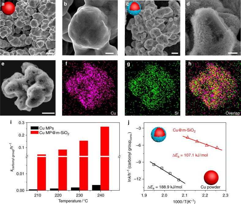
To create Cu–O–SiOx interfaces, a non-continuous layer of m-SiO2 was deposited onto the surface of commercial Cu microparticles (MPs) with diameter of 2–3 µm by hydrolysis of tetraethoxysilane (TEOS) in the presence of cetyltrimethylammonium bromide (CTAB) (See 'Methods' section). Scanning electron microscopy (SEM) and energy dispersive spectroscopy (EDS) analysis (Fig. 1a–h, Supplementary Fig. 1) revealed the successful deposition of a downy layer of SiO2 on Cu MPs. The mesoporous nature of the SiO2 layer deposited on Cu MPs was confirmed by the N2 adsorption and desorption isotherm at 77 K (Supplementary Fig. 2). It should also be noted that the m-SiO2 layer was not continuously grown on Cu, resulting in the exposure of partial Cu sites on the as-obtained hybrid of Cu-MP@m-SiO2.
Fig. 1.
Demonstration of Cu–O–SiOX interface effect in DMO hydrogenation. a–d SEM images of Cu microparticle before (a, b) and after (c, d) coating mesoporous silica; e–h EDX mapping images of mesoporous silica coated Cu microparticles (Cu@m-SiO2); i, j Catalytic performance and the apparent activation energy (Ea) of Cu microparticles before and after coating mesoporous silica for the selective hydrogenation of DMO, respectively; Reaction conditions were as follows: H2/DMO = 80 mol/mol, P (H2) = 3.0 MPa. Scale bars in a, c and e are 2 µm. Scale bars in b and d are 500 nm
To evaluate the Cu–O–SiOx interfacial effect, we chose the gas-phase hydrogenation of DMO.27–29 As shown in Fig. 1i, Cu MPs without SiO2 coating displayed a negligible activity in the hydrogenation of DMO at the temperature below 250 °C. In comparison, Cu MPs coated with m-SiO2 exhibited a significant activity even at the temperature of 210 °C. The turnover rate of carbonyl groups (kcarbonyl group) over Cu-MP@m-SiO2 was approximately 80 times higher than that on uncoated Cu MPs at the temperature between 200 and 240 °C. In this comparison, the kcarbonyl group was calculated based on the hydrogenation rate of carbonyl groups over the total amount of Cu in the catalysts. The hydrogenation activity of the Cu-MP@m-SiO2 catalyst was increased with the temperature. Considering that less Cu sites were exposed on Cu-MP@m-SiO2, the catalytic enhancement induced by the Cu–O–SiOx interfaces was tremendous. Moreover, the apparent activation energy (Ea) over Cu-MP@m-SiO2 was measured to be 107.1 kJ mol−1 (Fig. 1j), almost only half of that on Cu MPs (188.9 kJ mol−1), indicating the as-built Cu-O-SiOx interfaces would completely alter the hydrogenation mechanism.
Hydrogenation mechanism over the Cu–O–SiOx interface
The promotional effect of the Cu–O–SiOx interface on the catalytic hydrogenation of DMO was studied by using DFT calculations. In this work, structural models of periodic Cu(111) with/without SiO2 coating were built to simulate the modified and unmodified Cu MPs, respectively. Until now, it is still a great challenge to identify the interfacial structure between metal and silica since the SiO2 deposition could present as various crystalline or vitreous films. To simplify the interface model, we assumed that the [SiO4] tetrahedra could be stacked on Cu(111) in a two-dimensional ordered network with a composition of SiO2.5, in which every Si has one Si–O–Cu bond and three Si–O–Si bonds (Supplementary Fig. 3). According to our DFT calculations, the proposed model was calculated to be exothermic by 0.66 eV/Si with respective to Cu(111), α-quartz SiO2 and gaseous O2. Bader charge analysis showed that surface Cu atoms, which were directly bonded with O-SiO3, would carry significantly positive charge, implying that the SiO2 overgrowth would lead to the formation of Cuδ+ species (Supplementary Fig. 3). Interestingly, similar silica films with c(2 × 2) structures have been demonstrated to form on Mo(112) and Ru(0001) single crystal surfaces30–32. As suggested previously, the adsorption energy of oxygen atoms plays an important role in determining whether silica monolayer film can be stabilized on the metal surface or not. In our case, the dissociative adsorption energy of O2 on Cu(111) was calculated as −3.13 eV, just between those of Mo(112) (−5.64 eV) and Ru(0001) (−2.28 eV). In addition, Cu(111) has lattice constant of 2.556 Å, matching well with the silica film with a 5.2~5.3 Å periodicity in the (2 × 2) manner. All these results indicated that the formation of monolayer SiO2 network over Cu(111) surfaces is reasonable. On the surface of Cu-MP@m-SiO2, there were still a large amount of uncovered Cu atoms so that the silica film should not be continuous. To account for the experimental observation, we extended our model to a (8 × 4) structure in which 50% [SiO4] tetrahedra were removed, and the as-generated Si–O dangling bonds were saturated by H atoms. Thus, the Cu–O–SiOx interface turned to be accessible for the substrate molecules. Hereafter, such a model is denoted as SiO2/Cu(111).
Hydrogenation on the heterogeneous catalysts usually follows Horiuti Polanyi mechanism, which consists of following steps: (i) dissociation of hydrogen; (ii) adsorption of unsaturated compounds; (iii) stepwise hydrogenation with H atoms. Supplementary Fig. 4 showed the dissociation of H2 on the two distinct surfaces, i.e., Cu(111) and SiO2/Cu(111). On Cu(111), H–H bond splitting occurred via a homolytic mechanism, generating two hydrogen atoms adsorbed on the three-fold sites. Alternatively, the presence of Cu–O–SiOx interface on SiO2/Cu(111) enable H2 activation in a heterolytic way, yielding Cu–H and interfacial SiO–H species simultaneously at the interface. From Cu(111) to SiO2/Cu(111), the calculated barrier for H2 dissociation does not change too much (0.28 eV vs. 0.30 eV), indicating that heterolytic dissociation could be competitive with the hemolytic one. However, from the viewpoint of thermodynamics, H2 dissociation on SiO2/Cu(111) was found to be more exothermic than that on Cu(111) (−0.81 eV vs. −0.47 eV) because the interfacial SiO–H bond is stronger than Cu–H bond. Therefore, even the H2 dissociation occurred via the hemolytic route, interfacial SiO–Hδ+ species would be generated through thermodynamics-driven hydrogen spillover. All these indicated that there existed abundant interfacial SiO–Hδ+ and Cu–Hδ− at the Cu–O–SiOx interfaces upon hydrogenation.
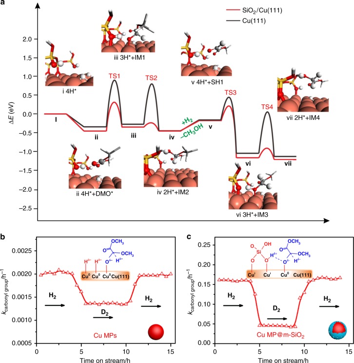
The DMO hydrogenation pathways on Cu(111) and SiO2/Cu(111) were compared, with the optimized structures of transition states (TS’) and important intermediates (IMs) illustrated in Fig. 2a, Supplementary Figs. 5 and 6. For clarity, only the lowest energy pathways were shown for these two surfaces. According to our DFT calculations, DMO was weakly adsorbed on both Cu(111) and SiO2/Cu(111) (from i to ii in Fig. 2a). In this regard, there are two possible mechanisms, namely hydroxyl mechanism and alkoxy mechanism. In the former case, the first hydrogen atom would attack the O end of C=O group, producing the hydroxyl intermediate, while in the latter case, C end of C=O group would be hydrogenated first, leading to the formation of alkoxy intermediate. It has been reported previously that hydroxyl intermediate was thermodynamically less favorable than alkoxy intermediate33,34. On both surfaces, the hydrogenation of DMO begins with the nucleophilic attack of Hδ− to the electron deficient carbon of ester group. On Cu(111), a 1.22 eV barrier (TS1) has to be surmounted when the initial hydrogenation takes place. In contrast, a relative low barrier of 0.77 eV (TS1) is required when the reaction occur at the Cu–O–SiOx interface. It should be noted that both of the reactions proceed with similar endothermicity of ~0.10 eV, indicating that the difference in the barrier should be attributed to the electronic effect (from ii to iii in Fig. 2a). Based on the Bader charge analysis, it was found that when Hδ− approaches, the adsorbed DMO would bear a ~ −0.5 a.u. charge. Such a negatively charged TS can be stabilized by the SiO–Hδ+ species, but repulsive with the co-adsorbed Cu–Hδ−. This finding nicely explained why the addition of ‘inert’ silica could significantly enhance the hydrogenation.
Fig. 2.
Mechanism of DMO hydrogenation on Cu–O–SiOx interface. a The DMO hydrogenation pathway on Cu(111) and SiO2/Cu(111); b, c The kinetic isotope effect of Cu MPs catalyst and Cu MPs@m-SiO2 catalyst in hydrogenation of DMO. Reaction conditions were as follows: H2/DMO = 80 mol/mol, P (H2) = 3.0 MPa, T = 230 °C
Next, the half-hydrogenated intermediate undergoes the second H addition to give alcohol species (iv in Fig. 2a IM2). On Cu(111), the TS2 occurs through a reductive elimination of CH3O(O=)C–CH(OCH3)Oδ− and Hδ−, yielding a barrier of 1.07 eV. Such a high barrier might be originated from the electrostatic repulsion between two negatively charged species. On the contrary, SiO-Hδ+ at the Cu–O–SiOx interface can provide a proton, which is quickly transferred to CH3O(O=)C–CH(OCH3)Oδ− to make a O–H bond by passing a small barrier of 0.11 eV (from iii to iv in Fig. 2a). From this result, the interfacial SiO–Hδ+ species should be very acidic since the Cu atoms at the interface can stabilize their deprotonated forms. The proposed mechanism nicely explains why the Cu–O–SiOx interface is a good choice for the hydrogenation reaction. On one hand, the Cu–OSi bond is not so strong to inhibit the dissociation of H2. On the other hand, moderate Cu–OSi bond would render interfacial SiO–H bond suitable strength, which not only stabilizes the charged TS’ but also releases a proton when necessary.
For IM2, the co-presence of OH and OMe groups on the same carbon atoms makes them unstable, which can be easily converted into adsorbed CH3O(O=)C–CHO (SH1) plus a methanol molecule (from iv to v in Fig. 2a). Subsequently, SH1 would undergo step-wise hydrogenation again, passing through TS3 and TS4, leading to formation of MG species (IM4). Again, the calculated barriers for TS3 and TS4 on SiO2/Cu(111) were much lower than those on Cu(111), and the hydrogenation of aldehyde groups was easier than that of the ester groups (from v to vii in Fig. 2a). Similarly, MG can further be hydrogenated into EG by consecutive H atoms addition. DFT calculations show that the hydrogenation of MG is harder to be conquered than that of DMO. Either on SiO2/Cu(111) or Cu(111), the energy gaps between TS5 and TS1 are nearly the same (0.42 eV ~0.45 eV) (from viii to ix in Supplementary Fig. 5). Structurally, DMO has two ester groups. When one of them is hydrogenated, the other serves as an electron-withdrawing group which can stabilize charged TS’ through electron delocalization. Unfortunately, the CH2OH group in MG lacks the ability to delocalize the negative charge upon ester hydrogenation. Thus, the rate determining step for the hydrogenation of DMO to EG is corresponding to the hydrogenation of MG.
The mechanism suggested by DFT calculations was verified by serial isotope-labeling experiments (Fig. 2b). The kinetic isotope effect (KIE) of Cu MPs@m-SiO2 catalyst (kH/kD = 3.5) is about two times higher than that of Cu MPs catalyst (kH/kD = 1.5), indicating the different hydrogenation mechanisms on the two catalysts. As shown in Supplementary Fig. 5, not only the Cu–H but also the SiO–H are involved in the TS1 state as well as TS5 on SiO2/Cu(111). It was expected that when hydrogen was replaced by deuterium, the vibrations of both Cu–D and SiO–D would contribute to the zero-point energy of TS’, leading to a large KIE. It is particularly interesting that, when Cu MPs@m-SiO2 was treated by a NaOH solution, Na+ ions pre-occupies the Cu–SiO sites so that the formation of SiO–H should be suppressed during the hydrogenation. As expected, the TOF for the NaOH-treated Cu MPs@m-SiO2 catalyst was decreased dramatically to a similar level to that of uncoated Cu MPs, and the KIE was also dropped back to 1.6 (Supplementary Fig. 7). With the combination of DFT calculation and isotope-labelling experiments, we conclude that the Cu–O–SiOx interface not only activates H2 molecules in the heterolytic way to form Cu–Hδ− and SiO–Hδ+, but also facilitates the hydrogenation of ester by stablizing the transition states.
Nanostructure engineering enriches the Cu–O–SiOx interfaces
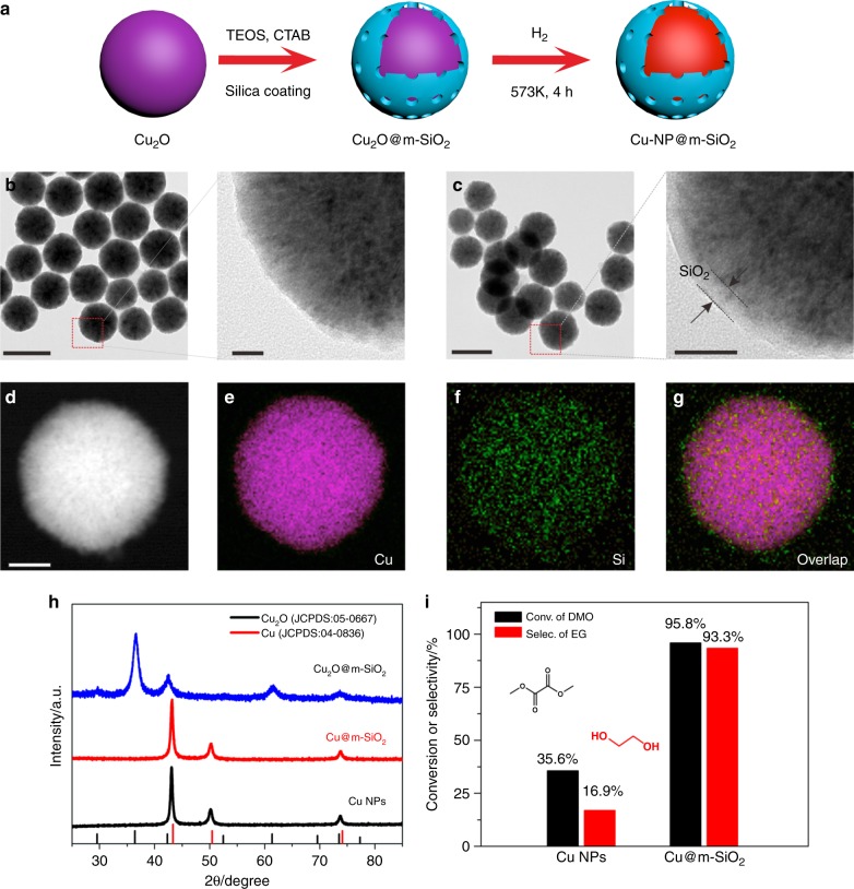
As the promotional effect induced by the Cu–O–SiOx interface, increasing Cu–O–SiOx interfaces should lead to further enhanced catalytic efficiency. Thus, reducing the size of Cu particles to the nanoscale would amplify the interfacial effect. To create Cu–SiO2 interface on Cu nanoparticles, Cu2O nanoparticles were first prepared and coated by m-SiO2 (Fig. 3a). The obtained Cu2O@m-SiO2 nanoparticles were then reduced under H2 atmosphere to convert into Cu@m-SiO2 nanoparticles (denoted as Cu-NP@m-SiO2). Comprehensive characterizations by TEM, EDS, XRD (Fig. 3b–h), and N2 adsorption/desorption isotherms (Supplementary Fig. 8) confirmed the core-shell structure of Cu-NP@m-SiO2. As expected, the as-prepared Cu-NP@m-SiO2 catalyst exhibited a much better catalytic performance in DMO hydrogenation than unmodified Cu NPs (Fig. 3i). With a liquid hourly space velocity (LHSV) of 2.4 h−1, Cu-NP@m-SiO2 exhibited both much higher DMO conversion (95.8%) and higher selectivity (93.3% to EG) at 200 °C. In contrast, when Cu NPs were used as the catalyst, only 35.6% of DMO was hydrogenated with 16.9% of selectivity to EG.
Fig. 3.
Creating Cu–O–SiOx interfaces on Cu nanoparticles. a Scheme for the synthesis of Cu@m-SiO2; b, c TEM images of as-prepared Cu2O nanoparticles and Cu2O@m-SiO2, respectively. d–g EDX mapping images of Cu2O@m-SiO2; h X-ray powder diffraction (XRD) pattern of Cu NPs, Cu2O@m-SiO2 and Cu@m-SiO2; i Catalytic performance of Cu NPs and Cu@m-SiO2 for the selective hydrogenation of DMO; Reaction conditions were as follows: H2/DMO = 80 mol/mol, P (H2) = 3.0 MPa, T = 200 °C, LHSV = 2.4 h−1. Scale bars in b (left) and c (left) are 200 nm. Scale bars in b (right) and c (right) are 20 nm. Scale bar in d is 50 nm
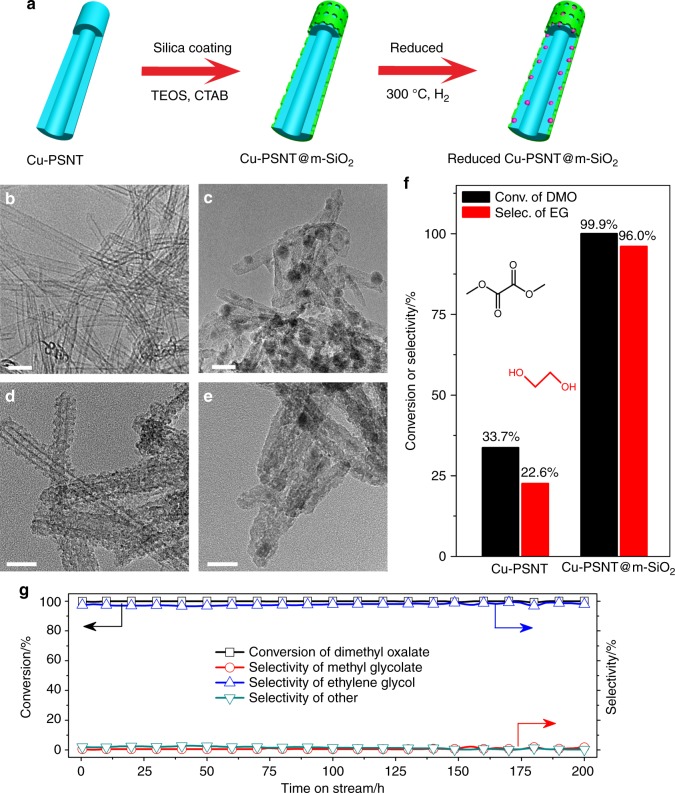
In term of Cu utilization, the core-shell overgrowth structure (Cu-NP@m-SiO2) demonstrated above was not the ideal structure for practical applications because most of Cu atoms in Cu-NP@m-SiO2 were not located on surface or Cu–O–SiOx interfaces. In this regard, encapsulating ultra-small Cu nanoparticles in a porous SiO2 matrix should be the most effective strategy to create highly active catalysts while maximizing the utilization of Cu. For this purpose, we chose copper phyllosilicate nanotubes as an alternative Cu precursor. Structurally, copper phyllosilicate has lamellar structure composed of alternate layers of SiO4 tetrahedra and discontinuous layers of CuO6 octahedra, in which Cu–O–SiOx moieties are readily available (Supplementary Fig. 9)35–39. By using a modified hydrothermal method, copper phyllosilicate nanotubes (Cu-PSNT) with sub-10 nm in diameter, 1–2 nm in wall thickness, and hundreds of nanometers in length were prepared (Fig. 4b). Both TEM and EDS analysis (Fig. 4c, Supplementary Fig. 10) confirmed the formation of metallic Cu nanoparticles which were embedded in SiO2 matrix after the H2 reduction. Some large Cu nanoparticles with size larger than 10 nm were also observed. Moreover, the BET analysis (Supplementary Fig. 11) revealed that the reduced Cu-PSNT had a BET surface area of 470.1 m2 g−1 and a pore volume of 1.47 cm3 g−1.
Fig. 4.
Confined growth strategy for maximizing Cu–O–SiOx interface. a Illustration of the synthetic strategy for the preparation of Cu-PSNT@m-SiO2; b–e TEM image of as-prepared Cu-PSNT, reduced Cu-PSNT, Cu-PSNT@m-SiO2 and reduced Cu-PSNT@m-SiO2, respectively; f Catalytic performance of reduced Cu-PSNT and reduced Cu-PSNT@ m-SiO2 for the selective hydrogenation of DMO to EG (LHSV = 7.8 h−1); g Catalytic performance of reduced Cu-PSNT@m-SiO2 catalyst as a function of time-on-stream (LHSV = 2.0 h−1). Reaction conditions were as follows: H2/DMO = 80 mol/mol, P (H2) = 3.0 MPa, T = 200 °C. Scale bars are 20 nm for (b–e)
To demonstrate the advantages of the reduced Cu-PSNT in catalysis, a Cu/SiO2 catalyst (denoted as Cu/SiO2-AE, Supplementary Fig. 12) was prepared by a reported ammonia-evaporation method for comparison38,40–42. The Cu/SiO2-AE catalyst represents the state-of-the-art Cu catalyst reported in the literature for the selective hydrogenation of DMO to EG29,43–45. Although Cu nanoparticles in the reduced Cu-PSNT had an average size larger than that in the reduced Cu/SiO2-AE (Supplementary Fig. 13), what particularly interesting is that, the reduced Cu-PSNT exhibited both much better activity and selectivity than the reduced Cu/SiO2-AE (Supplementary Fig. 14). At 200 °C, the reduced Cu-PSNT showed 99.8% conversion of DMO as well as 97.9% selectivity to EG with a LHSV as high as 4.2 h−1. In contrast, under the same reaction conditions, the reduced Cu/SiO2-AE catalyst gave only 76.2% conversion of DMO and 68.9% selectivity to EG. It should be noted that, even after long time (24 h) catalysis, Cu nanoparticles in the reduced Cu/SiO2-AE catalyst did not sinter much and were still smaller than those in the reduced Cu-PSNT catalyst (Supplementary Fig. 13).
The above catalysis comparison between Cu-PSNT and Cu/SiO2-AE clearly indicated that the particle size of Cu was not the predominant factor to determine the catalytic performance. The enhanced performance should be attributed to the presence of more abundant Cu–O–SiOx interfaces in the reduced Cu-PSNT than Cu/SiO2-AE. In principle, the presence of abundant Cu–O–SiOx interfaces should result in the presence of more Cuδ+ species in the reduced Cu-PSNT. While XPS measurements confirmed the reduction of Cu2+ in both reduced Cu-PSNT and Cu/SiO2-AE composite (Supplementary Fig. 15), the Cu LMM XAES studies demonstrated that the Cu+/Cu0 ratios were much different in the two catalysts. Two overlapping peaks at 914.1 eV and 917.8 eV were ascribed to Cu+ and Cu0, respectively.28,46,47 The ratio of Cu+/Cu0 was 0.65 (Table S1) for the reduced Cu-PSNT, 1.2 times higher than that of the reduced Cu/SiO2-AE catalyst (0.55). The high percentage of Cu+ confirmed the presence of more Cu–O–SiOx interfaces in the reduced Cu-PSNT catalyst, consistent with our proposal that the Cu–O–SiOx interface was the determining factor for the catalysis.
Maximizing both Cu–O–SiOx interfaces and Cu utilization
It should be noted that the simple reduction did not maximize the use of Cu due to the formation of some large Cu nanoparticles with size larger than 10 nm (Fig. 4c). There was still possibility to further improve the catalytic performance of Cu-PSNT if one could reduce the particle size of Cu nanoparticles during the H2 treatment. To achieve this goal, our strategy was to encapsulate Cu-PSNT with a thin layer of m-SiO2. The mesoporous layer SiO2 was used to prevent the sintering of Cu nanoparticles during the H2 treatment and also to create more Cu–O–SiOx interfaces. The coating of the m-SiO2 shell was carried out by hydrolysis of TEOS in the presence of CTAB (Fig. 4a, See Methods section)48. In the as-obtained core-shell material (denoted as Cu-PSNT@m-SiO2), the successful growth of a wormhole-like m-SiO2 shell on Cu-PSNT was revealed by TEM analysis (Fig. 4d), and also confirmed by N2 adsorption/desorption measurement (Supplementary Fig. 16). Compared with Cu-PSNT, the BET surface area of Cu-PSNT@m-SiO2 was increased to 605.5 m2 g−1.
As expected, the SiO2 coating on Cu-PSNT significantly prevented Cu nanoparticles from sintering during the H2 treatment as revealed by TEM and XRD studies (Fig. 4e, Supplementary Fig. 17a). After 4-h H2 treatment at 300 °C, the XRD and XPS results confirmed the reduction of Cu(II) in Cu-PSNT@m-SiO2 into fine fcc Cu nanoparticles (Supplementary Figs. 15 and 17a). The yielded Cu nanoparticles were even too small to be clearly detected by TEM and STEM (Fig. 4e and Supplementary Fig. 17b) due to the limited electronic contrast between Cu and SiO2. No formation of large Cu nanoparticles with size larger than 2 nm was observed in the reduced Cu-PSNT@m-SiO2 catalyst, dramatically different from the reduced Cu-PSNT. As determined by ICP-AES, the Cu content in Cu-PSNT@m-SiO2 was as high as 20.5 wt% (Supplementary Table 2). More importantly, the ratio of Cu+/Cu0 demonstrated by Cu LMM XAES spectra is 0.99 (Supplementary Fig. 15 and Supplementary Table 1), 1.5 and 1.8 times higher than those of Cu-PSNT and Cu/SiO2–AE catalysts. These results suggested that Cu in reduced Cu-PSNT@m-SiO2 were present mainly in the form of ultrafine nanoparticles confined in the SiO2 matrix. The confinement of fine Cu nanoparticles in porous SiO2 was expected to create abundant Cu–O–SiOx interfaces to boost the hydrogenation. In situ FT-IR measurements over the reduced Cu-PSNT@m-SiO2 catalyst under D2 atmosphere revealed the formation of SiO-D (Supplementary Fig. 18), further confirming the heterolytic activation pathway of D2 over the Cu–O–SiOx interfaces.
As compared to the reduced Cu-PSNT catalyst with the same amount of Cu, the catalytic performance of the reduced Cu-PSNT@m-SiO2 catalyst was greatly enhanced in both activity and selectivity for DMO hydrogenation to EG. As shown in Fig. 4f, a nearly 100 % conversion of DMO and a high selectivity of 96.0% to EG were achieved over the reduced Cu-PSNT@m-SiO2 catalyst even at a LHSV as high as 7.8 h−1 at 200 °C. Under the same catalytic conditions (high LHSV), the reduced Cu-PSNT only gave 33.7% conversion of DMO and 22.6% selectivity to EG. The TOF (Supplementary Table 2) of the reduced Cu-PSNT@m-SiO2 catalyst (40.62 h−1) was much higher than that of the reduced Cu-PSNT (23.08 h−1) or Cu/SiO2–AE (10.21 h−1) catalyst. More importantly, after catalysis studies at different LHSVs, no formation of large Cu nanoparticles caused by sintering was observed over the reduced Cu-PSNT@m-SiO2 catalyst (Supplementary Fig. 19). The reduced Cu-PSNT@m-SiO2 also displayed excellent stability in the time-on-stream experiment (Fig. 4g). No decay in the activity and stability of the reduced Cu-PSNT@m-SiO2 catalyst was observed even after the 200 h time-on-stream (LHSV = 2.0 h−1) experiment. Additionally, the reduced Cu-PSNT@m-SiO2 also displayed excellent stability in ultra-high temperature and ultra-high LHSV (Supplementary Fig. 20). At 280 °C, the reduced Cu-PSNT@m-SiO2 exhibited ~95% conversion of DMO as well as ~90% selectivity of EG with a LHSV as high as 300 h−1 during the long time (16 h) catalysis. To the best of our knowledge, the catalytic activity of the reduced Cu-PSNT@m-SiO2 showed the best performance among reported copper-based catalysts for ester hydrogenation (Supplementary Table 3).
Although SiO2 has been long considered as an inert support to create active metal-support interface for promoting catalysis, we demonstrate in this work that Cu–O–SiOx is a very active interface in selective hydrogenation of DMO to EG. The activity of silica-coated Cu catalysts with Cu–O–SiOx interfaces in selective hydrogenation of DMO is approximately 80 times higher than that on pristine Cu at the temperature between 200 and 240 °C. With the combination of DFT calculations and isotope-labelling experiments, the catalytic mechanism on Cu–O–SiOx interface has been well clarified. The existence of Cu-Hδ− and SiO-Hδ+ at Cu–O–SiOx interfaces would facilitate the hydrogenation of ester by stablizing the hydrogenation transition states. Based on this mechanism, we develped a confined growth strategy to maximize Cu–O–SiOx interfaces and Cu utilization. By coating copper phyllosilicate nanotubes with mesoporous silica followed by hydrogen reduction, a practical Cu nanocatalyst was produced and possessed abundant Cu–O–SiOx interfaces and thus exhibited the best performance in the hydrogenation of DMO to EG among all reported Cu catalysts. We envision that the discovery of the active Cu–O–SiOx interface for promoting catalysis in this work will lead us to revisit the support effects of SiO2 and create pratical SiO2-supported metal nanocatalysts with enhanced catalytic activity, selectivity, and durability.
Methods
Materials
Colloidal silica (Ludox-HS 40, SiO2 40 wt % aqueous solution), methanol and tetraethyl orthosilicate were purchased from Alfa Aesar Chemical Reagent Co. Ltd. (Tianjin, China). Cu(NO3)2·3H2O, DMO, CuCl2·2H2O, ammonium chloride, ethanol, N-hexadecyltrimethylammonium bromide and ammonia aqueous solution (25%~28%) were purchased from Sinopharm Chemical Reagent Co. Ltd. (Shanghai, China). Cu powder was purchased from Tianjin Guangfu Fine Chemical Co., Ltd. Deuterium gas (99.999%) was purchased from Chengdu Keyuan Gas Co. Ltd. All reagents were used as received without further purification. Water used in the studies was ultrapure water (Millipore, ≥18 MΩ cm).
Synthesis of cuprous oxide nanoparticles (Cu2O NPs)
Cu2O NPs were prepared by using the high-temperature ploy-mediated methods reported by Zeng49. In a typical synthesis of Cu2O NPs, 0.5 mmol of Cu(NO3)2·3H2O and 1.0 g of PVP were dissolved in 10 mL of DEG. The mixed solution was heated from room temperature to 190 °C in 0.5 h under argon atmosphere. The products were collected by centrifugation and washed with ethanol for several times. Finally, the products were dispersed in ethanol for further use.
Synthesis of Cu2O NPs coated with mesoporous silica
Cu2O@m-SiO2 were prepared by using the modified method reported by Fang50. In a typical synthesis of Cu2O@m-SiO2, 0.2 g of Cu2O NPs and 1 g of CTAB were added into 200 mL water in flask under argon atmosphere and then transferred to a 45 °C water bath. Then, 5 mL of ethanol and 0.4 mL of TEOS were added into above mixture and stirred for another 30 min. The products were collected by centrifugation and washed with ethanol several times. The method of extraction was used to remove CTAB from the products. Briefly, the products were dispersed in 100 mL of acetone and refluxed at 80 °C for 8 h. The extraction was repeated three times to fully remove CTAB. Finally, the products were collected by centrifugation and washed with ethanol several times.
Synthesis of copper phyllosilicate nanotubes
Copper phyllosilicate nanotubes (Cu-PSNT) were prepared by a hydrothermal method. Typically, 6.5 mmol of copper (II) salt (CuCl2·2H2O or Cu(NO3)2·3H2O) and 26 mmol of NH4Cl were dissolved in 60 mL water, into which 5 mL of NH3·H2O was added to form a blue solution. Then, 1 g of silica colloidal (SiO2 40 wt %) was added into above solution. Subsequently, the mixture was transferred into 100 mL capacity Teflon-lined stainless steel autoclave and then the autoclave was put in an oven at 200 °C for 48 h. The blue products were collected by centrifugation and washed with water for several times. Finally, the blue products were dried in a vacuum oven at 60 °C for 12 h.
Synthesis of Cu-PSNT coated with mesoporous silica
Cu-PSNT@m-SiO2 was prepared using the modified method reported by Fang50. For a typical synthesis of Cu-PSNT@m-SiO2, 0.6 g of copper phyllosilicate nanotubes was dispersed into 200 mL water containing 1 g of CTAB in the flask. The mixture was transferred to a 45 °C water bath and then ethanol (5 mL) and TEOS (2 mL) were added. After 30 min, the products were collected by centrifugation and washed with ethanol several times. Subsequently, the products were dried in a vacuum oven at 60 °C for 12 h. Calcination was used to remove CTAB and the products were heated to 500 °C (2 °C/min) for 2 h in air.
Synthesis of the Cu/SiO2-AE catalyst
Cu/SiO2-AE was prepared by ammonia evaporation method38. The ammonia evaporation method was described as follows: 3.05 g of Cu(NO3)2·3H2O was dissolved in a mixture of ultrapure water (75 mL) and ammonia aqueous (5 mL). Then, 20 g of silica colloidal (SiO2 40 wt %) was added into above copper ammonia complex solution. Subsequently, the mixed solution was heated in an 85 °C water bath to evaporate ammonia. As the process continues, the pH value of the mixture decreased slowly. When the pH value decreased below 7.0, the products were collected by centrifugation and washed with water for several times. Finally, the products were dried in an oven at 60 °C for 12 h and then were heated to 500 °C (2 °C per min) for 2 h in air.
Characterizations
Transmission electron microscopy (TEM) images were taken on a TECNAI F-30 high-resolution transmission electron microscope operating at 300 kV. The conventional and in situ X-ray powder diffraction (XRD) were performed with PANalytical X’pert PRO diffractometer using Cu Kα radiation (λ = 0.15418 nm), operating at 40 kV and 30 mA. For the in situ XRD measurement, the samples were put in an in situ chamber and 5%H2-95%N2 mixture gas was introduced to the system at a flow rate of 50 mL per min. Then, the sample was heated to 573 K (2 °C per min) for 4 h. When the temperature of sample cooled to room temperature, the XRD patterns were collected. N2 adsorption–desorption measurements were carried on a Micrometrics ASAP 2020 system. Pore size distributions were calculated from desorption branch by the Barrett-Joyner-Halenda (BJH) method. The total pore volume depended on the desorption N2. X-ray photoelectron spectroscopy (XPS) and X-ray induced Auger electron spectroscopy (XAES) were obtained using a PHI Quantum 2000 Scanning ESCA Microprobe instrument (physical Electronics) equipped with an Al Kα X-ray source (hν = 1486.6 eV) and binding energies referenced to C 1 s (284.8 eV). For quasi-in situ XPS measurement, the samples were treated with 5%H2-95%N2 (573K-4 h) in an in situ chamber, and then evacuated to obtain a high vacuum environment. Finally, the reduced samples were transferred from in situ chamber to testing chamber under vacuum conditions. The precise copper content of sample was determined by the inductively coupled plasma atomic emission spectroscopy (ICP-AES, Baird PS-4). The copper dispersions of the samples were measured by N2O titration on a Micromeritics Autochem II 2920 apparatus with a TCD. Typical steps as follows: (1) Samples were reduced in a 5%H2-95%N2 atmosphere (50 mL per min) at 573 K for 4 h (hydrogen consumption was denoted as A1) and then cooled down to 333 K in argon atmosphere. (2) The surface copper atoms were oxidized to Cu2O by N2O (30 mL/min) for 0.5 h and then the argon was introduced to the system for 0.5 h to remove the N2O. (3) The reduction of surface Cu2O to copper was carried out by a 5%H2-95%N2 mixture gas (50 mL per min) at 773 K for 2 h (hydrogen consumption was denoted as A2). The dispersion (D) of copper was calculated by D = (2A2/A1)*100%.
Catalytic performance tests
The catalytic performance for DMO hydrogenation was evaluated by using a fixed-bed microreactor. Typically, 200 mg of the catalyst was placed in the middle of the quartz tube and packed with quartz powders in the top side. The quartz tube was then loaded into the stainless steel tubular reactor. The catalyst was reduced under a 5%H2-95%N2 flow (50 mL per min) at 573 K (2 °C per min) for 4 h. The catalyst was cooled to desired reaction temperature (473 K). Subsequently, 10 wt % DMO in methanol and H2 were fed into the reactor at a H2/DMO molar ratio of 80 under a system pressure of 3.0 MPa. The liquid hourly space velocity of DMO was varied by changing the amount of feedstock. The outlet stream was sampled by an automatic Valco 6-ports valve system and analyzed by an online gas chromatograph (GC-9790, FuLi) with a flame ionization detector and a KB-Wax capillary column (30 m × 0.45 mm × 0.85 μm) at intervals of 0.5 h.
Computational details
DFT calculations are carried out by using the Vienna ab initio simulation package (VASP)51,52. Exchange and correlation were treated within the Perdew-Burke-Ernzerhof (PBE) generalized gradient approximation (GGA)43.The valence electrons are described by plane wave basis sets with a cut off energy of 400 eV, and the core electrons are replaced by the projector augmented wave pseudopotentials53,54. For a clean Cu(111) surface, a (5 × 3) supercell with five layer slabs was used, which for the SiO2/Cu(111), the Cu(111) substrate was extended into the (8 × 4) structure. During structural optimization, the bottom two layer slabs were fixed at a bulk truncated position, while the surface layers and the adsorbates were fully relaxed. For all of the calculations, the vacuum regions between the slabs were more than 10 Å, and Monkhurst-Pack k-point sampling with approximately 0.05 × 2π Å−1 spacing in a reciprocal lattice was utilized. The minimum energy reaction pathways were calculated using the nudged elastic band method. The final transition state structures were refined using a quasi-Newton algorithm until the Hellman-Feynman forces on each ion were lower than 0.03 eV Å−1. The adsorption energies (ΔEads) were calculated using Eq. 1, in which Ead/surf, Ead, and Esurf were the total energies of the optimized adsorbate/surface system, the adsorbate in the gas phase, and the surface respectively. For the DMO hydrogenation, we assumed that there already existed four adsorbed H atoms on the Cu(111) surface and SiO2/Cu(111) interface.
| 1 |
Data availability
The data that support the findings of this study are available from the corresponding author upon reasonable request.
Electronic supplementary material
Acknowledgements
We thank the National Key R&D Program of China (2017YFA0207302, 2017YFA0207303, 2017YFA0206801), the NNSF of China (21731005, 21420102001, 21721001, 21333008, 21373167, 21573178), and the Fundamental Research Funds for the Central Universities (20720160046) for financial support.
Author contributions
C. X. and G. C. conceived and carried out experiments, analyzed data, and wrote the paper. C. X. and G. C. contributed equally to this work. N. Z. designed the study, supervised the project, analyzed data, and wrote the paper. P. L., Y. Z. and G. F. carried out the density functional calculations, analyzed result and wrote the paper. L. G. carried out the STEM measurements. X. D. and Y. Y. carried out catalytic tests. All the authors contributed to the paper revision.
Competing interests
The authors declare no competing interests.
Footnotes
Publisher's note: Springer Nature remains neutral with regard to jurisdictional claims in published maps and institutional affiliations.
These authors contributed equally: Chaofa Xu, Guangxu Chen.
Contributor Information
Gang Fu, Email: gfu@xmu.edu.cn.
Youzhu Yuan, Email: yzyuan@xmu.edu.cn.
Nanfeng Zheng, Email: nfzheng@xmu.edu.cn.
Electronic supplementary material
Supplementary Information accompanies this paper at 10.1038/s41467-018-05757-6.
References
- 1.Fu Q, et al. Interface-confined ferrous centers for catalytic oxidation. Science. 2010;875:26–29. doi: 10.1126/science.1188267. [DOI] [PubMed] [Google Scholar]
- 2.Qiao B, et al. Single-atom catalysis of CO oxidation using Pt1/FeOx. Nat. Chem. 2011;3:634–641. doi: 10.1038/nchem.1095. [DOI] [PubMed] [Google Scholar]
- 3.Chen GX, et al. Interfacial effects in iron-nickel hydroxide-platinum nanoparticles enhance catalytic oxidation. Science. 2014;344:495–499. doi: 10.1126/science.1252553. [DOI] [PubMed] [Google Scholar]
- 4.Fu Q, Yang F, Bao XH. Interface-confined oxide nanostructures for catalytic oxidation reactions. Acc. Chem. Res. 2013;46:1692–1701. doi: 10.1021/ar300249b. [DOI] [PubMed] [Google Scholar]
- 5.Park JB, et al. High catalytic activity of Au/CeOx/TiO2(110) controlled by the nature of the mixed-metal oxide at the nanometer level. Proc. Natl Acad. Sci. 2009;106:4975–4980. doi: 10.1073/pnas.0812604106. [DOI] [PMC free article] [PubMed] [Google Scholar]
- 6.Rodriguez JA, et al. Activity of CeOx and TiOx nanoparticles grown on Au(111) in the water-gas shift reaction. Science. 2007;318:1757–1760. doi: 10.1126/science.1150038. [DOI] [PubMed] [Google Scholar]
- 7.Fu Q, Saltsburg H, Flytzani-Stephanopoulos M. Active nonmetallic Au and Pt species on ceria-based water-gas shift catalysts. Science. 2003;301:935–938. doi: 10.1126/science.1085721. [DOI] [PubMed] [Google Scholar]
- 8.Subbaraman R, et al. Trends in activity for the water electrolyzer reactions on 3d M(Ni,Co,Fe,Mn) hydro(oxy)oxide catalysts. Nat. Mater. 2012;11:550–557. doi: 10.1038/nmat3313. [DOI] [PubMed] [Google Scholar]
- 9.Subbaraman R, et al. Enhancing hydrogen evolution activity in water splitting by tailoring Li+-Ni(OH)2-Pt interfaces. Science. 2011;334:1256–1260. doi: 10.1126/science.1211934. [DOI] [PubMed] [Google Scholar]
- 10.Danilovic N, et al. Enhancing the alkaline hydrogen evolution reaction activity through the bifunctionality of Ni(OH)2 /metal catalysts. Angew. Chem. Int. Ed. 2012;124:12663–12666. doi: 10.1002/ange.201204842. [DOI] [PubMed] [Google Scholar]
- 11.Strmcnik D, et al. Improving the hydrogen oxidation reaction rate by promotion of hydroxyl adsorption. Nat. Chem. 2013;5:300–306. doi: 10.1038/nchem.1574. [DOI] [PubMed] [Google Scholar]
- 12.Tauster SJ, Fung SC, Garten RL. Strong interactions in supported-metal catalysts. Science. 1981;211:1121–1125. doi: 10.1126/science.211.4487.1121. [DOI] [PubMed] [Google Scholar]
- 13.Tauster SJ. Strong metal-support interactions. Acc. Chem. Res. 1987;20:389–394. doi: 10.1021/ar00143a001. [DOI] [Google Scholar]
- 14.Tauster SJ, et al. Strong metal-support interactions. Group 8 noble metals supported on titanium dioxide. J. Am. Chem. Soc. 1978;100:170–175. doi: 10.1021/ja00469a029. [DOI] [Google Scholar]
- 15.Matsubu JC, et al. Adsorbate-mediated strong metal-support interactions in oxide-supported Rh catalysts. Nat. Chem. 2017;9:120–127. doi: 10.1038/nchem.2607. [DOI] [PubMed] [Google Scholar]
- 16.Rodriguez JA, et al. Ceria-based model catalysts: fundamental studies on the importance of the metal-ceria interface in CO oxidation, the water-gas shift, CO2 hydrogenation, and methane and alcohol reforming. Chem. Soc. Rev. 2017;46:1824–1841. doi: 10.1039/C6CS00863A. [DOI] [PubMed] [Google Scholar]
- 17.Graciani J, et al. Highly active copper-ceria and copper-ceria-titania catalysts for methanol synthesis from CO2. Science. 2014;345:546–550. doi: 10.1126/science.1253057. [DOI] [PubMed] [Google Scholar]
- 18.Zhang Q, et al. Core-shell nanostructured catalysts. Acc. Chem. Res. 2013;46:1816–1824. doi: 10.1021/ar300230s. [DOI] [PubMed] [Google Scholar]
- 19.Zaera F. Nanostructured materials for applications in heterogeneous catalysis. Chem. Soc. Rev. 2013;42:2746–2762. doi: 10.1039/C2CS35261C. [DOI] [PubMed] [Google Scholar]
- 20.Joo SH, et al. Thermally stable Pt/mesoporous silica core-shell nanocatalysts for high-temperature reactions. Nat. Mater. 2009;8:126–131. doi: 10.1038/nmat2329. [DOI] [PubMed] [Google Scholar]
- 21.Dai YQ, et al. A sinter-resistant catalytic system based on platinum nanoparticles supported on TiO2 nanofibers and covered by porous silica. Angew. Chem. Int. Ed. 2010;49:8165–8168. doi: 10.1002/anie.201001839. [DOI] [PubMed] [Google Scholar]
- 22.Hansen TW, et al. Sintering of catalytic nanoparticles: particle migration or Ostwald ripening? Acc. Chem. Res. 2013;46:1720–1730. doi: 10.1021/ar3002427. [DOI] [PubMed] [Google Scholar]
- 23.Wu SH, Mou CY, Lin HP. Synthesis of mesoporous silica nanoparticles. Chem. Soc. Rev. 2013;42:3862–3875. doi: 10.1039/c3cs35405a. [DOI] [PubMed] [Google Scholar]
- 24.Gawande MB, et al. Core-shell nanoparticles: synthesis and applications in catalysis and electrocatalysis. Chem. Soc. Rev. 2015;44:7540–7590. doi: 10.1039/C5CS00343A. [DOI] [PubMed] [Google Scholar]
- 25.Yamada Y, et al. Nanocrystal bilayer for tandem catalysis. Nat. Chem. 2011;3:372–376. doi: 10.1038/nchem.1018. [DOI] [PubMed] [Google Scholar]
- 26.Berne BJ, et al. Nitriles at silica interfaces resemble supported lipid bilayers. Acc. Chem. Res. 2016;49:1605–1613. doi: 10.1021/acs.accounts.6b00169. [DOI] [PubMed] [Google Scholar]
- 27.Yue HR, et al. A copper-phyllosilicate core-sheath nanoreactor for carbon-oxygen hydrogenolysis reactions. Nat. Commun. 2013;4:2339. doi: 10.1038/ncomms3339. [DOI] [PubMed] [Google Scholar]
- 28.Gong JL, et al. Synthesis of ethanol via syngas on Cu/SiO2 catalysts with balanced Cu0-Cu+ sites. J. Am. Chem. Soc. 2012;134:13922–13925. doi: 10.1021/ja3034153. [DOI] [PubMed] [Google Scholar]
- 29.Yue HR, et al. Ethylene glycol: properties, synthesis, and applications. Chem. Soc. Rev. 2012;41:4218–4244. doi: 10.1039/c2cs15359a. [DOI] [PubMed] [Google Scholar]
- 30.Löffler D, et al. Growth and structure of crystalline silica sheet on Ru (0001) Phys. Rev. Lett. 2010;105:146104. doi: 10.1103/PhysRevLett.105.146104. [DOI] [PubMed] [Google Scholar]
- 31.Chen MS, Santra AK, Goodman DW. Structure of thin SiO2 films grown on Mo (112) Phy. Rev. B. 2004;69:155404. doi: 10.1103/PhysRevB.69.155404. [DOI] [Google Scholar]
- 32.Shaikhutdinov S, Freund HJ. Ultrathin silica films on metals: the long and winding road to understanding the atomic structure. Acc. Chem. Res. 2013;25:49–67. doi: 10.1002/adma.201203426. [DOI] [PubMed] [Google Scholar]
- 33.Konda SSM, et al. Computational insights into the role of metal and acid sites in bifunctional metal/zeolite catalysts: a case study of acetone hydrogenation to 2-propanol and subsequent dehydration to propene. ACS Catal. 2015;6:123–133. doi: 10.1021/acscatal.5b01686. [DOI] [Google Scholar]
- 34.Sinha NK, Neurock M. A first principles analysis of the hydrogenation of C1-C4 aldehydes and ketones over Ru(0001) J. Catal. 2012;295:31–44. doi: 10.1016/j.jcat.2012.07.018. [DOI] [Google Scholar]
- 35.Toupance T, Kermarec M, Louis C. Metal particle size in silica-supported copper catalysts. Influence of the conditions of preparation and of thermal pretreatments. J. Phys. Chem. B. 2000;104:965–972. doi: 10.1021/jp993399q. [DOI] [Google Scholar]
- 36.Wang, Y. Q., et al. One-pot synthesis of nanotube-based hierarchical copper silicate hollow spheres. Chem. Commun. 0, 6555-6557 (2008). [DOI] [PubMed]
- 37.Wang X, et al. Thermally stable silicate nanotubes. Angew. Chem. Int. Ed. 2004;43:2017–2020. doi: 10.1002/anie.200353507. [DOI] [PubMed] [Google Scholar]
- 38.Chen LF, et al. Cu/SiO2 catalysts prepared by the ammonia-evaporation method: texture, structure, and catalytic performance in hydrogenation of dimethyl oxalate to ethylene glycol. J. Catal. 2008;257:172–180. doi: 10.1016/j.jcat.2008.04.021. [DOI] [Google Scholar]
- 39.Toupance T, et al. Conditions of formation of copper phyllosilicates in silica-supported copper catalysts prepared by selective adsorption. J. Phys. Chem. B. 2002;106:2277–2286. doi: 10.1021/jp013153x. [DOI] [Google Scholar]
- 40.Zhao S, et al. Chemoselective synthesis of ethanol via hydrogenation of dimethyl oxalate on Cu/SiO2: Enhanced stability with boron dopant. J. Catal. 2013;297:142–150. doi: 10.1016/j.jcat.2012.10.004. [DOI] [Google Scholar]
- 41.Wang ZQ, et al. High-performance and long-lived Cu/SiO2 nanocatalyst for CO2 hydrogenation. ACS Catal. 2015;5:4255–4259. doi: 10.1021/acscatal.5b00682. [DOI] [Google Scholar]
- 42.Ye RP, et al. A new low-cost and effective method for enhancing the catalytic performance of Cu-SiO2 catalysts for the synthesis of ethylene glycol via the vapor-phase hydrogenation of dimethyl oxalate by coating the catalysts with dextrin. J. Catal. 2017;350:122–132. doi: 10.1016/j.jcat.2017.02.018. [DOI] [Google Scholar]
- 43.Yue HR, Ma XB, Gong JL. An alternative synthetic approach for efficient catalytic conversion of syngas to ethanol. Acc. Chem. Res. 2014;47:1483–1492. doi: 10.1021/ar4002697. [DOI] [PubMed] [Google Scholar]
- 44.He Z, et al. Effect of boric oxide doping on the stability and activity of a Cu-SiO2 catalyst for vapor-phase hydrogenation of dimethyl oxalate to ethylene glycol. J. Catal. 2011;277:54–63. doi: 10.1016/j.jcat.2010.10.010. [DOI] [Google Scholar]
- 45.Huang Y, et al. Silver-modulated SiO2-supported copper catalysts for selective hydrogenation of dimethyl oxalate to ethylene glycol. J. Catal. 2013;307:74–83. doi: 10.1016/j.jcat.2013.07.006. [DOI] [Google Scholar]
- 46.Yin AY, et al. The nature of active copper species in Cu-HMS catalyst for hydrogenation of dimethyl oxalate to ethylene glycol: new insights on the synergetic effect between Cu and Cu+ J. Phys. Chem. C. 2009;113:11003–11013. doi: 10.1021/jp902688b. [DOI] [Google Scholar]
- 47.Chusuei CC, Brookshier MA, Goodman DW. Correlation of relative X-ray photoelectron spectroscopy shake-up intensity with CuO particle size. Langmuir. 1999;15:2806–2808. doi: 10.1021/la9815446. [DOI] [Google Scholar]
- 48.Fang WJ, et al. Photo- and pH-triggered release of anticancer drugs from mesoporous silica-coated Pd@Ag nanoparticles. Adv. Funct. Mater. 2012;22:842–848. doi: 10.1002/adfm.201101960. [DOI] [Google Scholar]
- 49.Xiong SL, Zeng HC. Serial ionic exchange for the synthesis of multishelled copper sulfide hollow spheres. Angew. Chem. Int. Ed. 2012;51:949–952. doi: 10.1002/anie.201106826. [DOI] [PubMed] [Google Scholar]
- 50.Fang WJ, et al. Pd nanosheet-covered hollow mesoporous silica nanoparticles as a platform for the chemo-photothermal treatment of cancer cells. Small. 2012;8:3816–3822. doi: 10.1002/smll.201200962. [DOI] [PubMed] [Google Scholar]
- 51.Kresse G, Furthmüller J. Efficient iterative schemes for ab initio total-energy calculations using a plane-wave basis set. Phy. Rev. B. 1996;54:11169–11186. doi: 10.1103/PhysRevB.54.11169. [DOI] [PubMed] [Google Scholar]
- 52.Kresse G, Furthmüller J. Efficiency of ab initio total energy calculations for metals and semiconductors using a plane-wave basis set. Comput. Mater. Sci. 1996;6:15–50. doi: 10.1016/0927-0256(96)00008-0. [DOI] [PubMed] [Google Scholar]
- 53.Blöchl PE. Projector augmented-wave method. Phy. Rev. B. 1994;50:17953–17979. doi: 10.1103/PhysRevB.50.17953. [DOI] [PubMed] [Google Scholar]
- 54.Kresse G, Joubert D. From ultrasoft pseudopotentials to the projector augmented-wave method. Phys. Rev. B. 1999;59:1758–1775. doi: 10.1103/PhysRevB.59.1758. [DOI] [Google Scholar]
Associated Data
This section collects any data citations, data availability statements, or supplementary materials included in this article.
Supplementary Materials
Data Availability Statement
The data that support the findings of this study are available from the corresponding author upon reasonable request.


