Abstract
Salmonella enterica serotype Typhimurium (S. Typhimurium) is one of the most frequent causes of food-borne illness in humans and usually associated with acute self-limiting gastroenteritis. However, in immunocompromised patients, the pathogen can disseminate and lead to severe systemic diseases. S. Typhimurium are facultative intracellular bacteria. For uptake and intracellular life, Salmonella translocate numerous effector proteins into host cells using two type-III secretion systems (T3SS), which are encoded within Salmonella pathogenicity islands 1 (SPI-1) and 2 (SPI-2). While SPI-1 effectors mainly promote initial invasion, SPI-2 effectors control intracellular survival and proliferation. Here, we elucidate the mode of action of Salmonella SPI-2 effector SseI, which is involved in control of systemic dissemination of S. Typhimurium. SseI deamidates a specific glutamine residue of heterotrimeric G proteins of the Gαi family, resulting in persistent activation of the G protein. Gi activation inhibits cAMP production and stimulates PI3-kinase γ by Gαi-released Gβγ subunits, resulting in activation of survival pathways by phosphorylation of Akt and mTOR. Moreover, SseI-induced deamidation leads to non-polarized activation of Gαi and, thereby, to loss of directed migration of dendritic cells.
Author summary
Salmonella Typhimurium is one of the most common causes of gastroenteritis in humans. In immunocompromised patients, the pathogen can cause systemic infections. Crucial virulence factors are encoded on two Salmonella pathogenicity islands SPI-1 and SPI-2. While SPI-1 encodes virulence factors essential for host cell invasion, intracellular proliferation of the pathogen depends mainly on SPI-2 effectors. Here, we elucidate the mode of action of Salmonella SPI-2 effector SseI. SseI activates heterotrimeric G proteins of the Gαi family by deamidation of a specific glutamine residue. Deamidation blocks GTP hydrolysis by Gαi, resulting in a persistently active G protein. Gi activation inhibits cAMP production and stimulates PI3Kγ by Gαi-released Gβγ subunits, resulting in activation of survival pathways by phosphorylation of Akt and mTOR. Moreover, deamidation of Gαi leads to a loss of directed migration in dendritic cells. The data offers a new perspective in the understanding of the actions of SseI.
Introduction
Salmonella enterica serovars are pathogenic bacteria that cause severe diseases ranging from enteric fever (e.g. by Salmonella Typhi) to gastroenteritis and bacteraemia caused by non-typhoidal Salmonella (NTS). Salmonella Typhimurium, the model organism of NTS infection, has a broad host spectrum and is one of the most frequent causes of food-borne illness in humans and other vertebrates including food-producing animals. S. Typhimurium infection is usually associated with acute self-limiting gastroenteritis in immunocompetent individuals. However, in immunocompromised patients, S. Typhimurium can disseminate and lead to severe systemic diseases [1–4].
S. Typhimurium are facultative intracellular bacteria, which exploit uptake by phagocytic intestinal cells but are also able to force their uptake into non-phagocytic epithelial cells [5]. Inside host cells, Salmonella reside and proliferate in a specific membrane compartment defined as Salmonella-containing vacuole (SCV). Uptake and intracellular life of Salmonella depends on two type-III secretion systems (T3SS) that are encoded within Salmonella pathogenicity islands 1 (SPI-1) and 2 (SPI-2). These T3SSs act as molecular syringes that translocate > 40 Salmonella effector proteins into the host cell cytosol. While initial invasion is mainly promoted by SPI-1 T3SS, intracellular survival and proliferation largely depends on SPI-2 T3SS effectors [6–9].
At least 28 effectors are secreted by the SPI-2 T3SS into host cells. A “core” subset of effectors (e.g., SseF, SseG, SifA, and PipB2) appear to be involved in organization and maturation of Salmonella containing vacuoles (SCV) [9]. Other effectors play major roles in suppression of innate immune signaling pathways or modulate adaptive immune responses [9–12]. Recently, the SPI-2 effector SseI (also known as SrfH) has attracted increased attention, because it inhibits directed migration of dendritic cells and is involved in long-term systemic infection [13]. Moreover, pseudogenization of the effector gene sseI confers rapid systemic hyperdissemination of S. Typhimurium (sequence type) ST313, which commonly causes systemic bacteremia in children and immunocompromised adults in sub-Saharan Africa [14]. SseI consists of 322 amino acids and its N-terminal part is similar to other SPI-2 effectors, suggesting a role in translocation and membrane localization. In fact, cysteine-9 of SseI has been shown to be palmitoylated in host cells to achieve membrane binding [15]. Until now, however, the molecular mechanism of SseI has remained unknown. Because crystallographic studies suggested that the 37 kDa SseI effector protein exhibits structural similarity with the catalytic domain of the deamidating Pasteurella multocida toxin (PMT) [16], we studied whether SseI possesses deamidase activity.
Deamidation is a post-translational modification, which is exploited by various bacterial exotoxins and effectors [17, 18]. A prototype of these exotoxins is PMT [17, 19, 20]. This exotoxin is a 145 kDa protein that is responsible for atrophic rhinitis in pigs. The toxin activates osteoclast differentiation, while differentiation of osteoblasts is blocked [21, 22]. The underlying molecular mechanism of the action of PMT is the activation of heterotrimeric G proteins by deamidation [23]. PMT deamidates a specific glutamine residue in the α-subunits of Gq/11, Gi/o and G12/13 proteins, which plays a crucial role in hydrolysis of GTP and in inactivation of heterotrimeric G proteins [24]. Thus, deamidation of the glutamine residue by PMT freezes the G protein in its active state.
Here, we elucidated the molecular mode of action of SseI. We report that the Salmonella SPI-2 T3SS effector deamidates heterotrimeric G proteins of the Gi family in vitro and in vivo. Thereby, SseI is responsible for the activation of Akt kinase after target cell invasion and increases cell survival during Salmonella infection. Moreover, our studies reveal a pivotal role of SseI-induced deamidation of Gαi in the inhibition of dendritic cell migration.
Results
Characterization of the deamidation of G proteins by SseI in vitro
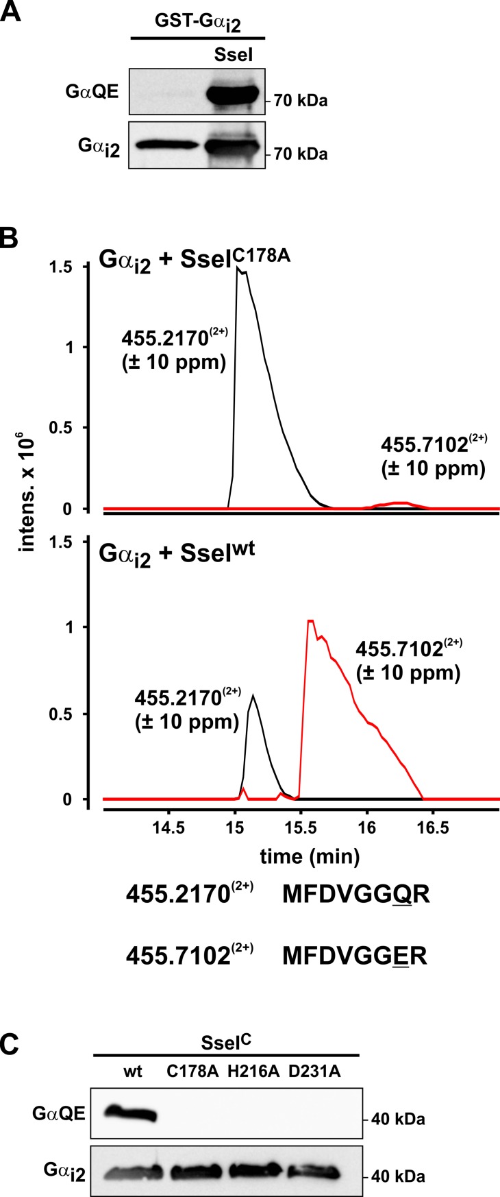
The comparison of the amino acid sequence of the deamidase domain of PMT (residues 1144–1240) with SseI revealed high sequence similarity (S1A Fig). More importantly, amino acids known to be crucial for deamidase activity of PMT are conserved in SseI (e.g., C178, H216, and D231) [19]. Therefore, we utilized a previously described method to detect deamidation of G proteins by bacterial effectors [23, 24]. To this end, SseI was coexpressed in E. coli with the α-subunit of Gi2. Deamidation of Gαi2 was determined by immunoblot analysis, utilizing a monoclonal antibody (GαQE) that specifically recognizes Gα after deamidation of a specific glutamine residue in the switch II region [25]. The GαQE antibody detected Gαi2 coexpressed with SseI in E. coli but not the solely expressed Gαi2, indicating a deamidation activity of SseI (Fig 1A). Purified Gαi2 was subjected to mass spectrometric (MS) analysis. MS analysis identified a tryptic peptide of Gαi2 (m/z 455.21702+) corresponding to amino acid residues 199–206. This peptide includes the glutamine residue (Gln-205) essential for the hydrolysis of GTP. Additionally, a second peptide was identified with a mass shift of 1 Da (m/z 455.71022+). Tandem MS analysis revealed that Gln-205 was deamidated, resulting in a glutamic acid residue at this position. No relevant deamidation occurred when Gαi2 was coexpressed with mutant SseI (SseI-C178A), which lacks deamidation activity (Fig 1B and S1B and S1C Fig). Similarly, recombinantly expressed wild type (wt) SseI (amino acids 137–322, SseIC), but not the C178A, H216A, or D231A mutants, caused deamidation of purified Gαi2 (Fig 1C).
Fig 1. SseI deamidates an essential glutamine residue in the switch II region of Gαi2.
(A) Western blot analysis of Gαi2. GST-Gαi2 was coexpressed without (control) and with SseI in E. coli. Purified GST-Gαi2 was immunoblotted and detected by the Gαi2-specific antibody (Gαi2) and by the deamidation-specific antibody (GαQE). (B) Gαi2 coexpressed with SseI was analyzed by HPLC-MS/MS spectrometry. Combined extracted ion chromatograms for m/z 455.2(2+) and 455.7(2+), corresponding to the tryptic peptides MFDVGGQR and MFDVGGER (amino acids 199–206) of Gαi2, are shown (see also S1B and S1C Fig). (Upper panel) Gαi2 coexpressed with inactive SseI-C178A. (Lower panel) Gαi2 coexpressed with active wt SseI. (C) Immunoblot analysis of recombinantly expressed Gαi2 incubated with wild type C-terminal part of SseIC (wt) or with 3 different mutant SseIC (C178A, H216A and D231A).
Construction of a PMT-SseI chimeric toxin
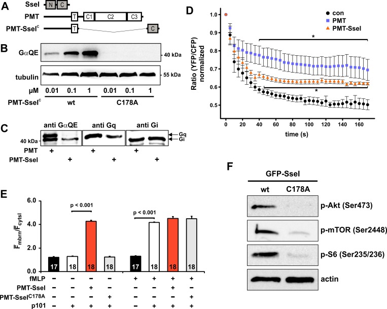
The effector SseI is secreted by a type III secretion system of S. Typhimurium. Therefore, SseI is not taken up by eukaryotic cells as compared to AB-type bacterial exotoxins like PMT. To enable cellular uptake of SseI, we utilized the receptor binding and translocation domain of PMT [26]. The deamidation domain of SseI (SseIC) was fused C-terminally to the N-terminal part of PMT (amino acids 1–505) (Fig 2A). This chimera, PMT-SseIC, was recombinantly expressed, purified and tested for cellular activity. Treatment of HEK-293 cells with increasing concentrations of PMT-SseIC led to a deamidation of Gα proteins as determined by immunoblot analysis using the GαQE antibody. Treatment of cells with the C178A mutant of PMT-SseIC exhibited no deamidation (Fig 2B). In a next step, we compared the activity of PMT-SseIC with that of PMT. HEK-293 cells were intoxicated either with PMT or with PMT-SseIC and cell lysates were tested for deamidated Gα proteins. PMT led to two deamidation signals migrating at the molecular mass of the Gαq and Gαi proteins (Fig 2C). However, PMT-SseIC only induced one deamidation signal migrating at the molecular mass of Gαi.
Fig 2. Cell permeable SseI activates Gαi-dependent signal transduction pathways.
(A) Schematic representation of the cell permeable PMT-SseIC chimera. The C-terminal domain of SseI, encompassing amino acids 137–322 (SseIC), was fused to the receptor binding and translocation domain of PMT (PMT, amino acids 1–505). (B) Immunoblot analyses of HEK-293 cells incubated with indicated concentrations of PMT-SseIC or PMT-SseIC-C178A for 16 h. RIPA buffer lysates were prepared and immunoblots were performed to detect Gα deamidation, using the GαQE antibody. Equal loading was verified by detection of tubulin. (C) Comparison of PMT- and PMT-SseIC-induced Gα deamidation. PMT treatment of cells (1 nM, 16 h) led to two signals of deamidated Gα proteins in immunoblot analysis, migrating at the same size of Gαi and Gαq. PMT-SseIC (100 nM, 16 h) induced one deamidation signal at the size of Gαi. (D) PMT-SseIC blocks stimulation of the adenylyl cyclase (AC) activity. HeLa cells were transfected overnight (16 h) with the FRET sensor construct EPAC2-camps. Cells were left untreated (con) or were incubated with PMT (1 nM) or PMT-SseIC (100 nM) for 4 h. AC was stimulated with forskolin (40 μM, added at time point 0) and FRET measurement was performed. cAMP increase is depicted as normalized ratio of YFP/CFP of the sensor. (E) PMT-SseIC stimulates the PI3Kγ. HEK-293 cells were transfected with the PIP3 sensor GFP-Grp1PH and the PI3-kinase subunit p110γ. In addition, HEK-293 cells were transfected with the non-catalytic PI3-kinase γ subunit p101 as indicated. Cells were stimulated with PMT-SseIC or the inactive C178A mutant of PMT-SseIC (each 100 nM). After baseline measurement for 1 min fMLP (1 μM) was added and the measurement was continued for 5 min. Histogram shows the quantification of the membrane translocation of GFP-Grp1PH. Data depicted represent the mean ±SEM from 3 independent sets of experiments analyzing 17 or 18 cells in total. (F) Immunoblot analysis of BMDMs transiently transfected with GFP-SseI or GFP-SseI-C178A. Cells were incubated for 24 h, followed by serum starvation for 4 h. Cells were lysed and subjected to Western blot analysis with indicated antibodies. Representative immunoblots from one experiment are shown. See also S2 Fig.
Activation of Gi proteins by the chimeric toxin PMT-SseIC
Deamidation of the essential glutamine residue in the switch II region impairs the GTP hydrolysis by the α-subunit resulting in a permanent active phenotype of the G protein [23]. Activated Gαi family members inhibit adenylyl cyclase leading to decreased cAMP levels. We utilized a FRET-based approach to determine how cAMP levels respond to SseI treatment. The FRET sensor Epac2-camps [27] exhibits a decreased FRET ratio, when cAMP is increased. When HeLa cells, expressing the FRET sensor, were treated with the adenylyl cyclase activator forskolin, the FRET ratio declined, indicating increased cAMP levels. In line with an activation of Gαi, pretreatment with PMT-SseIC or PMT attenuated the effect of forskolin, indicating an inhibitory effect on adenylyl cyclase (Fig 2D). Similar results were obtained by direct measurements of cAMP levels (see ELISA assay S2A Fig).
Activation of heterotrimeric G proteins leads to dissociation into Gα and Gβγ-subunits [28]. Both subunits can interact with their specific effectors and stimulate specific signaling cascades. Therefore, we studied the impact of SseI on phosphoinositol-3-kinase (PI3K)γ, a prototypical Gβγ effector [29–31]. Activation of Gβγ was monitored by translocation of the phosphatidylinositol-3, 4, 5-triphosphate (PIP3) sensor protein GFP-Grp1PH from the cytosol to the membrane after additional ectopic expression of p110γ without and with p101 (Fig 2E and S2B Fig). As a control, we used N-formylmethionine-leucyl-phenylalanine (fMLP), which stimulates Gi-coupled GPCRs (G protein-coupled receptors). fMLP induced translocation of Grp1PH to the membrane in the presence of complete PI3Kγ (p101 with p110γ) (S2Bb Fig). However, in the absence of complete PI3Kγ (p101 without p110γ), fMLP did not redistribute Grp1PH, indicating the insensitivity of endogenously expressed PI3Ks (S2Ba Fig). Treatment of transfected cells with PMT-SseIC, but not with the inactive mutant, strongly stimulated the redistribution of Grp1PH to the plasma membrane (Fig 2E and S2Bc/d Fig). This effect was not further stimulated by fMLP incubation, indicating an activation of the PI3K independent of the stimulation by GPCRs.
Activation of Akt signaling by SseI
Various recent studies suggested that Gαi/o proteins and PI3-kinases play crucial roles in regulation of immune cell signaling and cell survival in infection [32–36]. Therefore, we studied the downstream signaling of Gβγ subunits and of PI3Kγ in more detail. To this end, we transfected bone marrow-derived macrophages (BMDMs) or RAW264.7 macrophages with a GFP expression vector either harboring SseI or the catalytically inactive mutant (SseI-C178A). After starvation of cells for 4 h, phosphorylation of Akt, mTOR and the mTOR-effector S6 ribosomal protein was analyzed by immunoblotting. Transfection with SseI strongly increased phosphorylation of the PI3K downstream effectors Akt, mTOR, and S6 ribosomal protein in BMDMs (Fig 2F and S2C Fig). Similar results were obtained in RAW264.7 macrophages (S2D and S2E Fig).
Role of SseI in infection of macrophages by Salmonella Typhimurium
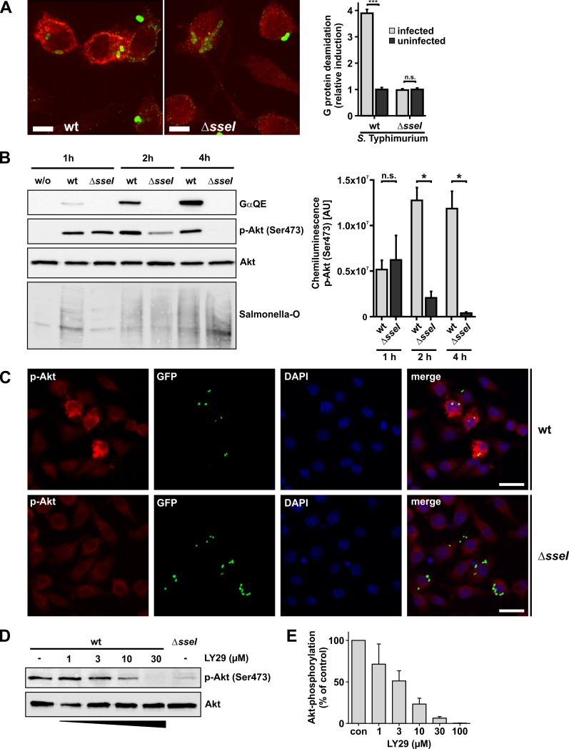
To study the role of SseI in infection of RAW264.7 macrophages and BMDMs, we employed wt- or ΔsseI-Salmonella Typhimurium strains and used immunofluorescence microscopy. As depicted in Fig 3A, S. Typhimurium were identified by Salmonella O-antigen antiserum inside and outside of macrophages. Notably, we observed deamidated G proteins, determined by the GαQE antibody, only in macrophages infected with wt-Salmonella. Moreover, deamidation occurred strictly after internalization of bacteria (Fig 3A, S3A and S3B Fig).
Fig 3. Cellular effects of SseI during infection.
(A) Fluorescence microscopy of fixed cells. RAW264.7 cells were infected with wild type (wt) S. Typhimurium or a ΔsseI-strain at a MOI of 1 for 5 h. Deamidation of Gα was detected utilizing the GαQE antibody and an Alexa 568-conjugated secondary antibody. Salmonella were identified by Salmonella O antiserum and an Alexa 488-conjugated secondary antibody. Deamidated Gα is depicted in red and Salmonella in green. Z planes showing Salmonella were maximum projected into one image. Scale bars = 5 μm. Quantification of images (right panel). Gα protein deamidation of Salmonella-infected or uninfected cells was calculated by determining the average intensity of Alexa 568 fluorescence of the whole cell area in the Z plane with internalized Salmonella. Significance was determined by two-tailed Student’s t-test. Data are means ±SEM. (n = 10 cells). (B-D) Serum-starved RAW264.7 cells were infected with a MOI of 30 for 30 min. (B) Time-resolved immunoblot analysis. At indicated times p.i., cells were lysed and processed for immunoblotting. Blotting membranes were treated with indicated antibodies (GαQE, p-Akt and Akt). Representative blots of 3 independent experiments are shown. Equal loading was verified by detection of Akt, presence of Salmonella was verified by Salmonella-O antigen staining. Right panel shows the quantification of p-Akt (Ser473) labeling over time after infection with wt- and ΔsseI-Salmonella strains from 3 independent experiments. Chemiluminescence intensity, given as area units [AU], was determined for each band. Statistical significance was assessed by Mann Whitney test. (C) Only macrophages infected with wild type (wt) Salmonella show strong phosphorylation of Akt. Cells were infected with pEGFP expressing wt- or ΔsseI-Salmonella Typhimurium for 30 min. 5 h p.i. cells were fixed and processed for immunofluorescence microscopy. Cells were stained for phospho-Akt Ser473 (red) and nuclei were stained with DAPI (blue). Scale bars = 20 μm. (D) Addition of the PI3K inhibitor LY29 leads to concentration dependent inhibition of Akt phosphorylation in wild type (wt) Salmonella-infected macrophages. 1.5 h p.i. cells were treated with the indicated concentrations of LY29 for 3.5 h. Thereafter, cells were lysed. Lysates were processed for immunoblotting with indicated antibodies (Akt, p-Akt(Ser473)). (E) Quantification of p-Akt (Ser473) immunoblots of infected cells treated with increased concentrations of LY29. Experiments were performed as depicted in (D). Intensity of labeled bands was normalized to untreated (con) cells. Values are means ±SEM from at least 3 independent experiments.
Next, we studied the activation of the PI3K/Akt pathway by SseI during infection of RAW264.7 macrophages with wt- or ΔsseI-Salmonella strains. Immunoblot analysis revealed G protein deamidation as early as ~2 h post infection (p.i.) with subsequent increase over time (Fig 3B). Infection with wt- and ΔsseI-Salmonella strains led to phosphorylation of Akt at 1 h p.i.. Hereafter, phosphorylation of Akt decreased over time in macrophages infected with ΔsseI-Salmonella. In wt-Salmonella-infected macrophages, phosphorylation of Akt remained longer than in the ΔsseI-Salmonella-infected cells (Fig 3B). Immunofluorescence microscopic studies revealed that 5 h p.i. an increase in Akt-phosphorylation was seen in wt-Salmonella-infected cells, but not in non-infected or ΔsseI-infected cells (Fig 3C and S3C Fig). In line with an essential role of PI3K, LY294002 (LY29), a specific PI3K inhibitor, decreased phosphorylation of Akt in a concentration-dependent manner (Fig 3D and 3E).
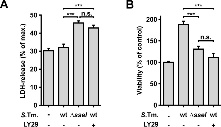
Several publications have shown that activation of Akt leads to anti-apoptotic and pro-survival outcomes in various mammalian cell types [33, 36]. Therefore, we were prompted to investigate, whether the infection with wt- or ΔsseI-Salmonella has an effect on the survival of host cells. To this end, we measured cell death of infected cells by determination of LDH release from infected RAW264.7 macrophages. This study revealed that infection with ΔsseI-Salmonella caused higher amounts of LDH release than with wt-Salmonella (Fig 4A). Accordingly, cell viability measured by the metabolic capacity of the cells, was increased after infection with wt-Salmonella as compared to ΔsseI-infected cells (Fig 4B). Inhibition of PI3K with LY29 diminished the effects of wt-Salmonella (Fig 4A and 4B).
Fig 4. Effects of SseI on host cell survival.
(A and B) RAW264.7 macrophages were infected with wild type (wt) or ΔsseI-Salmonella (MOI = 30; 30 min). 1.5 h p.i., cells were starved for 3.5 h in Earle’s balanced salt solution. The PI3K inhibitor LY29 (30 μM) was added 1.5 h p.i. where indicated. (A) The amount of LDH released into the medium was measured fluorometrically 5 h p.i.. ΔsseI-infected macrophages showed higher amounts of released LDH compared to wt-Salmonella infected cells. Data show means ±SEM from 3 independent experiments. Maximum LDH-release was obtained in the presence of 1% Triton X-100. Statistical significance was assessed using ANOVA (Bonferroni post-test). (B) Viability of RAW264.7 macrophages was fluorometrically measured 5 h p.i. by the ability of cells to reduce Resorufin. Experiments were performed in triplicates. Values are means ±SEM from at least 3 independent experiments. Statistical significance was assessed using ANOVA (Bonferroni post-test).
Effect of SseI on migration of dendritic cells
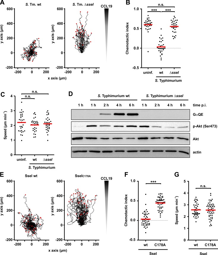
Recent studies from the Monack laboratory showed that SseI inhibits directed cell migration [10, 13]. Therefore, we were prompted to study the role of G proteins in the action of SseI in more detail. To this end, we investigated the migration of dendritic cells in a 3D collagen model. We infected bone-marrow derived mature dendritic cells (DCs) with wt- or ΔsseI-Salmonella, seeded the cells into collagen gels and applied a CCL19 chemokine gradient. ΔsseI-infected cells migrated with comparable speed and directness towards the chemokine compared to non-infected cells. In contrast, wt-infected DCs completely lost their ability to migrate in a directed manner, confirming previous results [10, 13] (Fig 5A–5C; S1 Video). As found for macrophages, time-resolved Western blot analysis revealed deamidation of Gα-subunits by wt-Salmonella in DCs (Fig 5D). Moreover, very similar as observed in macrophages, downstream signaling of the PI3K pathway was elevated in wt-Salmonella-infected DCs, exhibiting increase in Akt phosphorylation (Fig 5D).
Fig 5. Influence of SseI on directed migration of dendritic cells.
(A-C) DCs were infected with GFP-expressing wild type (wt)- or ΔsseI-Salmonella (MOI = 30; 30 min). Then, cells were transferred into a collagen matrix. (A) A CCL19 gradient was applied and directed migration for 4 h was determined via time-lapse microscopy. Red dots mark the end of the migration tracks. Every track starts at x = 0 / y = 0. (B) Tracks of n = 30 randomly selected DCs for each condition from 3 independent experiments were quantified and the chemotactic index was determined with Chemotaxis and Migration tool V2.0 (Ibidi). (C) Similarly, the speed of migration was quantified. Statistical significance was assessed using ANOVA with Bonferroni post-test. (D) DCs infected with wt- or ΔsseI-Salmonella (MOI = 30; 30 min) were lysed at the indicated times and immunoblot analysis was performed with indicated antibodies (GαQE, p-Akt, Akt and actin). (E-G) Migration of DCs after ectopical expression of wild type (wt) SseI or SseI-C178A (C178A). Cells were transferred into a collagen matrix, a CCL19 gradient was applied, and directed migration (E) was determined by time-lapse microscopy (wt SseI, n = 43; SseI-C178A, n = 49). Cells were randomly selected from 3 independent experiments. (F, G) Quantification of the chemotactic index (F) and speed (G) of DCs. Statistical significance was assessed using unpaired, two-tailed Students t-test.
To investigate whether the inhibition of migration observed with wt-Salmonella was due to the enzymatic activity of SseI, we ectopically expressed wt SseI or the inactive C178A-SseI mutant in DCs. As shown in Fig 5E and 5F (see also S2 Video), transfection with wt sseI led to a similar loss of chemotaxis as compared to infection with wt-Salmonella, while cells transfected with SseI-C178A mutant migrated normally. Interestingly, we did not detect a difference in the speed of migration between wt- and inactive sseI-transfected cells (Fig 5G).
Persistent Gαi activation inhibits directed migration of dendritic cells
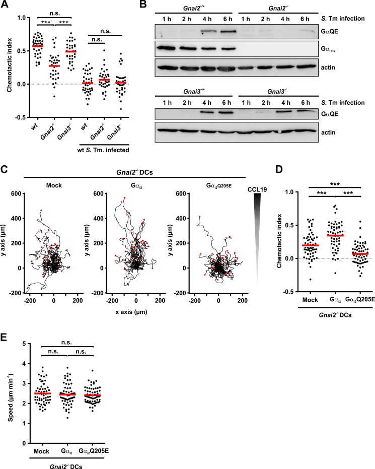
To gain further evidence, whether deamidation of Gαi-subunits is the cause for impaired chemotaxis, we investigated the directed migration of DCs derived from Gnai deleted mice. The 3 Gαi isoforms (Gαi1,2,3) are encoded by Gnai1, 2 and 3. Relevant for DCs are Gαi2,3 (see Discussion section). Gαi1 is only poorly or not at all expressed in dendritic cells [37] (http://www.immgen.org/databrowser/index.html). We purified Gαi2 and Gαi3 as recombinant proteins and performed in vitro deamidation assays. Both isoforms of the Gαi-subunit are deamidated in a comparable manner (S4A Fig). Because double Gnai2-/- / Gnai3-/- mice are not viable [38], we obtained DCs from Gnai2-/- or Gnai3-/- C57BL/6 mice. In uninfected cells (controls), depletion of Gαi2 (Gnai2-/-) impaired chemotaxis as compared to wild type DCs, while Gαi3 depletion (Gnai3-/-) had no major effect on migration (Fig 6A). Thus, as reported for macrophages [39], our data indicated a crucial role of Gαi2 but not of Gαi3 in directed migration of DCs. Infection with wt-Salmonella impaired chemotaxis of control, Gαi2- and Gαi3-depleted DCs (Fig 6A), while migration speed was not significantly altered (S4B Fig). As expected, immunoblot analysis with anti-Gαi1,2 antibody revealed deletion of Gαi2 in Gnai2-/- DCs (Fig 6B). While infection of control Gnai2+/+ cells with wt-Salmonella resulted in strong labeling of deamidated G proteins after 6 h, almost no labeling was observed in Gnai2-/- DCs (Fig 6B). In Gnai3-/- DCs deamidation was observed in a similar intensity as in Gnai3+/+ cells (Fig 6B). This indicates that Gαi2 is the predominant SseI substrate in mouse DCs. To mimic SseI-induced deamidation, we expressed mutant Gαi2Q205E or wt Gαi2 in Gnai2-/- DCs. Expression was monitored by immunoblotting (S4C Fig). Ectopic expression of wt Gαi2 partially rescued the impaired chemotaxis of Gnai2-/- DCs, while expression of mutant Gαi2Q205E strongly inhibited directed migration of DCs (Fig 6C and 6D, and S3 Video). Again, the migration speed was not changed by expression of Gαi2, or Gαi2Q205E (Fig 6E).
Fig 6. Effects of G protein deamidation on chemotaxis.
(A) Wild type (wt)-, Gnai2-/-—and Gnai3-/-—DCs were infected with wt-Salmonella (wt S. Tm) (MOI = 30; 30 min) and the chemotactic indices were determined from at least n = 34 cells for each condition from 3 independent experiments as in Fig 5. Statistical significance was assessed using ANOVA and Bonferroni post-test. (B) Time-resolved immunoblot analysis of DCs derived from Gnai2-/—(upper panel), Gnai3-/—(lower panel) mice or wild type littermates (Gnai2+/+, upper panel; Gnai3+/+, lower panel) after infection with wild type Salmonella (S. Tm infection) (MOI = 30, 30 min) for indicated times. Proteins were analyzed by specific antibodies (GαQE, Gαi1+2 and actin). (C-E) Gnai2-/- DCs were transfected with Gαi2, Gαi2Q205E or empty vector. Chemotaxis in a collagen matrix (C) with CCL19 gradient was measured by cell tracking using time-lapse microscopy. Chemotactic index (D) and migration speed (E) were calculated as described in Fig 5. Statistical significance was assessed using ANOVA (Bonferroni post-test).
Discussion
Our studies elucidate the molecular mechanism of the S. Typhimurium SPI-2 T3SS effector SseI. This effector protein plays a crucial role in S. Typhimurium infection after invasion of the pathogen and modulates the immune responses of the host [9]. Here, we show that SseI deamidates a specific glutamine residue (Gln205 in Gαi2) in the α-subunits of heterotrimeric G proteins, which is involved in GTP hydrolysis, thereby the G protein is persistently activated. Thus, T3SS effector SseI exhibits the same mode of action as exotoxin PMT from Pasteurella multocida. This finding is in line with the structural similarity of both toxins, although the primary sequence identity of the catalytic domains is only ~ 20% [16]. Exchange of the conserved catalytic amino acid residues C178, H216 and D231 inhibited the deamidase activity of SseI. Because SseI is per se not able to enter target cells, we constructed a chimeric toxin (PMT-SseIC), consisting of the N-terminal binding and translocation domain of PMT and the deamidating domain of SseI. Additionally, we transfected mammalian cells with a GFP-SseI construct. These experiments confirmed our in vitro results in intact cells, showing that SseI acts as a deamidase on heterotrimeric G proteins.
While PMT deamidates several G proteins, including Gi/o, Gq/11 and G12/13 [23, 24], our data indicate that in intact cells, SseI is specific for Gi proteins. In line with activation of Gi, SseI decreased cAMP levels in cells. Besides inhibition of adenylyl cyclase, Gi is crucially involved in activation of PI3 kinase via release of Gβγ subunits [29, 40]. Accordingly, we observed the translocation of a PIP3 sensor protein (GFP-GrpPH) to the cell membrane after treatment with PMT-SseIC. Many reports describe increase in Akt/mTOR signaling in Salmonella-host interactions [33, 36, 41]. We found that SseI increased phosphorylation and activation of Akt, its target mTOR and S6 ribosomal protein. These effects depended on the active SseI protein. The inactive mutant SseI-C178A had no effect.
To study the role of SseI in infection, we employed S. Typhimurium, in which SseI was deleted. These studies confirmed that after infection of macrophages with wt-S. Typhimurium, Gi proteins were deamidated, while this was not observed with ΔsseI-Salmonella. Importantly, deamidation of G proteins was detected only after uptake of Salmonella into host cells, indicating the importance of invasion for activation of SPI-2 T3SS and translocation of the effector SseI. wt-Salmonella strongly activated the pro-survival kinase Akt in macrophages and DCs. Notably, also infection with ΔsseI-Salmonella resulted in Akt phosphorylation. However, the phosphorylation of Akt, observed in the absence of SseI, diminished with time, whereas wt-Salmonella caused longer lasting Akt phosphorylation. Short-term Akt activation by ΔsseI-Salmonella is probably caused by Salmonella outer protein B (SopB), which is an effector of SPI-1 T3SS and a strong activator of Akt [42, 43]. Activation of Akt by SopB differs from SseI and might be independent of class I PI3 kinase [44, 45]. In contrast, we observed that activation of Akt by SseI is inhibited by the class I PI3K inhibitor LY29. In line with the activation of the PI3K-Akt signaling pathway, we observed increased survival of host cells after infection with wt-Salmonella, as compared to ΔsseI-infection.
Previously, it was reported that SseI inhibits the directed motility of macrophages and DCs towards a chemokine gradient [13]. Using an infection model, we confirmed that wt-Salmonella but not ΔsseI-Salmonella inhibited directed migration of DCs. Until recently, the molecular mechanism by which SseI affects directed migration was largely enigmatic. Our data indicate that the SseI-catalyzed deamidation of Gi proteins is responsible for this effect. Concomitantly with inhibition of directed migration, infection with wt-Salmonella but not with ΔsseI-Salmonella caused deamidation of Gi in DCs.
It is widely accepted that Gi proteins play a pivotal role in chemotaxis and most, if not all, chemokine receptors are coupled to Gi proteins [37, 46]. Three Gi proteins (Gi1-3) exist, which share 85–95% amino acid sequence identity in their Gα subunits. While Gi1 is only poorly expressed in leukocytes, Gi2 and Gi3 are highly expressed [37] (http://www.immgen.org/databrowser/index.html). Especially, Gi2 appears to be essential for immune cell signaling. Several studies showed that Gi2 plays an important role in directed migration and homing of B and T-cells [47–50] and in macrophage migration [39]. Similarly, we show that deletion of Gαi2 inhibits the migration of DCs towards a CCL19 gradient. In line with recent publications, deletion of Gαi3 had minor effects on directed DC migration [39, 51]. However, it is notable that wt-Salmonella infection was still able to inhibit directed migration even in Gnai2-/- DCs, possibly indicating that Gαi3 is able to substitute in part for Gαi2. When wild type Gαi2 was re-expressed in Gnai2-/- DCs, directed migration was partially rescued. Importantly, expression of the mutant Gαi2-Q205E that mimics the action of SseI, inhibited DC migration. Moreover, our studies reveal that SseI-induced deamidation of Gi proteins inhibits directed migration but does not affect the migration speed. Also pertussis toxin was shown to inhibit directed migration of DCs without affecting migration speed [52]. However, in contrast to SseI, pertussis toxin inactivates Gi proteins by blocking the interaction with GPCRs.
Thus, how might SseI, which activates Gi proteins, inhibit directed cell migration? McLaughlin and coworkers demonstrated that SseI binds to the scaffold protein IQGAP1 (Ras GTPase-activating-like protein) [13], which interacts with numerous proteins (e.g. Rho proteins) involved in regulation of actin dynamics [53]. Because IQGAP1 also binds to the inactive SseI mutant, this cannot explain the action of SseI [13]. Earlier studies, which were mainly based on two-hybrid screen analyses, found interactions of SseI with the actin-binding protein filamin and the LIM domain fragment of TRIP6 [54, 55]. However, these studies did not consider an enzyme activity of SseI. We propose that inhibition of directed migration by SseI is caused by blockade of the GTPase cycle and the non-polarized persistent activation of Gi. SseI blocks rapid deactivation and activation of Gi, which is probably essential to follow a chemoattractant gradient. In line with this hypothesis are recent findings about RGS (regulators of G protein signaling) proteins in immune cells. RGS proteins decrease the activity of G proteins by accelerating GTP hydrolysis and, thereby, accelerate G protein inactivation but also its activation [56–58], which appears to be essential for chemotaxis [8, 37]. It was shown that immune cells, harboring the Gαi2 G184S mutant, which causes resistance to RGS proteins, exhibit major alteration in chemotaxis [8]. The active state of the G184S Gαi2 mutant is prolonged, a condition mimicking the effects of SseI that persistently activates Gi proteins. Thus, a similar scenario as seen with the G184S Gαi2 mutant but even stronger, might be induced by SseI, resulting in insufficient downstream signaling from GPCRs-involved in chemotaxis and loss of polarization necessary for directed migration. In line with this hypothesis are also previous findings obtained with constitutively activated PI3Kγ, which was fused with the CAAX box of K-Ras resulting in persistent cell membrane localization and non-polarized enhancement of PIP3 production [59]. Leukocytes with constitutively activated PI3Kγ lost cell polarity and failed to follow a chemotactic gradient but they did not show differences in mean migration velocity. Surve and coworkers reported that selective activation of Gβγ by compound 12155 causes directed migration of neutrophils [60]. This effect was inhibited by Wortmannin, underlining the essential involvement of PI3K. PI3K is able to activate Rac, which is crucially involved in polarity control during neutrophil chemotaxis [61]. A recent study performed with optogenetic activation of PI3K sheds some light on the complexity of signaling pathways involved in directed migration downstream of PIP3 [62]. The authors showed that PI3K activates Rac mainly via the guanine nucleotide exchange factor P-Rex-1. However, Rac activation by chemoattractants (involving GPCRs and not only PI3K) depended on multiple Rac regulators indicating involvement of a signaling network.
Taken together our data elucidate the molecular mode of action of the SPI-2 effector SseI by deamidation of a crucial glutamine residue in the α-subunits of Gi proteins of the host, which results in inhibition of the turn-off mechanism of the G protein. The finding that Gi proteins are persistently activated by SseI will offer a new perspective in studies on the biological effects of this SPI-2 T3SS effector and may help to identify further signal factors involved in chemotaxis, acting downstream of Gi proteins.
Materials and methods
Materials
PCR primers were from Apara (Denzlingen, Germany). All other reagents were of analytical grade and purchased from commercial sources. All Antibodies used in this study are listed in S1 Table.
Bacterial strains
S. enterica serovar Typhimurium NCTC12023 (ATCC 14028) was used as parent strain for the ΔsseI strain, which was created by deletion of the SseI coding sequence [63].
Mouse primary cell cultures
Murine BMDMs and DCs were isolated and cultivated as described before [64, 65] with adjustments. 6–12 weeks old wt (Charles River), Gnai2-/-, and Gnai3-/- (Dr. B. Nürnberg, maintained at animal facility, Medizinische Fakultät und ICePhA, University of Tübingen) mice (C57BL/6N, female, [66]) were euthanized and femurs and tibiae were extracted. Bones were cut open and bone marrows were flushed with sterile, ice-cold PBS. Cells and matrix were resuspended and pipetted through a 70 μm cell strainer. Cell suspension was centrifuged (300 x g, 5 min). Supernatants were discarded, pellets were resuspended in RPMI-1640 with 10% fetal calf serum (FCS) and 1% penicillin/streptomycin (P/S).
Dendritic cells: Cell concentration was adjusted to 2.5 x 10⁵ cells/ml and 10 ml suspension was added into 10 cm bacterial petri dishes. GM-CSF was added for a final concentration of 20 ng/ml. At day 3, 10 ml medium with 20 ng/ml GM-CSF was added. At day 6, 10 ml medium were carefully removed and 10 ml fresh medium with 20 ng/ml GM-CSF were added. At day 8–9, non-adherent cells were collected and centrifuged (300 x g, 5 min). Pellets were resuspended in 10 ml fresh medium with 20 ng/ml GM-CSF without P/S and transferred into 6 cm cell culture dishes. LPS (200 ng/ml) were added for 24 h for further maturation of the DCs, when needed. Only non-adherent cells were used for experiments.
Bone marrow-derived macrophages: Cell concentration was adjusted to 5–7 x 10⁵ cells/ml and 10 ml suspension was added into 10 cm tissue culture dishes. M-CSF was added for a final concentration of 20 ng/ml. At day 2, 10 ml fresh medium with 20 ng/ml M-CSF were added. At day 4, 10 ml medium were removed and exchanged by 10 ml fresh medium with 20 ng/ml M-CSF. At day 6–8, medium and non-adherent cells were discarded, adherent cells were detached with trypsin/EDTA and plated in medium with 20 ng/ml M-CSF without P/S at desired concentrations for experiments.
Cell lines
HEK-293 (female; ACC 305 from DSMZ, Germany) and HeLa (female, ATCC CCL-2) cells were cultured in Dulbecco's modified Eagle's medium (DMEM) supplemented with 10% FCS and 1% P/S in an atmosphere of 5% CO2 at 37°C. RAW264.7 cells (male, ATCC TIB-71) were cultured in DMEM supplemented with 5% FCS and 1% P/S.
Plasmids
SseI encoding DNA was amplified from S. enterica serovar Typhimurium NCTC12023 and cloned into the bacterial expression vector pCOLDII and mammalian expression vectors pEGFP-C1 and pcDNA3.1 by standard cloning techniques. Additionally, a C-terminal fragment of SseI harboring the homology domain to PMT-C3, starting with amino acid 137 was cloned into pCOLDII. Chimeric PMT-SseIC consists of the N-terminal portion (amino acid 1–505) of PMT and the PMT-C3 homology domain of SseI (amino acids 137–322). PMT-SseIC was cloned in accordance to the method described before for other PMT fusion proteins into the pGEX2T vector [26]. The biological inactive mutant of SseI (C178A) was constructed by site-directed mutagenesis. Gαi2 DNA (with optimized codon usage for prokaryotic expression) was ordered as gBlock (Integrated DNA Technology, IDT) and cloned into pCOLDII. All oligonucleotides used in this study are listed in S2 Table.
Transfection, infection
HeLa cells were transfected using PEI as described previously [67]. Transfection of RAW264.7 cells was performed utilizing lipofectamin2000 (Thermo Fisher Scientific) according to the manufacturer’s instructions. Transfection of BMDMs and BMDCs was done using amaxa nucleofector kits (Lonza) according to manufacturer’s instructions. For in vitro infection studies S. enterica serovar Typhimurium NCTC12023 and the isogenic variant MvP393ΔsseI were used. BMDMs or RAW 264.7 cells were infected with Salmonella with indicated MOIs for 30 min. Then cells were washed 3 times with warm PBS and incubated with DMEM (RAW264.7) or RPMI-1640 (BMDMs) supplemented with 100 μg/ml gentamicin for 60 min. For the remaining time of the experiment, the medium was changed to DMEM or RPMI supplemented with 10 μg/ml gentamicin. For immunofluorescence detection of deamidation in RAW264.7 macrophages, these cells were infected with Salmonella for 5 h (MOI = 1). Dendritic cells were infected with a MOI = 30 for 30 min. Due to non-adherence of stimulated DCs, cells were resuspended in infection-medium and centrifuged (300 x g / 5 min). Pellets were resuspended in RPMI-1640 containing 100 μg/ml gentamicin and further treatment of the cells was similar to treatment of BMDMs.
For lysates cells were treated with RIPA buffer (1 mM EDTA, 25 mM Tris, 150 mM NaCl, 1% (v/v) Triton X-100, and 1% (m/v) sodium deoxycholate, pH 7.4), containing complete protease inhibitor (Roche) and phosphatase inhibitor (phosphatase inhibitor cocktail 2/3, Sigma) for 20 min on ice with occasional vortexing. Lysates were then centrifuged at 4°C (10,000 rpm for 10 min).
FRET measurements
cAMP was measured by fluorescence resonance energy transfer (FRET) using a single chain cAMP sensor as described before [27]. The construct EPAC2-camps was kindly provided by Dr. Viacheslav O. Nikolaev (University of Göttingen, Germany).
cAMP measurements
HEK-293 cells were incubated with wildtype PMT-SseI or inactive mutant (PMT-SseI-C178A) for 8 h. Then forskolin (10 μM) and 3-isobutyl-1-methylxanthin (IBMX 100 μM) were added to the medium to induce adenylyl-cyclase activity and cells were incubated for further 45 min. Medium was discarded and cells were lysed with lysis buffer from the cAMP Parameter Kit (Biotechne). Further treatment of the samples was performed according to the manufacturer’s instructions (cAMP Parameter Kit/R&D Systems Biotechne).
Confocal Laser Scanning Microscopy of GFP-Grp1PH translocation
The subcellular distribution of GFP-Grp1PH reflects the activity of PI3Ks, i.e. enhancement of membrane-associated fluorescence was taken as an indicator of PI3K activation. Assay was performed as previously described [31].
Fluorescence microscopy
For fluorescence microscopy, cells were prepared as described before [26]. Fixed samples were analyzed with an inverted Axiovert 200 M microscope (Carl Zeiss) with a Yokogawa (Tokyo, Japan) CSU-X1 spinning disc equipped with an emission filter wheel and 405 nm / 488 nm / 561 nm laser lines.
Protein expression
SseI and Gαi2(His) were expressed as N-terminal His6-tagged proteins and purified by affinity chromatography via a Ni-NTA column. Expressions of PMT or chimeric constructs of PMT and SseI were performed as glutathione-S-transferase fusion proteins in accordance with the method previously described [67]. Coexpression of SseI or the inactive mutant (SseI-C178A) with GST-Gαi2 was done as previously described [23]. G proteins used for the in vitro deamidation assay in S4A Fig were purified as GST-fusion proteins. The GST-tag was cleaved with Thrombin.
In vitro deamidation assay
Purified Gαi2 or Gαi3 (1 μM) was preincubated in deamidase buffer (50 mM sodium HEPES (pH 7.5), 50 mM NaCl, 1 mM EDTA, 5 mM MgCl2 and 0.1% NP-40) together with Guanosine diphosphate (GDP, 5 μM) for 15 min at 30°C. SseI-C deamidase domain (100 nM) was added and reaction was performed at 16°C for 16 h. Reaction was stopped by the addition of 5x Lämmli buffer and shock freezing in liquid N2, followed by immunoblot analysis using the deamidation specific GαQE antibody.
Immunoblot analysis
For immunoblotting, samples were subjected to SDS-PAGE and transferred onto polyvinylidene difluoride-membrane. Deamidation specific antibody anti-Gαq Q209E (3G3) was used as described before [25]. Binding of the appropriate horseradish peroxidase-coupled secondary antibody was detected by using enhanced chemiluminescent detection reagent (New England Biolabs) and the imaging system LAS-3000 (Fujifilm).
Mass spectrometric analysis
Gel bands were destained for 10 min using 30% acetonitrile / 70% 0.1 M NH4HCO3, followed by 10 min washing with 0.1 M NH4HCO3; this procedure was repeated twice. Prior to drying in a Vacuum Concentrator (Concentrator 5301, Eppendorf), gel bands were shrunk with 100% acetonitrile. By applying 0.1 μg trypsin per gel band, proteolytic digest was performed overnight at 37°C in 0.1 M NH4HCO3. Peptides were extracted from the gel matrix with 5% formic acid. Peptides were analyzed by LC/MS on a Q-TOF mass spectrometer (Agilent 6520, Agilent Technologies) coupled to a 1200 Agilent nanoflow system via a HPLC-Chip cube electrospray ionization interface carrying a ProtID-II-Chip (Agilent Technologies). For peptide separation, a linear gradient ranging from 3% acetonitrile (ACN) to 30% ACN running for 30 min at a flow rate of 300 nl/min was used. MS spectra were acquired from m/z 50 to m/z 3000 while operating the Q-TOF in the 2 GHz extended dynamic range mode. Internal mass calibration was enabled. Three multiple charged peptides were selected in a data-dependent manner from each survey MS-scan for acquisition of fragment mass spectra. For peak list generation, raw data files were processed using Mascot Daemon2.4.0 with default settings for the Agilent Q-TOF. Using Mascot Server 2.4.1 database searches were performed with tryptic specificity and two missed cleavages against a small database comprised of known contaminants and the target sequence. Mass tolerances of 50 ppm and 0.05 Da were allowed for peptide and fragment masses, respectively. As variable modifications deamidation of glutamine and phosphorylation of serine and threonine were selected in addition to Carbamidomethyl (C), Gln-> pyroGlu (N-term. Q) and oxidation (M). Extracted Ion Chromatograms were generated using the Qualitative Analysis module within the MassHunter B04.00 software.
LDH-release/cell viability
RAW264.7 macrophages were infected with a MOI of 30. 1.5 h p.i. medium was exchanged by Earle’s balanced salt solution (EBSS) containing 10 μg/ml gentamicin and LY29 (30 μM), where indicated. Further treatment of the cells was performed according to the manufacturer’s instructions (Promega CytoTox-ONE/CellTiter-Blue). In-plate fluorescence was acquired with the Tecan Infinite M200 plate reader.
3D dendritic cell migration
Chemotactic migration assays of dendritic cells were performed as described before [64]. Briefly, pre-treated dendritic cells were mixed with bovine collagen (1.5 mg/ml) and incubated in self-built migration chambers at 37°C, 5% CO2 until the collagen network was polymerized. The collagen-cell mixture was covered with medium, containing CCL19 (500 ng/ml), which diffuses into the gel matrix and thus forming the chemokine gradient. Cell migration was monitored with a Lionheart FX automated microscope. Cell tracks were analyzed with ImageJ and the Manual Tracking plugin, followed by evaluation of the data with Ibidi chemotaxis and migration tool. Calculation of the chemotactic indices:
Statistics
Results are presented as means ±SEM, unless otherwise stated. Statistical analyses were performed using GraphPad PRISM Version 5.04. Significance was assessed by Student`s t-test and Mann-Whitney test, depending on whether the data was normally distributed. p values < 0.05 were considered statistically significant (* = p < 0.05; ** = p < 0.01;*** = p < 0,001; ns, not significant). Multiple group comparisons were analyzed by ANOVA and Bonferroni post-tests.
Ethics statement
All animal experiments were performed in compliance with the German animal protection law (TierSchG). The animals were housed and handled in accordance with good animal practice as defined by FELASA (www.felasa.eu) and the national animal welfare body GV-SOLAS (www.gv-solas.de). The animal welfare committees of the University of Freiburg as well as the local authorities (Regierungspräsidium Freiburg, licenses X-13/03A and X-17/01F) approved all animal experiments.
Supporting information
(A) Alignment of the deamidase domain of PMT with SseI (UniProt accession no.: PMT, P17452; SseI, Q8ZQ79). Alignment was performed with ClustalO (Sievers et al., 2011). Catalytic triad of PMT deamidase domain is highlighted by blue boxes. (B, C) Electron-transfer dissociation (ETD) tandem MS/MS spectrum of (B) m/z 455.2170(2+) and of (C) m/z 455.7102(2+) showing a shift of 1 Dalton from y2 upwards, indicating a deamidation of glutamine-205 to glutamic acid.
(TIF)
(A) PMT-SseIC blocks forskolin-induced cAMP accumulation. HEK-293 cells were incubated with 100 nM PMT-SseIC or the inactive mutant (C178A) for 8 h. Cells were then incubated with forskolin (10 μM) and IBMX (100 μM) for 45 min. Cells were lysed and cAMP levels determined by cAMP Parameter Assay (Biotechne). Shown are data as means ±SEM from 4 independent experiments. Significance was assessed by Student`s t-test. (B) PI3Kγ activation by PMT-SseIC. All HEK-293 cells were transfected with the PI3K subunit p110γ, transfection with the PI3K subunit p101 was as indicated. Shown are confocal images of representative cells before (-fMLP) and 4 min after stimulation with fMLP (+fMLP). (panel a) Transfection without p101. (panels b-d) transfection with p101. Incubation of cells with PMT-SseIC (panel c) or the inactive C178A mutant of PMT-SseIC (PMT-SseICC178A) (panel d, each 100 nM). The lower panels show the fluorescence line scan through cells representing the membrane translocation of the PIP3 sensor GFP-Grp1PH upon treatment without and with PMT-SseIC, inactive PMT-SseICC178A and fMLP (gray curve, before fMLP; black curve, after fMLP). (C) Densitometric quantification and statistical analysis of n = 3 immunoblots from cell lysates treated as described in Fig 2F. Statistical significance was assessed using one sample t-test, with *p<0.05, **p<0.01. (D) Influence of GFP-SseI transfection of RAW264.7 cells on phosphorylation of Akt (p-Akt) and S6 ribosomal protein (p-S6). p-Akt, p-S6 and actin were detected by specific antibodies after transfection of cells with wt GFP-SseI or inactive mutant GFP-SseI-C178A. (E) Quantification of the amount of p-S6 from S2D Fig. Phosphorylation of p-S6 after transfection with GFP-SseI was set 100, phosphorylation after GFP-SseI-C178A transfection is given as percent of maximum. Shown are means ±SEM of 3 independent experiments.
(TIF)
(A) Fluorescence microscopy of fixed BMDMs infected with wild type (wt-) or ΔsseI-S. Typhimurium. Macrophages were infected for 60 min with a MOI of 15 and washed. Cells were fixed and stained for deamidation (GαQE—green) and Salmonella O antigen (red) 5 h p.i.. Orthogonal views, cutting the z-stacks, show intracellular localization of the Salmonella. (B) Quantification of fluorescence intensity of deamidation of n = 30 cells without and with infection by wild type (wt)- or ΔsseI-S. Typhimurium from 3 independent experiments. Cells were treated as described in (A). Gα protein deamidation of Salmonella-infected or uninfected cells was calculated by determining the average intensity of Alexa 568 fluorescence of the whole cell area in the Z plane with internalized Salmonella. Means ±SD are shown. Significance was assessed by ANOVA. (C) Quantification of p-Akt immunofluorescence. RAW264.7 cells were infected with wt- or ΔsseI-S. Typhimurium or remained non-infected as shown in Fig 3C. Fluorescence intensity of n = 45 cells for each condition from three independent experiments was measured. Statistical significance was assessed using ANOVA.
(TIF)
(A) In vitro deamidation of G protein isoforms Gαi2 and Gαi3. Immunoblot analysis of the recombinantly expressed G proteins incubated with wild type C-terminal part of SseIC (wt) or mutant SseIC (C178A). (B) Quantification of the migratory speed of DCs obtained from wild type (wt)-, Gnai2-/- or Gnai3-/- mice. Cells were infected with wild type (wt S. Tm.) S. Typhimurium (MOI = 30, 30 min) and, thereafter, transferred into migration chambers. Infected and non-infected DCs from each condition (n≥34 from 3 independent experiments) were tracked and migratory speed was calculated. (C) Immunoblot analysis of transfected cells with indicated antibodies (GαQE, Gαi1+2 and actin). DCs were transfected with pcDNA constructs, encoding for wild type Gαi2 or mutant Gαi2Q205E. Mock indicates empty vector transfection. Lysates were prepared 16 h post transfection. Representative blots from n = 3 experiments are shown.
Statistical significance was assessed using ANOVA.
(TIF)
Arrows indicate 2 examples of infected migrating cells.
(AVI)
Tracks of migrating cells are shown.
(AVI)
Tracks of migrating cells are shown.
(AVI)
(XLSX)
(XLSX)
Acknowledgments
The excellent technical assistance by Silke Ludigkeit and Petra Bartholome is acknowledged. We thank Dr. Y. Horiguchi (Osaka University, Japan) for deamidation specific antibody.
Data Availability
All relevant data are within the paper and its Supporting Information files.
Funding Statement
The study was financially supported by the Deutsche Forschungsgemeinschaft Collaborative Research Centre 746 (project P17 to KA; http://www.dfg.de), the University of Freiburg, Faculty of Medicine (ORT924/13) and by BIOSS Centre for Biological Signalling Studies to KA (http://www.bioss.uni-freiburg.de). The funders had no role in study design, data collection and analysis, decision to publish, or preparation of the manuscript.
References
- 1.Feasey NA, Dougan G, Kingsley RA, Heyderman RS, Gordon MA. Invasive non-typhoidal salmonella disease: an emerging and neglected tropical disease in Africa. Lancet. 2012;379(9835):2489–99. 10.1016/S0140-6736(11)61752-2 [DOI] [PMC free article] [PubMed] [Google Scholar]
- 2.Haraga A, Ohlson MB, Miller SI. Salmonellae interplay with host cells. Nat Rev Microbiol. 2008;6(1):53–66. 10.1038/nrmicro1788 [DOI] [PubMed] [Google Scholar]
- 3.Keestra-Gounder AM, Tsolis RM, Baumler AJ. Now you see me, now you don't: the interaction of Salmonella with innate immune receptors. Nat Rev Microbiol. 2015;13(4):206–16. 10.1038/nrmicro3428 [DOI] [PubMed] [Google Scholar]
- 4.Kingsley RA, Msefula CL, Thomson NR, Kariuki S, Holt KE, Gordon MA, et al. Epidemic multiple drug resistant Salmonella Typhimurium causing invasive disease in sub-Saharan Africa have a distinct genotype. Genome Res. 2009;19(12):2279–87. 10.1101/gr.091017.109 [DOI] [PMC free article] [PubMed] [Google Scholar]
- 5.Santos RL, Baumler AJ. Cell tropism of Salmonella enterica. Int J Med Microbiol. 2004;294(4):225–33. 10.1016/j.ijmm.2004.06.029 [DOI] [PubMed] [Google Scholar]
- 6.Galan JE. Salmonella interactions with host cells: type III secretion at work. Annu Rev Cell Dev Biol. 2001;17:53–86. 10.1146/annurev.cellbio.17.1.53 [DOI] [PubMed] [Google Scholar]
- 7.Hume PJ, Singh V, Davidson AC, Koronakis V. Swiss Army Pathogen: The Salmonella Entry Toolkit. Front Cell Infect Microbiol. 2017;7:348 10.3389/fcimb.2017.00348 [DOI] [PMC free article] [PubMed] [Google Scholar]
- 8.Hwang IY, Park C, Harrison K, Kehrl JH. Normal Thymocyte Egress, T Cell Trafficking, and CD4+ T Cell Homeostasis Require Interactions between RGS Proteins and Gαi2. J Immunol. 2017;198(7):2721–34. 10.4049/jimmunol.1601433 [DOI] [PMC free article] [PubMed] [Google Scholar]
- 9.Jennings E, Thurston TLM, Holden DW. Salmonella SPI-2 Type III Secretion System Effectors: Molecular Mechanisms And Physiological Consequences. Cell Host Microbe. 2017;22(2):217–31. 10.1016/j.chom.2017.07.009 [DOI] [PubMed] [Google Scholar]
- 10.McLaughlin LM, Xu H, Carden SE, Fisher S, Reyes M, Heilshorn SC, et al. A microfluidic-based genetic screen to identify microbial virulence factors that inhibit dendritic cell migration. Integr Biol (Camb). 2014;6(4):438–49. [DOI] [PMC free article] [PubMed] [Google Scholar]
- 11.Rolhion N, Furniss RC, Grabe G, Ryan A, Liu M, Matthews SA, et al. Inhibition of Nuclear Transport of NF-kB p65 by the Salmonella Type III Secretion System Effector SpvD. PLoS Pathog. 2016;12(5):e1005653 10.1371/journal.ppat.1005653 [DOI] [PMC free article] [PubMed] [Google Scholar]
- 12.Yang Z, Soderholm A, Lung TW, Giogha C, Hill MM, Brown NF, et al. SseK3 Is a Salmonella Effector That Binds TRIM32 and Modulates the Host's NF-kappaB Signalling Activity. PLoS One. 2015;10(9):e0138529 10.1371/journal.pone.0138529 [DOI] [PMC free article] [PubMed] [Google Scholar]
- 13.McLaughlin LM, Govoni GR, Gerke C, Gopinath S, Peng K, Laidlaw G, et al. The Salmonella SPI2 effector SseI mediates long-term systemic infection by modulating host cell migration. PLoS Pathog. 2009;5(11):e1000671 10.1371/journal.ppat.1000671 [DOI] [PMC free article] [PubMed] [Google Scholar]
- 14.Carden SE, Walker GT, Honeycutt J, Lugo K, Pham T, Jacobson A, et al. Pseudogenization of the Secreted Effector Gene sseI Confers Rapid Systemic Dissemination of S. Typhimurium ST313 within Migratory Dendritic Cells. Cell Host Microbe. 2017;21(2):182–94. 10.1016/j.chom.2017.01.009 [DOI] [PMC free article] [PubMed] [Google Scholar]
- 15.Hicks SW, Galan JE. Exploitation of eukaryotic subcellular targeting mechanisms by bacterial effectors. Nat Rev Microbiol. 2013;11(5):316–26. 10.1038/nrmicro3009 [DOI] [PMC free article] [PubMed] [Google Scholar]
- 16.Bhaskaran SS, Stebbins CE. Structure of the catalytic domain of the Salmonella virulence factor SseI. Acta Crystallogr D Biol Crystallogr. 2012;68(Pt 12):1613–21. 10.1107/S0907444912039042 [DOI] [PMC free article] [PubMed] [Google Scholar]
- 17.Wilson BA, Ho M. Recent insights into Pasteurella multocida toxin and other G-protein-modulating bacterial toxins. Future Microbiol. 2010;5(8):1185–201. 10.2217/fmb.10.91 [DOI] [PMC free article] [PubMed] [Google Scholar]
- 18.Zhao J, Li J, Xu S, Feng P. Emerging Roles of Protein Deamidation in Innate Immune Signaling. J Virol. 2016;90(9):4262–8. 10.1128/JVI.01980-15 [DOI] [PMC free article] [PubMed] [Google Scholar]
- 19.Orth JH, Aktories K. Pasteurella multocida toxin activates various heterotrimeric G proteins by deamidation. Toxins (Basel). 2010;2(2):205–14. [DOI] [PMC free article] [PubMed] [Google Scholar]
- 20.Orth JH, Aktories K. Molecular Biology of Pasteurella multocida Toxin. Curr Top Microbiol Immunol. 2012;361:73–92. 10.1007/82_2012_201 [DOI] [PubMed] [Google Scholar]
- 21.Siegert P, Schmidt G, Papatheodorou P, Wieland T, Aktories K, Orth JH. Pasteurella multocida toxin prevents osteoblast differentiation by transactivation of the MAP-kinase cascade via the Gα(q/11)—p63RhoGEF—RhoA axis. PLoS Pathog. 2013;9(5):e1003385 10.1371/journal.ppat.1003385 [DOI] [PMC free article] [PubMed] [Google Scholar]
- 22.Strack J, Heni H, Gilsbach R, Hein L, Aktories K, Orth JH. Noncanonical G-protein-dependent modulation of osteoclast differentiation and bone resorption mediated by Pasteurella multocida toxin. MBio. 2014;5(6):e02190 10.1128/mBio.02190-14 [DOI] [PMC free article] [PubMed] [Google Scholar]
- 23.Orth JH, Preuss I, Fester I, Schlosser A, Wilson BA, Aktories K. Pasteurella multocida toxin activation of heterotrimeric G proteins by deamidation. Proc Natl Acad Sci U S A. 2009;106(17):7179–84. 10.1073/pnas.0900160106 [DOI] [PMC free article] [PubMed] [Google Scholar]
- 24.Orth JH, Fester I, Siegert P, Weise M, Lanner U, Kamitani S, et al. Substrate specificity of Pasteurella multocida toxin for alpha subunits of heterotrimeric G proteins. FASEB J. 2013;27(2):832–42. 10.1096/fj.12-213900 [DOI] [PMC free article] [PubMed] [Google Scholar]
- 25.Kamitani S, Ao S, Toshima H, Tachibana T, Hashimoto M, Kitadokoro K, et al. Enzymatic actions of Pasteurella multocida toxin detected by monoclonal antibodies recognizing the deamidated alpha subunit of the heterotrimeric GTPase G(q). FEBS J. 2011;278(15):2702–12. 10.1111/j.1742-4658.2011.08197.x [DOI] [PubMed] [Google Scholar]
- 26.Bergmann S, Jehle D, Schwan C, Orth JH, Aktories K. Pasteurella multocida toxin as a transporter of non-cell permeable proteins. Infect Immun. 2013;81(7):2459–67. 10.1128/IAI.00429-13 [DOI] [PMC free article] [PubMed] [Google Scholar]
- 27.Nikolaev VO, Bunemann M, Hein L, Hannawacker A, Lohse MJ. Novel single chain cAMP sensors for receptor-induced signal propagation. J Biol Chem. 2004;279(36):37215–8. 10.1074/jbc.C400302200 [DOI] [PubMed] [Google Scholar]
- 28.Hepler JR, Gilman AG. G proteins. Trends Biochem Sci. 1992;17(10):383–7. [DOI] [PubMed] [Google Scholar]
- 29.Brock C, Schaefer M, Reusch HP, Czupalla C, Michalke M, Spicher K, et al. Roles of Gβγ in membrane recruitment and activation of p110γ/p101 phosphoinositide 3-kinase γ. J Cell Biol. 2003;160(1):89–99. 10.1083/jcb.200210115 [DOI] [PMC free article] [PubMed] [Google Scholar]
- 30.Maier U, Babich A, Nurnberg B. Roles of non-catalytic subunits in Gβγ-induced activation of class I phosphoinositide 3-kinase isoforms β and γ. J Biol Chem. 1999;274(41):29311–7. [DOI] [PubMed] [Google Scholar]
- 31.Preuss I, Kurig B, Nürnberg B, Orth JH, Aktories K. Pasteurella multocida toxin activates Gβγ dimers of heterotrimeric G proteins. Cell Signal. 2009;21(4):551–8. 10.1016/j.cellsig.2008.12.007 [DOI] [PubMed] [Google Scholar]
- 32.Braun MC, Lahey E, Kelsall BL. Selective suppression of IL-12 production by chemoattractants. J Immunol. 2000;164(6):3009–17. [DOI] [PubMed] [Google Scholar]
- 33.Faherty CS, Maurelli AT. Staying alive: bacterial inhibition of apoptosis during infection. Trends Microbiol. 2008;16(4):173–80. 10.1016/j.tim.2008.02.001 [DOI] [PMC free article] [PubMed] [Google Scholar]
- 34.Fukao T, Koyasu S. PI3K and negative regulation of TLR signaling. Trends Immunol. 2003;24(7):358–63. [DOI] [PubMed] [Google Scholar]
- 35.He J, Gurunathan S, Iwasaki A, Ash-Shaheed B, Kelsall BL. Primary role for Gi protein signaling in the regulation of interleukin 12 production and the induction of T helper cell type 1 responses. J Exp Med. 2000;191(9):1605–10. [DOI] [PMC free article] [PubMed] [Google Scholar]
- 36.Knodler LA, Finlay BB, Steele-Mortimer O. The Salmonella effector protein SopB protects epithelial cells from apoptosis by sustained activation of Akt. J Biol Chem. 2005;280(10):9058–64. 10.1074/jbc.M412588200 [DOI] [PubMed] [Google Scholar]
- 37.Kehrl JH. The impact of RGS and other G-protein regulatory proteins on Gαi-mediated signaling in immunity. Biochem Pharmacol. 2016;114:40–52. 10.1016/j.bcp.2016.04.005 [DOI] [PMC free article] [PubMed] [Google Scholar]
- 38.Gohla A, Klement K, Piekorz RP, Pexa K, vom Dahl S, Spicher K, et al. An obligatory requirement for the heterotrimeric G protein Gi3 in the antiautophagic action of insulin in the liver. Proc Natl Acad Sci U S A. 2007;104(8):3003–8. 10.1073/pnas.0611434104 [DOI] [PMC free article] [PubMed] [Google Scholar]
- 39.Wiege K, Le DD, Syed SN, Ali SR, Novakovic A, Beer-Hammer S, et al. Defective macrophage migration in Gαi2- but not Gαi3-deficient mice. J Immunol. 2012;189(2):980–7. 10.4049/jimmunol.1200891 [DOI] [PubMed] [Google Scholar]
- 40.Stephens LR, Eguinoa A, Erdjument-Bromage H, Lui M, Cooke F, Coadwell J, et al. The Gβγ sensitivity of a PI3K is dependent upon a tightly associated adaptor, p101. Cell. 1997;89(1):105–14. [DOI] [PubMed] [Google Scholar]
- 41.Finn CE, Chong A, Cooper KG, Starr T, Steele-Mortimer O. A second wave of Salmonella T3SS1 activity prolongs the lifespan of infected epithelial cells. PLoS Pathog. 2017;13(4):e1006354 10.1371/journal.ppat.1006354 [DOI] [PMC free article] [PubMed] [Google Scholar]
- 42.Hernandez LD, Hueffer K, Wenk MR, Galan JE. Salmonella modulates vesicular traffic by altering phosphoinositide metabolism. Science. 2004;304(5678):1805–7. 10.1126/science.1098188 [DOI] [PubMed] [Google Scholar]
- 43.Marcus SL, Wenk MR, Steele-Mortimer O, Finlay BB. A synaptojanin-homologous region of Salmonella typhimurium SigD is essential for inositol phosphatase activity and Akt activation. FEBS Lett. 2001;494(3):201–7. [DOI] [PubMed] [Google Scholar]
- 44.Cooper KG, Winfree S, Malik-Kale P, Jolly C, Ireland R, Knodler LA, et al. Activation of Akt by the bacterial inositol phosphatase, SopB, is wortmannin insensitive. PLoS One. 2011;6(7):e22260 10.1371/journal.pone.0022260 [DOI] [PMC free article] [PubMed] [Google Scholar]
- 45.Roppenser B, Kwon H, Canadien V, Xu R, Devreotes PN, Grinstein S, et al. Multiple host kinases contribute to Akt activation during Salmonella infection. PLoS One. 2013;8(8):e71015 10.1371/journal.pone.0071015 [DOI] [PMC free article] [PubMed] [Google Scholar]
- 46.Thelen M, Stein JV. How chemokines invite leukocytes to dance. Nat Immunol. 2008;9(9):953–9. 10.1038/ni.f.207 [DOI] [PubMed] [Google Scholar]
- 47.Han SB, Moratz C, Huang NN, Kelsall B, Cho H, Shi CS, et al. Rgs1 and Gnai2 regulate the entrance of B lymphocytes into lymph nodes and B cell motility within lymph node follicles. Immunity. 2005;22(3):343–54. 10.1016/j.immuni.2005.01.017 [DOI] [PubMed] [Google Scholar]
- 48.Hornquist CE, Lu X, Rogers-Fani PM, Rudolph U, Shappell S, Birnbaumer L, et al. Gαi2-deficient mice with colitis exhibit a local increase in memory CD4+ T cells and proinflammatory Th1-type cytokines. J Immunol. 1997;158(3):1068–77. [PubMed] [Google Scholar]
- 49.Hwang IY, Park C, Kehrl JH. Impaired trafficking of Gnai2+/- and Gnai2-/- T lymphocytes: implications for T cell movement within lymph nodes. J Immunol. 2007;179(1):439–48. [DOI] [PubMed] [Google Scholar]
- 50.Rudolph U, Finegold MJ, Rich SS, Harriman GR, Srinivasan Y, Brabet P, et al. Ulcerative colitis and adenocarcinoma of the colon in Gαi2-deficient mice. Nat Genet. 1995;10(2):143–50. 10.1038/ng0695-143 [DOI] [PubMed] [Google Scholar]
- 51.Hwang IY, Park C, Luong T, Harrison KA, Birnbaumer L, Kehrl JH. The loss of Gnai2 and Gnai3 in B cells eliminates B lymphocyte compartments and leads to a hyper-IgM like syndrome. PLoS One. 2013;8(8):e72596 10.1371/journal.pone.0072596 [DOI] [PMC free article] [PubMed] [Google Scholar]
- 52.Ricart BG, John B, Lee D, Hunter CA, Hammer DA. Dendritic cells distinguish individual chemokine signals through CCR7 and CXCR4. J Immunol. 2011;186(1):53–61. 10.4049/jimmunol.1002358 [DOI] [PubMed] [Google Scholar]
- 53.Hedman AC, Smith JM, Sacks DB. The biology of IQGAP proteins: beyond the cytoskeleton. EMBO Rep. 2015;16(4):427–46. 10.15252/embr.201439834 [DOI] [PMC free article] [PubMed] [Google Scholar]
- 54.Miao EA, Brittnacher M, Haraga A, Jeng RL, Welch MD, Miller SI. Salmonella effectors translocated across the vacuolar membrane interact with the actin cytoskeleton. Mol Microbiol. 2003;48(2):401–15. [DOI] [PubMed] [Google Scholar]
- 55.Worley MJ, Nieman GS, Geddes K, Heffron F. Salmonella typhimurium disseminates within its host by manipulating the motility of infected cells. Proc Natl Acad Sci U S A. 2006;103(47):17915–20. 10.1073/pnas.0604054103 [DOI] [PMC free article] [PubMed] [Google Scholar]
- 56.Doupnik CA, Davidson N, Lester HA, Kofuji P. RGS proteins reconstitute the rapid gating kinetics of Gβγ-activated inwardly rectifying K+ channels. Proc Natl Acad Sci U S A. 1997;94(19):10461–6. [DOI] [PMC free article] [PubMed] [Google Scholar]
- 57.Woodard GE, Jardin I, Berna-Erro A, Salido GM, Rosado JA. Regulators of G-protein-signaling proteins: negative modulators of G-protein-coupled receptor signaling. Int Rev Cell Mol Biol. 2015;317:97–183. 10.1016/bs.ircmb.2015.02.001 [DOI] [PubMed] [Google Scholar]
- 58.Zhang Q, Dickson A, Doupnik CA. Gβγ-activated inwardly rectifying K(+) (GIRK) channel activation kinetics via Gαi and Gαo-coupled receptors are determined by Gα-specific interdomain interactions that affect GDP release rates. J Biol Chem. 2004;279(28):29787–96. 10.1074/jbc.M403359200 [DOI] [PubMed] [Google Scholar]
- 59.Costa C, Barberis L, Ambrogio C, Manazza AD, Patrucco E, Azzolino O, et al. Negative feedback regulation of Rac in leukocytes from mice expressing a constitutively active phosphatidylinositol 3-kinase γ. Proc Natl Acad Sci U S A. 2007;104(36):14354–9. 10.1073/pnas.0703175104 [DOI] [PMC free article] [PubMed] [Google Scholar]
- 60.Surve CR, Lehmann D, Smrcka AV. A chemical biology approach demonstrates G protein βγ subunits are sufficient to mediate directional neutrophil chemotaxis. J Biol Chem. 2014;289(25):17791–801. 10.1074/jbc.M114.576827 [DOI] [PMC free article] [PubMed] [Google Scholar]
- 61.Srinivasan S, Wang F, Glavas S, Ott A, Hofmann F, Aktories K, et al. Rac and Cdc42 play distinct roles in regulating PI(3,4,5)P3 and polarity during neutrophil chemotaxis. J Cell Biol. 2003;160:375–85. 10.1083/jcb.200208179 [DOI] [PMC free article] [PubMed] [Google Scholar]
- 62.Graziano BR, Gong D, Anderson KE, Pipathsouk A, Goldberg AR, Weiner OD. A module for Rac temporal signal integration revealed with optogenetics. J Cell Biol. 2017;216(8):2515–31. 10.1083/jcb.201604113 [DOI] [PMC free article] [PubMed] [Google Scholar]
- 63.Halici S, Zenk SF, Jantsch J, Hensel M. Functional analysis of the Salmonella pathogenicity island 2-mediated inhibition of antigen presentation in dendritic cells. Infect Immun. 2008;76(11):4924–33. 10.1128/IAI.00531-08 [DOI] [PMC free article] [PubMed] [Google Scholar]
- 64.Sixt M, Lammermann T. In vitro analysis of chemotactic leukocyte migration in 3D environments. Methods Mol Biol. 2011;769:149–65. 10.1007/978-1-61779-207-6_11 [DOI] [PubMed] [Google Scholar]
- 65.Zhang X, Goncalves R, Mosser DM. The isolation and characterization of murine macrophages. Curr Protoc Immunol. 2008;Chapter 14:Unit 14 1. [DOI] [PMC free article] [PubMed] [Google Scholar]
- 66.Devanathan V, Hagedorn I, Kohler D, Pexa K, Cherpokova D, Kraft P, et al. Platelet Gi protein Gαi2 is an essential mediator of thrombo-inflammatory organ damage in mice. Proc Natl Acad Sci U S A. 2015;112(20):6491–6. 10.1073/pnas.1505887112 [DOI] [PMC free article] [PubMed] [Google Scholar]
- 67.Busch C, Orth J, Djouder N, Aktories K. Biological activity of a C-terminal fragment of Pasteurella multocida toxin. Infect Immun. 2001;69:3628–34. 10.1128/IAI.69.6.3628-3634.2001 [DOI] [PMC free article] [PubMed] [Google Scholar]
Associated Data
This section collects any data citations, data availability statements, or supplementary materials included in this article.
Supplementary Materials
(A) Alignment of the deamidase domain of PMT with SseI (UniProt accession no.: PMT, P17452; SseI, Q8ZQ79). Alignment was performed with ClustalO (Sievers et al., 2011). Catalytic triad of PMT deamidase domain is highlighted by blue boxes. (B, C) Electron-transfer dissociation (ETD) tandem MS/MS spectrum of (B) m/z 455.2170(2+) and of (C) m/z 455.7102(2+) showing a shift of 1 Dalton from y2 upwards, indicating a deamidation of glutamine-205 to glutamic acid.
(TIF)
(A) PMT-SseIC blocks forskolin-induced cAMP accumulation. HEK-293 cells were incubated with 100 nM PMT-SseIC or the inactive mutant (C178A) for 8 h. Cells were then incubated with forskolin (10 μM) and IBMX (100 μM) for 45 min. Cells were lysed and cAMP levels determined by cAMP Parameter Assay (Biotechne). Shown are data as means ±SEM from 4 independent experiments. Significance was assessed by Student`s t-test. (B) PI3Kγ activation by PMT-SseIC. All HEK-293 cells were transfected with the PI3K subunit p110γ, transfection with the PI3K subunit p101 was as indicated. Shown are confocal images of representative cells before (-fMLP) and 4 min after stimulation with fMLP (+fMLP). (panel a) Transfection without p101. (panels b-d) transfection with p101. Incubation of cells with PMT-SseIC (panel c) or the inactive C178A mutant of PMT-SseIC (PMT-SseICC178A) (panel d, each 100 nM). The lower panels show the fluorescence line scan through cells representing the membrane translocation of the PIP3 sensor GFP-Grp1PH upon treatment without and with PMT-SseIC, inactive PMT-SseICC178A and fMLP (gray curve, before fMLP; black curve, after fMLP). (C) Densitometric quantification and statistical analysis of n = 3 immunoblots from cell lysates treated as described in Fig 2F. Statistical significance was assessed using one sample t-test, with *p<0.05, **p<0.01. (D) Influence of GFP-SseI transfection of RAW264.7 cells on phosphorylation of Akt (p-Akt) and S6 ribosomal protein (p-S6). p-Akt, p-S6 and actin were detected by specific antibodies after transfection of cells with wt GFP-SseI or inactive mutant GFP-SseI-C178A. (E) Quantification of the amount of p-S6 from S2D Fig. Phosphorylation of p-S6 after transfection with GFP-SseI was set 100, phosphorylation after GFP-SseI-C178A transfection is given as percent of maximum. Shown are means ±SEM of 3 independent experiments.
(TIF)
(A) Fluorescence microscopy of fixed BMDMs infected with wild type (wt-) or ΔsseI-S. Typhimurium. Macrophages were infected for 60 min with a MOI of 15 and washed. Cells were fixed and stained for deamidation (GαQE—green) and Salmonella O antigen (red) 5 h p.i.. Orthogonal views, cutting the z-stacks, show intracellular localization of the Salmonella. (B) Quantification of fluorescence intensity of deamidation of n = 30 cells without and with infection by wild type (wt)- or ΔsseI-S. Typhimurium from 3 independent experiments. Cells were treated as described in (A). Gα protein deamidation of Salmonella-infected or uninfected cells was calculated by determining the average intensity of Alexa 568 fluorescence of the whole cell area in the Z plane with internalized Salmonella. Means ±SD are shown. Significance was assessed by ANOVA. (C) Quantification of p-Akt immunofluorescence. RAW264.7 cells were infected with wt- or ΔsseI-S. Typhimurium or remained non-infected as shown in Fig 3C. Fluorescence intensity of n = 45 cells for each condition from three independent experiments was measured. Statistical significance was assessed using ANOVA.
(TIF)
(A) In vitro deamidation of G protein isoforms Gαi2 and Gαi3. Immunoblot analysis of the recombinantly expressed G proteins incubated with wild type C-terminal part of SseIC (wt) or mutant SseIC (C178A). (B) Quantification of the migratory speed of DCs obtained from wild type (wt)-, Gnai2-/- or Gnai3-/- mice. Cells were infected with wild type (wt S. Tm.) S. Typhimurium (MOI = 30, 30 min) and, thereafter, transferred into migration chambers. Infected and non-infected DCs from each condition (n≥34 from 3 independent experiments) were tracked and migratory speed was calculated. (C) Immunoblot analysis of transfected cells with indicated antibodies (GαQE, Gαi1+2 and actin). DCs were transfected with pcDNA constructs, encoding for wild type Gαi2 or mutant Gαi2Q205E. Mock indicates empty vector transfection. Lysates were prepared 16 h post transfection. Representative blots from n = 3 experiments are shown.
Statistical significance was assessed using ANOVA.
(TIF)
Arrows indicate 2 examples of infected migrating cells.
(AVI)
Tracks of migrating cells are shown.
(AVI)
Tracks of migrating cells are shown.
(AVI)
(XLSX)
(XLSX)
Data Availability Statement
All relevant data are within the paper and its Supporting Information files.


