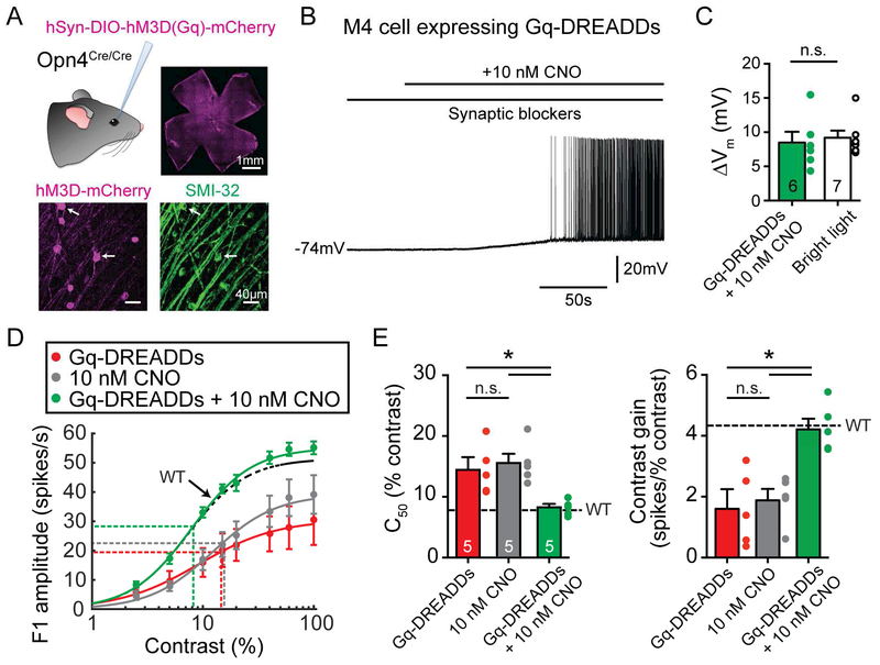
Figure 2. Activation of the Gq cascade using Gq-DREADDs rescues contrast sensitivity deficits in Opn4−/− M4 cells.
(A) Gq-DREADDs were expressed in ipRGCs of melanopsin null (Opn4Cre/Cre) mice via intravitreal injections of AAV2/hSyn-DIO-hM3D-mCherry (Gq-DREADDs). Top right panel shows successful transfection of ipRGCs across the retina. Bottom panels show mCherry immunostaining of Gq-DREADD positive ipRGCs (magenta) and SMI-32 immunostaining (green), which strongly labels alpha RGCs. White arrows indicate putative M4 cells expressing Gq-DREADDs. (B) Whole-cell current clamp recording of a Gq-DREADD-expressing M4 cell in an Opn4Cre/Cre retina exposed to 10 nM CNO in the dark in the presence of synaptic blockers. (C) 10 nM CNO elicited a membrane potential depolarization of 8.48 ± 1.58 mV in Opn4Cre/Cre M4 cells expressing Gq-DREADDs in the dark (green), similar to light-evoked depolarization of 9.20 ± 1.02 mV in WT M4 cells (white) in bright (12 log quanta/cm2/s) light (Figure 4C). (D) Contrast response functions of M4 cells under bright (12 log quanta/cm2/s) background illumination in melanopsin null (Opn4Cre/Cre) M4 cells infected with Gq-DREADDs but not exposed to CNO (red), not infected with Gq-DREADDs but exposed to 10 nM CNO (gray), or infected with DREADDs and exposed to 10 nM CNO (green). Dotted black line indicates contrast response function of WT M4 cells in bright (12 log quanta/cm2/s) background light (Figure 1B). (E) C50 and contrast gain of M4 cells in D. Black dotted line indicates WT values recorded in bright (12 log quanta/cm2/s) light from Figure 1C. All data are mean ± SEM. * P < 0.05. ** P < 0.01. n.s. not significant. CNO: clozapine-N-oxide.

