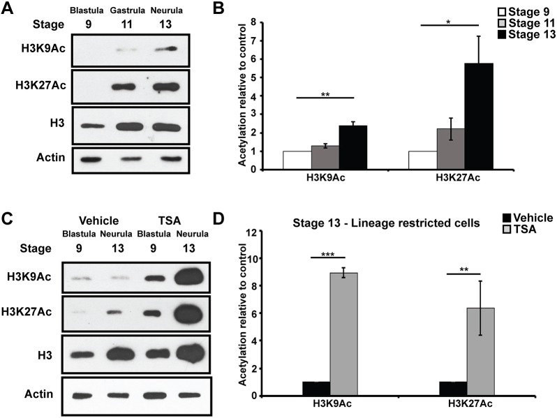
Fig. 5.
Histone acetylation increases as cells become lineage restricted. (A,B) Western blot analysis of lysates of aging animal caps examining H3K9Ac and H3K27Ac alongside total H3 levels via chemiluminescence (A) and quantified using Odyssey (B) (*P<0.05, **P<0.01). Explants were cultured alongside sibling embryos until late blastula (stage 9), mid-gastrula (stage 11) and neural plate (stage 13) stages. (C,D) Western blot analysis of lysates of aging animal caps treated with vehicle (DMSO) or inhibitor (500 nM TSA) examining H3K9Ac and H3K27Ac alongside total H3 and actin levels via chemiluminescence (C) and quantified using Odyssey (D) (**P<0.01, ***P<0.005).

