Abstract
[Purpose] To investigate changes in hamstring flexibility in relation to intensity of proprioceptive neuromuscular facilitation stretching and changes in pain over time, and examine the correlations between pain level and target intensity or flexibility gain. [Participants and Methods] Sixty-one healthy adults were randomly divided into 4 groups (100% [P100], 70% [P70], 40% [P40], and 10% [P10] of maximum voluntary isometric contraction) according to intensity of hold-relax stretching. Hamstring flexibility was measured with the active knee extension test, and pain was measured using the visual analogue scale. [Results] Concerning hamstring flexibility, P100 showed significant differences from P40 and P10, and P70 was significantly different from P10. At post-stretch, P100 significantly differed from P70, P40, and P10 in visual analogue scale. At 1 day, P100 significantly differed from P40 and P10. Although there was a significant correlation between post-stretch pain level and stretching intensity, there was no significant correlation between pain level and flexibility improvement. [Conclusion] Repetitive high-intensity stretching may cause heavy burden on muscle tissues, and pain caused by high-intensity stretching can hinder muscle performance. Moderate stretching intensity is recommended and considered conducive to maintaining the effects of stretching while ensuring its safety.
Key words: Intensity, Pain, Stretching
INTRODUCTION
Even well-trained athletes can incur injuries during exercise. To prevent such injuries and improve flexibility, stretching is recommended as a warm-up exercise. There are several types of stretching, including static, ballistic, and proprioceptive neuromuscular facilitation (PNF) stretching, and extensive research data comparing them are available. It is difficult to conclude that a specific stretching technique is superior over others, as results vary across studies depending on the study conditions. However, PNF is known to be more effective than other stretching techniques in that it increases both passive and active flexibility and improves range of motion in the short term1, 2). Multiple studies have attempted to investigate the effects of PNF in relation to duration, frequency, and repetition, to identify the ideal and most efficient stretching protocol. However, not many studies have investigated the effects of various stretching intensities and even fewer studies have examined PNF stretching requiring isometric contraction. The few studies that have investigated this issue mostly focused on the improvement of flexibility1, 3). High intensity stretching may lead to a greater increase in range of motion than low-intensity stretching by inflicting higher load on the musculotendinous units (MTUs). However, radically increasing flexibility in a short period with high-intensity stretching may excessively strain the MTUs. Intense stretching that exceeds the physiologic range and beyond the yield point may cause pain and muscle injury4, 5). Therefore, it would be important to identify the submaximal intensity that would maintain the efficacy of stretching on flexibility without posing physical burden such as pain. In the present study, we aimed to investigate the improvement in flexibility after the application of different intensities of PNF stretching and the change in pain level over time, as well as to examine how pain after stretching is correlated with individual target intensities and with flexibility improvement after stretching.
ParticipantS AND METHODS
A total of 61 adults (27 males and 34 females, age 22.4 ± 1.8 years, height 165.6 ± 14.1 cm, weight 64.4 ± 19.8 kg) were enrolled in this study. The study protocol was approved by the Institutional Review Board of Woosong University (1041549-161115-SB-34). All participants provided informed consent.
Participants were randomly divided into groups according to 4 varying intensities of PNF stretching (100% [P100], 70% [P70], 40% [P40], and 10% [P10] of the maximum voluntary isometric contraction [MVIC]) and measured the improvement in flexibility by using the active knee extension (AKE) test, and the change in pain level immediately after stretching (post-stretch). In addition, we measured the pain level at 1 day (day 1) and 2 days (day 2) after stretching to examine the changes in pain level over time. For the AKE test, the participants were placed in the supine position on the treatment table with their hip and knee joints flexed at 90°. To maintain and assist with the hip and knee position, a custom-made metal structure was placed on the table, which supported the hip flexion at 90°. Before stretching, AKE was performed 6 times as a warm-up, and the AKE value at the 6th trial was recorded as the pre-stretch value. The AKE test was performed once more after stretching, and the value was recorded as the post-stretch value.
The PNF stretching intensity was measured by connecting a wireless tension dynamometer (Re-live Inc., Kimhae, Korea) with an LCD to the sling system. The tension dynamometer was connected between the sling rope attached to the ceiling and a sling strap where the participant’s ankle was placed. The LCD provided real-time feedback to the participants to let them adjust the stretching intensity by themselves. The position of the sling was adjusted such that the sling wire hanging from the ceiling was at a 90° angle with the lower extremity of the participant. After fixing the lower extremity of the participant to the sling system, the hold-relax PNF technique was performed 3 times (10-s for each trial with 5-s rest between trials). After a sufficient rest period, each group additionally performed 6 trials of hold-relax PNF stretching at their designated stretching intensity (100%, 70%, 40%, and 10% of the MVIC), based on the mean MVIC value of the previous 3 trials.
The Kruskal-Wallis test with Dunn’s post hoc test was used to compare the visual analogue scale (VAS) scores at each time point between groups, and to compare the differences in AKE value before and after stretching (ΔAKE) between groups. The correlations between the VAS score at post-stretch and the target intensity or ΔAKE were analyzed using Spearman’s rank order correlation coefficients. Data analysis was performed using IBM SPSS Statistics 23 (IBM Corp., Armonk, NY, USA). Statistical significance was set at p<0.05 for all tests. All values are presented as the mean ± standard deviation unless otherwise noted.
RESULTS
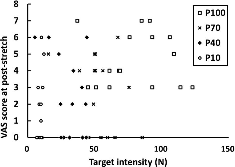
The Kruskal-Wallis test, which was used to compare VAS scores between groups, showed that the groups significantly differed at post-stretch (p=0.002) and day 1 (p=0.006) but not at day 2 (p=0.927). When analyzed in a pairwise manner, the P100 group significantly differed in VAS scores from the P70 (p=0.000), P40 (p=0.012), and P10 (p=0.036) groups at post-stretch (Table 1). On day 1, the P100 group significantly differed from the P40 (p=0.021) and P10 (p=0.001) groups only. There were no significant differences in VAS scores across groups on day 2. With regard to improvement in flexibility, the P100 group significantly differed from the P40 (p=0.004) and P10 (p<0.000) groups, whereas the P70 group significantly differed from the P10 (p=0.004) group (Table 2). The P100 group did not significantly differ from the P70 (p=0.291) group. There was a significant correlation between the pain level after stretching and the individual stretching target intensity (p=0.006); however, it was a weak positive correlation (rho=0.349) (Fig. 1). There was no significant correlation between the pain level after stretching and the improvement in flexibility.
Table 1. Changes in visual analogue scale (VAS) scores over time for each stretching intensity.
| Group | P100 (n=16) | P70 (n=15) | P40 (n=15) | P10 (n=15) |
| Post-stretch | 4.63 ± 1.63 | 2.80 ± 2.24* | 2.53 ± 2.36* | 1.47 ± 2.00* |
| Day 1 | 2.13 ± 1.89 | 1.93 ± 2.40 | 0.54 ± 0.78* | 0.27 ± 0.80* |
| Day 2 | 0.46 ± 0.88 | 0.45 ± 1.04 | 0.27 ± 0.47 | 0.33 ± 0.82 |
Values are expressed as mean ± standard deviation. *Significance against the P100 group.
Table 2. Increase in flexibility (ΔAKE) after stretching.
| Group | P100 (n=16) | P70 (n=15) | P40 (n=15) | P10 (n=15) |
| −13.88 ± 6.59 | −11.40 ± 6.50 | −6.80 ± 5.72* | −5.00 ± 4.77*,# |
ΔAKE: difference in active knee extension value before and after stretching; Values are expressed as mean ± standard deviation. *Significance against the P100 group, #Significance against the P70 group.
Fig. 1.
Correlation between individual target intensity and visual analogue scale (VAS) score.
P100, P70, P40, and P10 are groups that performed stretching at 100%, 70%, 40%, and 10% of the maximum voluntary isometric contraction, respectively.
DISCUSSION
PNF stretching is one of the most widely used stretching techniques, and its effects have been extensively documented in the literature. The concept of neuromuscular facilitation and inhibition, which is the basis of PNF, was first introduced by Sherrington in 1909, after which it has been clinically implemented by Kabat6, 7). However, the GTO, which has been believed to respond only to maximal contraction, has also been found to respond to a very low force, and it has been observed that there is very weak or no GTO activity after contraction8, 9). Furthermore, reciprocal inhibition is assumed to be realized through a complex mechanism; however, electromyography or the Hoffmann reflex method, which are widely used to identify and understand the mechanisms of reciprocal inhibition, are limited in that they are easily influenced by other factors and that they can only explain a part of the complex mechanism10). In addition to the characteristics of PNF stretching, the general features of static stretching are also involved in PNF stretching. The viscous and elastic properties of the MTUs, involving stress relaxation and elongated MTUs (creep) as a result of prolonged stretching, play an important role in increasing flexibility. However, the reduced passive resistance of the MTUs after stretching is only temporary and does not persist in the long term11).
Although the exact underlying mechanism of PNF stretching remains incompletely understood, its efficacy has been demonstrated in experimental studies. Numerous studies have investigated the efficacy of PNF stretching in relation to the duration and frequency of PNF, and several recent studies have examined the effects of PNF according to stretching intensity. Some studies found that not only high-intensity but also low-intensity stretching significantly increased flexibility with no significant differences between the 2 groups. In the present study, we assessed the pain level after stretching as well as the differences in hamstring flexibility before and after stretching in relation to stretching intensity. First, the P100 (high-intensity) group showed greater improvement of flexibility than did the P40 (low- to moderate-intensity) and P10 (low-intensity) groups, and the P70 (high- to moderate-intensity) group also showed greater improvement of flexibility than did the P10 group. In other words, in our study, high-intensity stretching led to a greater increase in flexibility. Similarly, Sheard and Paine’s study found that low-intensity stretching (20% of MVIC) led to a smaller increase of flexibility than that shown by the high-intensity group12). In some studies, there were no differences in flexibility improvement between 20% and 100% of MVIC1, 3). Although most previous studies set low-intensity stretching to 20% of MVIC, we set the low-intensity group to 10% of MVIC, maybe the minimum intensity that can cause a significant increase. Our results showed that high-intensity stretching was superior in terms of increasing flexibility; however, there were some concerns about the increase in pain level as determined using the VAS. The P100 group showed significantly higher VAS scores than the P70, P40, and P10 groups immediately after stretching, and still significantly higher VAS scores than those of the P40 and P10 groups even on day 1 after stretching. In contrast, the P70 group did not show a significant difference in pain level from the P40 or P10 groups. Furthermore, all participants in the P100 (100%) group complained of pain (VAS score>0) at post-stretch, whereas a considerable number of participants in the P70 (33.33%), P40 (26.67%), and P10 (56.25%) groups did not complain of pain (VAS score 0) after stretching. Considering that high-intensity stretching (P100) caused pain that persisted until the day after stretching, repeated high-intensity stretching before recovery may lead to muscle tissue injury4). Excessively intense stretching exceeding the physiological limit would induce microtears in muscle tissues, which may cause pain. In fact, T2-weighted magnetic resonance images related to inflammation showed an elevation of signal intensity after intense eccentric or isometric exercise that inflicts high load on the MTUs13, 14).
The VAS scores were weakly correlated with the individual target intensities but were not correlated with flexibility changes. In other words, high-intensity stretching may cause pain, but severe pain during stretching does not translate to an increase in flexibility. Among various scales that are clinically used to measure pain, VAS is superior in terms of its psychometric characteristic and quick and easy to measure. Thus it is preferred over other scales15). However, the fact that this scale can be influenced by the subjective characteristics of participants must not be neglected. Since the study by Zborowski16), there has been a large research interest in pain from a sociological perspective. Pain can be classified into different types (e.g., cold-pressor, contact heat, acute post-surgical pain, and low back pain); however, those types also have individual differences. For instance, pain threshold and pain tolerance may differ with race, gender, genetic factors, and environmental factors17). Perceived pain may actually differ from the actual degree of pain18), and there is always a chance that the pain level may be overestimated or underestimated depending on the characteristics of the person feeling the pain. Overall, pain level was significantly higher in the high-intensity group; however, the pain level measured in our experiment would encompass other factors in addition to the actual load delivered to the MTUs of the hamstring.
PNF is often performed at high intensity in clinical practice in order to maximize the improvement in flexibility. However, when repeatedly performed, it may induce muscle tissue injury. As shown in our experiment, high-intensity stretching can cause severe pain in muscle tissue, and this may cause discomfort during exercise and consequently hinder muscle performance. Moderate-intensity stretching is recommended to maintain the effects of stretching while ensuring its safety.
Funding
This research was supported by 2018 Woosong University Academic Research Funding and a Basic Science Research Program through the National Research Foundation of Korea (NRF) funded by the Ministry of Education (NRF-2017R1C1B5076885).
Conflict of interest
The author has no conflicts of interest to disclose.
REFERENCES
- 1.Feland JB, Marin HN: Effect of submaximal contraction intensity in contract-relax proprioceptive neuromuscular facilitation stretching. Br J Sports Med, 2004, 38: E18. [DOI] [PMC free article] [PubMed] [Google Scholar]
- 2.Feland JB, Myrer JW, Merrill RM: Acute changes in hamstring flexibility: PNF versus static stretch in senior athletes. Phys Ther Sport, 2001, 2: 186–193. [Google Scholar]
- 3.Khodayari B, Dehghani Y: The investigation of mid-term effect of different intensity of PNF stretching on improve hamstring flexibility, 4th World Conference on Educational Sciences (WCES-2012) 02–05 February 2012 Barcelona, Spain. Procedia Soc Behav Sci, 2012, 46: 5741–5744. [Google Scholar]
- 4.Askling C, Lund H, Saartok T, et al. : Self-reported hamstring injuries in student-dancers. Scand J Med Sci Sports, 2002, 12: 230–235. [DOI] [PubMed] [Google Scholar]
- 5.Kim JW, Kwon OY, Kim MH: Differentially expressed genes and morphological changes during lengthened immobilization in rat soleus muscle. Differentiation, 2007, 75: 147–157. [DOI] [PubMed] [Google Scholar]
- 6.Kabat H: Studies of neuromuscular dysfunction; treatment of chronic multiple sclerosis with neostigmine and intensive muscle re-education. Perm Found Med Bull, 1947, 5: 1–14. [PubMed] [Google Scholar]
- 7.Sherrington CS: On plastic tonus and proprioceptive reflexes. Exp Physiol, 1909, 2: 109–156. [Google Scholar]
- 8.Smith JL, Hutton RS, Eldred E: Postcontraction changes in sensitivity of muscle afferents to static and dynamic stretch. Brain Res, 1974, 78: 193–202. [DOI] [PubMed] [Google Scholar]
- 9.Gollhofer A, Schöpp A, Rapp W, et al. : Changes in reflex excitability following isometric contraction in humans. Eur J Appl Physiol Occup Physiol, 1998, 77: 89–97. [DOI] [PubMed] [Google Scholar]
- 10.Zehr EP: Considerations for use of the Hoffmann reflex in exercise studies. Eur J Appl Physiol, 2002, 86: 455–468. [DOI] [PubMed] [Google Scholar]
- 11.Magnusson SP, Simonsen EB, Aagaard P, et al. : Mechanical and physical responses to stretching with and without preisometric contraction in human skeletal muscle. Arch Phys Med Rehabil, 1996, 77: 373–378. [DOI] [PubMed] [Google Scholar]
- 12.Sheard PW, Paine TJ: Optimal contraction intensity during proprioceptive neuromuscular facilitation for maximal increase of range of motion. J Strength Cond Res, 2010, 24: 416–421. [DOI] [PubMed] [Google Scholar]
- 13.Foley JM, Jayaraman RC, Prior BM, et al. : MR measurements of muscle damage and adaptation after eccentric exercise. J Appl Physiol 1985, 1999, 87: 2311–2318. [DOI] [PubMed] [Google Scholar]
- 14.Weidman ER, Charles HC, Negro-Vilar R, et al. : Muscle activity localization with 31P spectroscopy and calculated T2-weighted 1H images. Invest Radiol, 1991, 26: 309–316. [DOI] [PubMed] [Google Scholar]
- 15.Price DD, Bush FM, Long S, et al. : A comparison of pain measurement characteristics of mechanical visual analogue and simple numerical rating scales. Pain, 1994, 56: 217–226. [DOI] [PubMed] [Google Scholar]
- 16.Zborowski M: People in pain. San Francisco: Jossey-Bass, 1969. [Google Scholar]
- 17.Nielsen CS, Stubhaug A, Price DD, et al. : Individual differences in pain sensitivity: genetic and environmental contributions. Pain, 2008, 136: 21–29. [DOI] [PubMed] [Google Scholar]
- 18.Lim W, Park H: No significant correlation between the intensity of static stretching and subject’s perception of pain. J Phys Ther Sci, 2017, 29: 1856–1859. [DOI] [PMC free article] [PubMed] [Google Scholar]


